Schwann cell

Last updated
Schwann cell
1210 Glial Cells of the PNS.jpg
The PNS has satellite cells and Schwann cells.
Identifiers
MeSH D012583
FMA 62121
Anatomical terms of neuroanatomy

Schwann cells or neurolemmocytes (named after German physiologist Theodor Schwann) are the principal glia of the peripheral nervous system (PNS). Glial cells function to support neurons and in the PNS, also include satellite cells, olfactory ensheathing cells, enteric glia and glia that reside at sensory nerve endings, such as the Pacinian corpuscle. The two types of Schwann cells are myelinating and nonmyelinating. [1] Myelinating Schwann cells wrap around axons of motor and sensory neurons to form the myelin sheath. The Schwann cell promoter is present in the downstream region of the human dystrophin gene that gives shortened transcript that are again synthesized in a tissue-specific manner.

Contents

During the development of the PNS, the regulatory mechanisms of myelination are controlled by feedforward interaction of specific genes, influencing transcriptional cascades and shaping the morphology of the myelinated nerve fibers. [2]

Schwann cells are involved in many important aspects of peripheral nerve biology—the conduction of nervous impulses along axons, nerve development and regeneration, trophic support for neurons, production of the nerve extracellular matrix, modulation of neuromuscular synaptic activity, and presentation of antigens to T-lymphocytes.

Charcot–Marie–Tooth disease, Guillain–Barré syndrome (acute inflammatory demyelinating polyradiculopathy type), schwannomatosis, chronic inflammatory demyelinating polyneuropathy, and leprosy are all neuropathies involving Schwann cells.

Structure

Schwann cells wrapped around an axon

Schwann cells are a variety of glial cells that keep peripheral nerve fibres (both myelinated and unmyelinated) alive. In myelinated axons, Schwann cells form the myelin sheath. The sheath is not continuous. Individual myelinating Schwann cells cover about 1 mm of an axon [3] —equating to about 1000 Schwann cells along a 1-m length of the axon. The gaps between adjacent Schwann cells are called nodes of Ranvier.

9-O-Acetyl GD3 ganglioside is an acetylated glycolipid which is found in the cell membranes of many types of vertebrate cells. During peripheral nerve regeneration, 9-O-acetyl GD3 is expressed by Schwann cells. [4]

Function

The vertebrate nervous system relies on the myelin sheath for insulation and as a method of decreasing membrane capacitance in the axon. The action potential jumps from node to node, in a process called saltatory conduction, which can increase conduction velocity up to 10 times, without an increase in axonal diameter. In this sense, Schwann cells are the PNS's analogues of the central nervous system's oligodendrocytes. However, unlike oligodendrocytes, each myelinating Schwann cell provides insulation to only one axon (see image). This arrangement permits saltatory conduction of action potentials with repropagation at the nodes of Ranvier. In this way, myelination greatly increases speed of conduction and saves energy. [5]

Nonmyelinating Schwann cells are involved in maintenance of axons and are crucial for neuronal survival. Some group around smaller axons (External image here) and form Remak bundles.

Myelinating Schwann cells begin to form the myelin sheath in mammals during fetal development and work by spiraling around the axon, sometimes with as many as 100 revolutions. A well-developed Schwann cell is shaped like a rolled-up sheet of paper, with layers of myelin between each coil. The inner layers of the wrapping, which are predominantly membrane material, form the myelin sheath, while the outermost layer of nucleated cytoplasm forms the neurilemma. Only a small volume of residual cytoplasm allows communication between the inner and outer layers. This is seen histologically as the Schmidt-Lantermann incisure.

Regeneration

Schwann cells are known for their roles in supporting nerve regeneration. [6] Nerves in the PNS consist of many axons myelinated by Schwann cells. If damage occurs to a nerve, the Schwann cells aid in digestion of its axons (phagocytosis). Following this process, the Schwann cells can guide regeneration by forming a type of tunnel that leads toward the target neurons. This tunnel is known as band of Büngner, a guidance track for the regenerating axons, which behaves like an endoneural tube. The stump of the damaged axon is able to sprout, and those sprouts that grow through the Schwann-cell "tunnel" do so at the rate around 1 mm/day in good conditions. The rate of regeneration decreases with time. Successful axons can, therefore, reconnect with the muscles or organs they previously controlled with the help of Schwann cells, but specificity is not maintained and errors are frequent, especially when long distances are involved. [7] Because of their ability to impact regeneration of axons, Schwann cells have been connected to preferential motor reinnervation, as well. If Schwann cells are prevented from associating with axons, the axons die. Regenerating axons will not reach any target unless Schwann cells are there to support them and guide them. They have been shown to be in advance of the growth cones.

Schwann cells are essential for the maintenance of healthy axons. They produce a variety of factors, including neurotrophins, and also transfer essential molecules across to axons.

A Schwann cell in culture. Cultured schwann cell.jpg
A Schwann cell in culture.

Genetics

Schwann cell formation

Sox10

SOX10 is a transcription factor active during embryonic development and abundant evidence indicates that it is essential for the generation of glial lineages from trunk crest cells. [8] [9] When SOX10 is inactivated in mice, satellite glia and Schwann cell precursors fail to develop, though neurons are generated normally without issue. [8] In the absence of SOX10, neural crest cells survive and are free to generate neurons, but glial specification is blocked. [9] SOX10 might influence early glial precursors to respond to neuregulin 1 [8] (see below).

Neuregulin 1

Neuregulin 1 (NRG1) acts in a number of ways to both promote the formation and ensure the survival of immature Schwann cells. [10] During embryonic development, NRG1 inhibits the formation of neurons from neural crest cells, instead contributing to neural crest cells being led down a path to gliogenesis. NRG1 signaling is not, however, required for glial differentiation from the neural crest. [11]

NRG1 plays important roles in the development of neural crest derivatives. It is required for neural crest cells to migrate past the site of dorsal root ganglia to find the ventral regions of sympathetic gangliogenesis. [12] It is also an essential axon-derived survival factor and a mitogen for Schwann cell precursors. [13] It is found in the dorsal root ganglion and motor neurons at the point in time that Schwann cell precursors begin to populate spinal nerves and therefore influences Schwann cell survival. [11] In embryonic nerves, the transmembrane III isoform likely is the primary variant of NRG1 responsible for survival signals. In mice that lack the transmembrane III isoform, Schwann cell precursors are eventually eliminated from spinal nerves. [14]

Formation of myelin sheath

P0

Myelin protein zero (P0) is a cell-adhesion molecule belonging to the immunoglobulin superfamily and is the major component of peripheral myelin, constituting over 50% of the total protein in the sheath. [15] [16] P0 has been shown to be essential for the formation of compact myelin, as P0 null mutant (P0-) mice showed severely aberrant peripheral myelination. [17] Although myelination of large caliber axons was initiated in P0- mice, the resulting myelin layers were very thin and poorly compacted. Unexpectedly, P0- mice also showed degeneration of both axons and their surround myelin sheaths, suggesting that P0 plays a role in maintaining the structural integrity of both myelin formation and the axon with which it is associated. P0- mice developed behavioral deficits around 2 weeks of age when mice began to show signs of slight trembling. Gross incoordination also arose as the animals developed, while trembling became more severe and some older mice developed convulsing behaviors. Despite the array of impaired motor behavior, no paralysis was observed in these animals. P0 is also an important gene expressed early within the Schwann cell lineage, expressed in Schwann cell precursors after differentiating from migrating neural crest cells within the developing embryo. [18]

Krox-20

Several important transcription factors are also expressed and involved at various stages in development changing the features on the Schwann cells from an immature to mature state. One indispensable transcription factor expressed during the myelination process is Krox-20. It is a general zinc-finger transcription factor and is expressed in the rhombomeres 3 and 5.

Krox-20 is considered one of the master regulators of PNS myelination and is important in driving transcription of specific structural proteins in the myelin. It has been shown to control a set of genes responsible for interfering with this feature in the axon changing it from a pro-myelinating to myelinating state. [19] In this way, in Krox-20 double knock out mice, it has been recorded that hindbrain segmentation is affected as well as myelination of Schwann cell associated axons. Indeed, in these mice, the Schwann cells are not able to perform their myelination properly as they only wrap their cytoplasmic processes one and half turn around the axon and despite the fact that they still express the early myelin marker, late myelin gene products are absent. In addition, recent studies have also proven the importance of this transcription factor in maintaining the myelination phenotype (and requires the co-expression of Sox 10) as its inactivation leads to dedifferentiation of the Schwann cells. [2]

Clinical significance

Charcot–Marie–Tooth disease (CMT), Guillain–Barré syndrome (GBS, acute inflammatory demyelinating polyradiculopathy type), schwannomatosis, and chronic inflammatory demyelinating polyneuropathy (CIDP), leprosy, and Zika Virus are all neuropathies involving Schwann cells. [20]

Transplantation

A number of experimental studies since 2001 have implanted Schwann cells in an attempt to induce remyelination in multiple sclerosis-afflicted patients. [21] In the past two decades, many studies have demonstrated positive results and potential for Schwann cell transplantation as a therapy for spinal cord injury, both in aiding regrowth and myelination of damaged CNS axons. [22] Schwann cell transplants in combination with other therapies such as Chondroitinase ABC have also been shown to be effective in functional recovery from spinal cord injury. [23]

See also

Related Research Articles

<span class="mw-page-title-main">Axon</span> Long projection on a neuron that conducts signals to other neurons

An axon or nerve fiber is a long, slender projection of a nerve cell, or neuron, in vertebrates, that typically conducts electrical impulses known as action potentials away from the nerve cell body. The function of the axon is to transmit information to different neurons, muscles, and glands. In certain sensory neurons, such as those for touch and warmth, the axons are called afferent nerve fibers and the electrical impulse travels along these from the periphery to the cell body and from the cell body to the spinal cord along another branch of the same axon. Axon dysfunction can be the cause of many inherited and acquired neurological disorders that affect both the peripheral and central neurons. Nerve fibers are classed into three types – group A nerve fibers, group B nerve fibers, and group C nerve fibers. Groups A and B are myelinated, and group C are unmyelinated. These groups include both sensory fibers and motor fibers. Another classification groups only the sensory fibers as Type I, Type II, Type III, and Type IV.

<span class="mw-page-title-main">Central nervous system</span> Brain and spinal cord

The central nervous system (CNS) is the part of the nervous system consisting of the brain and spinal cord, the retina and optic nerve, and the olfactory nerve and epithelia. The CNS is so named because the brain integrates the received information and coordinates and influences the activity of all parts of the bodies of bilaterally symmetric and triploblastic animals—that is, all multicellular animals except sponges and diploblasts. It is a structure composed of nervous tissue positioned along the rostral to caudal axis of the body and may have an enlarged section at the rostral end which is a brain. Only arthropods, cephalopods and vertebrates have a true brain, though precursor structures exist in onychophorans, gastropods and lancelets.

<span class="mw-page-title-main">Myelin</span> Fatty substance that surrounds nerve cell axons to insulate them and increase transmission speed

Myelin is a lipid-rich material that surrounds nerve cell axons to insulate them and increase the rate at which electrical impulses pass along the axon. The myelinated axon can be likened to an electrical wire with insulating material (myelin) around it. However, unlike the plastic covering on an electrical wire, myelin does not form a single long sheath over the entire length of the axon. Rather, myelin ensheaths the axon segmentally: in general, each axon is encased in multiple long sheaths with short gaps between, called nodes of Ranvier. At the nodes of Ranvier, which are approximately one thousandth of a mm in length, the axon's membrane is bare of myelin.

<span class="mw-page-title-main">Nervous tissue</span> Main component of the nervous system

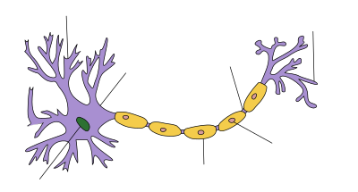
Nervous tissue, also called neural tissue, is the main tissue component of the nervous system. The nervous system regulates and controls body functions and activity. It consists of two parts: the central nervous system (CNS) comprising the brain and spinal cord, and the peripheral nervous system (PNS) comprising the branching peripheral nerves. It is composed of neurons, also known as nerve cells, which receive and transmit impulses, and neuroglia, also known as glial cells or glia, which assist the propagation of the nerve impulse as well as provide nutrients to the neurons.

<span class="mw-page-title-main">Oligodendrocyte</span> Neural cell type

Oligodendrocytes, also known as oligodendroglia, are a type of neuroglia whose main functions are to provide support and insulation to axons within the central nervous system (CNS) of jawed vertebrates. Their function is similar to that of Schwann cells, which perform the same task in the peripheral nervous system (PNS). Oligodendrocytes accomplish this by forming the myelin sheath around axons. Unlike Schwann cells, a single oligodendrocyte can extend its processes to cover around 50 axons, with each axon being wrapped in approximately 1 μm of myelin sheath. Furthermore, an oligodendrocyte can provide myelin segments for multiple adjacent axons.

<span class="mw-page-title-main">Glia</span> Support cells in the nervous system

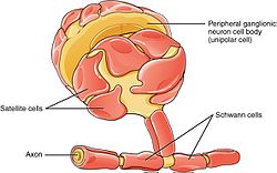
Glia, also called glial cells (gliocytes) or neuroglia, are non-neuronal cells in the central nervous system and the peripheral nervous system that do not produce electrical impulses. The neuroglia make up more than one half the volume of neural tissue in our body. They maintain homeostasis, form myelin in the peripheral nervous system, and provide support and protection for neurons. In the central nervous system, glial cells include oligodendrocytes, astrocytes, ependymal cells and microglia, and in the peripheral nervous system they include Schwann cells and satellite cells.

<span class="mw-page-title-main">Node of Ranvier</span> Gaps between myelin sheaths on the axon of a neuron

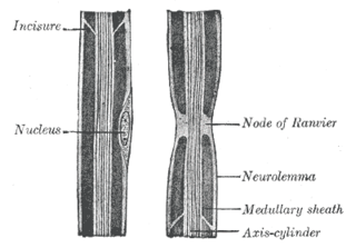
In neuroscience and anatomy, nodes of Ranvier, also known as myelin-sheath gaps, occur along a myelinated axon where the axolemma is exposed to the extracellular space. Nodes of Ranvier are uninsulated and highly enriched in ion channels, allowing them to participate in the exchange of ions required to regenerate the action potential. Nerve conduction in myelinated axons is referred to as saltatory conduction due to the manner in which the action potential seems to "jump" from one node to the next along the axon. This results in faster conduction of the action potential.

<span class="mw-page-title-main">Wallerian degeneration</span> Biological process of axonal degeneration

Wallerian degeneration is an active process of degeneration that results when a nerve fiber is cut or crushed and the part of the axon distal to the injury degenerates. A related process of dying back or retrograde degeneration known as 'Wallerian-like degeneration' occurs in many neurodegenerative diseases, especially those where axonal transport is impaired such as ALS and Alzheimer's disease. Primary culture studies suggest that a failure to deliver sufficient quantities of the essential axonal protein NMNAT2 is a key initiating event.

<span class="mw-page-title-main">Myelin incisure</span>

Myelin incisures are small pockets of cytoplasm left behind during the Schwann cell myelination process.

<span class="mw-page-title-main">Myelin-associated glycoprotein</span> Protein-coding gene in the species Homo sapiens

Myelin-associated glycoprotein is a type 1 transmembrane protein glycoprotein localized in periaxonal Schwann cell and oligodendrocyte membranes, where it plays a role in glial-axonal interactions. MAG is a member of the SIGLEC family of proteins and is a functional ligand of the NOGO-66 receptor, NgR. MAG is believed to be involved in myelination during nerve regeneration in the PNS and is vital for the long-term survival of the myelinated axons following myelinogenesis. In the CNS MAG is one of three main myelin-associated inhibitors of axonal regeneration after injury, making it an important protein for future research on neurogenesis in the CNS.

<span class="mw-page-title-main">Satellite glial cell</span> SINGLE CELL SOMATA

Satellite glial cells, formerly called amphicytes, are glial cells that cover the surface of neuron cell bodies in ganglia of the peripheral nervous system. Thus, they are found in sensory, sympathetic, and parasympathetic ganglia. Both satellite glial cells (SGCs) and Schwann cells are derived from the neural crest of the embryo during development. SGCs have been found to play a variety of roles, including control over the microenvironment of sympathetic ganglia. They are thought to have a similar role to astrocytes in the central nervous system (CNS). They supply nutrients to the surrounding neurons and also have some structural function. Satellite cells also act as protective, cushioning cells. Additionally, they express a variety of receptors that allow for a range of interactions with neuroactive chemicals. Many of these receptors and other ion channels have recently been implicated in health issues including chronic pain and herpes simplex. There is much more to be learned about these cells, and research surrounding additional properties and roles of the SGCs is ongoing.

<span class="mw-page-title-main">Nerve injury</span> Damage to nervous tissue

Nerve injury is an injury to a nerve. There is no single classification system that can describe all the many variations of nerve injuries. In 1941, Seddon introduced a classification of nerve injuries based on three main types of nerve fiber injury and whether there is continuity of the nerve. Usually, however, nerve injuries are classified in five stages, based on the extent of damage to both the nerve and the surrounding connective tissue, since supporting glial cells may be involved.

Neuroregeneration involves the regrowth or repair of nervous tissues, cells or cell products. Neuroregenerative mechanisms may include generation of new neurons, glia, axons, myelin, or synapses. Neuroregeneration differs between the peripheral nervous system (PNS) and the central nervous system (CNS) by the functional mechanisms involved, especially in the extent and speed of repair. When an axon is damaged, the distal segment undergoes Wallerian degeneration, losing its myelin sheath. The proximal segment can either die by apoptosis or undergo the chromatolytic reaction, which is an attempt at repair. In the CNS, synaptic stripping occurs as glial foot processes invade the dead synapse.

<span class="mw-page-title-main">Neuregulin 1</span> Protein-coding gene in the species Homo sapiens

Neuregulin 1, or NRG1, is a gene of the epidermal growth factor family that in humans is encoded by the NRG1 gene. NRG1 is one of four proteins in the neuregulin family that act on the EGFR family of receptors. Neuregulin 1 is produced in numerous isoforms by alternative splicing, which allows it to perform a wide variety of functions. It is essential for the normal development of the nervous system and the heart.

Myelinogenesis is the formation and development of myelin sheaths in the nervous system, typically initiated in late prenatal neurodevelopment and continuing throughout postnatal development. Myelinogenesis continues throughout the lifespan to support learning and memory via neural circuit plasticity as well as remyelination following injury. Successful myelination of axons increases action potential speed by enabling saltatory conduction, which is essential for timely signal conduction between spatially separate brain regions, as well as provides metabolic support to neurons.

<span class="mw-page-title-main">Olfactory ensheathing cell</span> Type of macroglia that ensheath unmyelinated olfactory neurons

Olfactory ensheathing cells (OECs), also known as olfactory ensheathing glia or olfactory ensheathing glial cells, are a type of macroglia found in the nervous system. They are also known as olfactory Schwann cells, because they ensheath the non-myelinated axons of olfactory neurons in a similar way to which Schwann cells ensheath non-myelinated peripheral neurons. They also share the property of assisting axonal regeneration.

Anti-MAG peripheral neuropathy is a specific type of peripheral neuropathy in which the person's own immune system attacks cells that are specific in maintaining a healthy nervous system. As these cells are destroyed by antibodies, the nerve cells in the surrounding region begin to lose function and create many problems in both sensory and motor function. Specifically, antibodies against myelin-associated glycoprotein (MAG) damage Schwann cells. While the disorder occurs in only 10% of those afflicted with peripheral neuropathy, people afflicted have symptoms such as muscle weakness, sensory problems, and other motor deficits usually starting in the form of a tremor of the hands or trouble walking. There are, however, multiple treatments that range from simple exercises in order to build strength to targeted drug treatments that have been shown to improve function in people with this type of peripheral neuropathy.

Perisynaptic schwann cells are neuroglia found at the Neuromuscular junction (NMJ) with known functions in synaptic transmission, synaptogenesis, and nerve regeneration. These cells share a common ancestor with both Myelinating and Non-Myelinating Schwann Cells called Neural Crest cells. Perisynaptic Schwann Cells (PSCs) contribute to the tripartite synapse organization in combination with the pre-synaptic nerve and the post-synaptic muscle fiber. PSCs are considered to be the glial component of the Neuromuscular Junction (NMJ) and have a similar functionality to that of Astrocytes in the Central Nervous System. The characteristics of PSCs are based on both external synaptic properties and internal glial properties, where the internal characteristics of PSCs develop based on the associated synapse, for example: the PSCs of a fast-twitch muscle fiber differ from the PSCs of a slow-twitch muscle fiber even when removed from their natural synaptic environment. PSCs of fast-twitch muscle fibers have higher Calcium levels in response to synapse innervation when compared to slow-twitch PSCs. This balance between external and internal influences creates a range of PSCs that are present in the many Neuromuscular Junctions of the Peripheral Nervous System.

Preferential motor reinnervation (PMR) refers to the tendency of a regenerating axon in the peripheral nervous system (PNS) to reinnervate a motor pathway as opposed to a somatosensory pathway. PMR affects how nerves regenerate and reinnervate within the PNS after surgical procedures or traumatic injuries. It is important to understand in order to further develop axonal regrowth surgical techniques. Further research of preferential motor reinnervation will lead to a better understanding of peripheral nervous system function in the human body regarding cell roles and abilities.

<span class="mw-page-title-main">Outline of the human nervous system</span> Overview of and topical guide to the human nervous system

The following diagram is provided as an overview of and topical guide to the human nervous system:

References

  1. Bhatheja, K; Field, J (2006). "Schwann cells: origins and role in axonal maintenance and regeneration". The International Journal of Biochemistry & Cell Biology. 38 (12): 1995–9. doi:10.1016/j.biocel.2006.05.007. PMID   16807057.
  2. 1 2 Topilko, Piotr; Schneider-Maunoury, Sylvie; Levi, Giovanni; Baron-Van Evercooren, Anne; Chennoufi, Amina Ben Younes; Seitanidou, Tania; Babinet, Charles; Charnay, Patrick (1994-10-27). "Krox-20 controls myelination in the peripheral nervous system". Nature. 371 (6500): 796–799. Bibcode:1994Natur.371..796T. doi:10.1038/371796a0. PMID   7935840. S2CID   4333028.
  3. Tortora, Gerard J. (2017). Principles of Anatomy and Physiology (15th ed.). USA: Wiley. p. 412. ISBN   978-1-119-32064-7.
  4. Túlio Ribeiro-Resende, Victor; Lopes, Michelle (2010). "Involvement of 9-O-Acetyl GD3 Ganglioside in Mycobacterium leprae Infection of Schwann Cells". J. Biol. Chem. 285 (44): 34086–34096. doi: 10.1074/jbc.M110.147272 . PMC   2962507 . PMID   20739294.
  5. Kalat, James W. Biological Psychology, 9th ed. USA: Thompson Learning, 2007.[ page needed ]
  6. Bhatheja, Kanav; Field, Jeffrey (2006). "Schwann cells: Origins and role in axonal maintenance and regeneration". The International Journal of Biochemistry & Cell Biology. 38 (12): 1995–9. doi:10.1016/j.biocel.2006.05.007. PMID   16807057.
  7. Carlson, Neil R. Physiology of Behavior, 9th ed. USA: Pearson Education, Inc., 2007.[ page needed ]
  8. 1 2 3 Britisch, S.; et al. (2001). "The transcription factor Sox10 is a key regulator of peripheral glial development". Genes Dev. 15 (1): 66–78. doi:10.1101/gad.186601. PMC   312607 . PMID   11156606.
  9. 1 2 Paratore, C., Goerich, D. E., Suter, U., Wegner, M. & Sommer, L. (2001). "Survival and glial fate acquisition of neural crest cells are regulated by an interplay between the transcription factor Sox10 and extrinsic combinatorial signalling". Development. 128 (20): 3949–3961. doi:10.1242/dev.128.20.3949. PMID   11641219.{{cite journal}}: CS1 maint: multiple names: authors list (link)
  10. Shah, N. M.; et al. (1994). "Glial growth factor restricts mammalian neural crest stem cells to glial fate". Cell. 77 (3): 349–360. doi:10.1016/0092-8674(94)90150-3. PMID   7910115. S2CID   20297598.
  11. 1 2 Jessen, K. R. & Misky, R. (2005). "The origin and development of glial cells in peripheral nerves". Nature Reviews Neuroscience. 6 (9): 671–682. doi:10.1038/nrn1746. PMID   16136171. S2CID   7540462.
  12. Britisch, S.; et al. (1998). "The ErbB2 and ErbB3 receptors and their ligand, neuregulin-1 are essential for development of the sympathetic nervous system". Genes Dev. 12 (12): 1825–1836. doi:10.1101/gad.12.12.1825. PMC   316903 . PMID   9637684.
  13. Dong, Z.; et al. (1995). "NDF is a neuron-glia signal and regulates survival, proliferation, and maturation of rat Schwann cell precursors". Neuron. 15 (3): 585–596. doi: 10.1016/0896-6273(95)90147-7 . PMID   7546738. S2CID   15332720.
  14. Wolpowitz, D.; et al. (2000). "Cysteine-rich domain isoforms of the neuregulin-1 gene are required for maintenance of peripheral synapses". Neuron. 25 (1): 79–91. doi: 10.1016/s0896-6273(00)80873-9 . PMID   10707974. S2CID   16187922.
  15. Greenfield, S.; Brostoff, S.; Eylar, E. H.; Morell, P. (1973). "Protein composition of myelin of the peripheral nervous system". Journal of Neurochemistry. 20 (4): 1207–1216. doi:10.1111/j.1471-4159.1973.tb00089.x. PMID   4697881. S2CID   30385476.
  16. Lemke, G. (1988). "Unwrapping the genes of myelin". Neuron. 1 (7): 535–543. doi:10.1016/0896-6273(88)90103-1. PMID   2483101. S2CID   27086229.
  17. Geise, K.; Martini, R.; Lemke, G; Soriano, P.; Schachner, M. (1992). "Mouse P0 Gene Disruption Leads to Hypomyelination, Abnormal Expression of Recognition Molecules, and Degeneration of Myelin and Axons". Cell. 71 (4): 565–576. doi:10.1016/0092-8674(92)90591-y. PMID   1384988. S2CID   41878912.
  18. Jessen, K.; Mirsky, R. (2005). "The origin and development of glial cells in peripheral nerves". Nature Reviews Neuroscience. 6 (9): 671–682. doi:10.1038/nrn1746. PMID   16136171. S2CID   7540462.
  19. Salzer, James (2015). "Schwann cell myelination". Cold Spring Harbor Perspectives in Biology. 7 (8): a020529. doi:10.1101/cshperspect.a020529. PMC   4526746 . PMID   26054742.
  20. Dhiman, Gaurav; Abraham, R.; Griffin, D. (2019). "Human Schwann cells are susceptible to infection with Zika and yellow fever viruses, but not dengue virus". Scientific Reports. 9 (1): 9951. Bibcode:2019NatSR...9.9951D. doi:10.1038/s41598-019-46389-0. PMC   6616448 . PMID   31289325.
  21. "First surgical transplant attempted to repair myelin". Inside MS. 2001. Archived from the original on 2007-03-11.
  22. Oudega, Martin; Xu, Xiao-Ming (2006). "Schwann Cell Transplantation for Repair of the Adult Spinal Cord". Journal of Neurotrauma. 23 (3–4): 453–67. doi:10.1089/neu.2006.23.453. PMID   16629629.
  23. Fouad, Karim; Lisa Schnell; Mary B. Bunge; Martin E. Schwab; Thomas Liebscher; Damien D. Pearse (2 February 2005). "Combining Schwann Cell Bridges and Olfactory-Ensheathing Glia Grafts with Chondroitinase Promotes Locomotor Recovery after Complete Transection of the Spinal Cord". The Journal of Neuroscience. 25 (5): 1169–78. doi: 10.1523/JNEUROSCI.3562-04.2005 . PMC   6725952 . PMID   15689553.