Group C nerve fiber

Last updated

Group C nerve fiber
Medulla spinalis - Substantia grisea - English.svg
C fiber not labeled, but substantia gelatinosa of Rolando is Rexed lamina II, labeled at upper left.
Details
Location Central nervous system and peripheral nervous system
Function nerve fiber
Anatomical terms of neuroanatomy

Group C nerve fibers are one of three classes of nerve fiber in the central nervous system (CNS) and peripheral nervous system (PNS). The C group fibers are unmyelinated and have a small diameter and low conduction velocity, whereas Groups A and B are myelinated. Group C fibers include postganglionic fibers in the autonomic nervous system (ANS), and nerve fibers at the dorsal roots (IV fiber). These fibers carry sensory information.

Contents

Damage or injury to nerve fibers causes neuropathic pain. Capsaicin activates C fibre vanilloid receptors, giving chili peppers a hot sensation.

Structure and anatomy

Location

C fibers are one class of nerve fiber found in the nerves of the somatic sensory system. [1] They are afferent fibers, conveying input signals from the periphery to the central nervous system. [2]

Structure

C fibers are unmyelinated unlike most other fibers in the nervous system. [1] This lack of myelination is the cause of their slow conduction velocity, which is on the order of no more than 2  m/s . [1] C fibers are on average 0.2-1.5 μm in diameter.

Remak bundles

C fiber axons are grouped together into what is known as Remak bundles. [3] These occur when a non-myelinating Schwann cell bundles the axons close together by surrounding them. [4] The Schwann cell keeps them from touching each other by squeezing its cytoplasm between the axons. [4] The condition of Remak bundles varies with age. [4] The number of C fiber axons in each Remak bundle varies with location. [3] For example, in a rat model, large bundles of greater than 20 axons are found exiting the L5 dorsal root ganglion, while smaller bundles of average 3 axons are found in distal nerve segments. [3] Multiple neurons contribute axons to the Remak bundle with an average ratio of about 2 axons contributed per bundle. [3] The cross sectional area of a Remak bundle is proportional to the number of axons found inside it. [3] Remak bundles in the distal peripheral nerve are clustered with other Remak bundles. [3] The Remak Schwann cells have been shown to be electrochemically responsive to action potentials of the axons contained within them. [3]

In experiments where nerve injury is caused but nearby C fibers remain intact, increased spontaneous activity in the C fibers is observed. [3] This phenomenon supports the theory that damaged nerve fibers may release factors that alter the function of neighboring undamaged fibers. [3] Study of Remak bundles has important implications in nerve regeneration after sustaining injury. [3] Currently, recovery of distal C fiber function takes months and may still only regain incomplete function. [3] This may result in abnormal sensory function or neuropathic pain. [3] Remak bundles are thought to release certain trophic factors that promote the regeneration of the damaged axons. [3]

Pathway

C fibers synapse to second-order projection neurons in the spinal cord at the upper laminae of the dorsal horn in the substantia gelatinosa. [5] The second-order projection neurons are of the wide dynamic range (WDR) type, which receive input from both nociceptive terminals as well as myelinated A-type fibers. [5] There are three types of second order projection neurons in the spinothalamic tract: wide dynamic range (WDR), high threshold (HT), and low threshold (LT). [6] These classifications are based on their responses to mechanical stimuli. [6] The second-order neurons ascend to the brain stem and thalamus in the ventrolateral, or anterolateral, quadrant of the contralateral half of the spinal cord, forming the spinothalamic tract. [1] The spinothalamic tract is the main pathway associated with pain and temperature perception, which immediately crosses the spinal cord laterally. [1] This crossover feature is clinically important because it allows for identification of the location of injury.

Function

Because of their higher conduction velocity owing to strong myelination and different activation conditions, Aδ fibers are broadly responsible for the sensation of a quick shallow pain that is specific on one area, termed as first pain. [1] They respond to a weaker intensity of stimulus. [1] C fibers respond to stimuli which have stronger intensities and are the ones to account for the slow, lasting and spread out second pain. [1] These fibers are virtually unmyelinated and their conduction velocity is, as a result, much slower which is why they presumably conduct a slower sensation of pain. [7]

C fibers are considered polymodal because they can react to various stimuli. They react to stimuli that are thermal, or mechanical, or chemical in nature. [1] C fibers respond to all kinds of physiological changes in the body. [8] For example, they can respond to hypoxia, hypoglycemia, hypo-osmolarity, the presence of muscle metabolic products, and even light or sensitive touch. [8] C fiber receptors include:

This variation of input signals calls for a variety of cells of the cortex in lamina 1 to have different modality-selectiveness and morphologies. [8] These varying neurons are responsible for the different feelings we perceive in our body and can be classified by their responses to ranges of stimuli. [8] The brain uses the integration of these signals to maintain homeostasis in the body whether it is temperature related or pain related. [8]

Vanilloid receptor

The vanilloid receptor (VR-1, TRPV1) is a receptor that is found on the free nerve endings of both C and Aδ fibers that responds to elevated levels of heat (>43 °C) and the chemical capsaicin. [10] Capsaicin activates C fibers by opening a ligand-gated ion channel and causing an action potential to occur. [10] Because this receptor responds to both capsaicin and heat, chili peppers are sensed as hot. [10] VR-1 is also able to respond to extracellular acidification and can integrate simultaneous exposure to all three sensory stimuli. [11] VR1 is essential for the inflammatory sensitization to noxious thermal stimuli. [11] A second type of receptor, a vanilloid-like receptor (TRPV2,VRL-1), has a higher threshold of activation regarding heat of about 52 °C and also responds to capsaicin and low pH. [1] Both types of receptors are transmembrane receptors that are closed during resting conditions. [1] When open, these receptors allow for an influx of sodium and calcium which initiates an action potential across the fibers. [1] Both receptors are part of a larger family of receptors called transient receptor potential (TRP) receptors. [1] If damage to these heat transducer receptors occurs, the result can be chronic neuropathic pain caused by lowering the heat pain threshold for their phosphorylation. [9] [12]

Role in neuropathic pain

Activation of nociceptors is not necessary to cause the sensation of pain. [12] Damage or injury to nerve fibers that normally respond to innocuous stimuli like light touch may lower their activation threshold needed to respond; this change causes the organism to feel intense pain from the lightest of touch. [12] Neuropathic pain syndromes are caused by lesions or diseases of the parts of the nervous system that normally signal pain. [13] There are four main classes:

After a nerve lesion of either C fibers or Aδ fibers, they become abnormally sensitive and cause pathological spontaneous activity. [5] This alteration of normal activity is explained by molecular and cellular changes of the primary afferent nociceptors in response to the nerve damage. [5] The abnormal activity of the damaged nerves is associated with the increased presence of mRNA for voltage-gated sodium channels. [14] Irregular grouping of these channels in sites of the abnormal activity may be responsible for lowering the activation threshold, thus leading to hyperactivity. [14]

Central sensitization

After nerve damage or repeated stimulation, WDR (wide dynamic range) neurons experience a general increase in excitability. [5] This hyper-excitability can be caused by an increased neuronal response to a noxious stimulus (hyperalgesia), a larger neuronal receptive field, or spread of the hyper-excitability to other segments. [5] This condition is maintained by C fibers. [5]

C fibers cause central sensitization of the dorsal horn in the spinal cord in response to their hyperactivity. [5] The mechanism underlying this phenomenon involves the release of glutamate by these pathologically sensitized C fibers. [5] The glutamate interacts with the postsynaptic NMDA receptors, which aids the sensitization of the dorsal horn. [5] Presynaptic neuronal voltage-gated N-calcium channels are largely responsible for the release of this glutamate as well as the neuropeptide, substance P. [5] The expression of presynaptic neuronal voltage-gated N-calcium channels increases after a nerve lesion or repeated stimulation. [5] NMDA receptor activation (by glutamate) enhances postsynaptic Nitric Oxide Synthase. Nitric Oxide is thought to migrate back to the presynaptic membrane to enhance the expression of the voltage-gated N-calcium channels resulting in a pain wind-up phenomenon. This abnormal central sensitization cycle results in increased pain (hyperalgesia) and pain responses from previously non-noxious stimuli (allodynia). [5]

Central sensitization of the dorsal horn neurons that is evoked from C fiber activity is responsible for temporal summation of "second pain" (TSSP). [15] This event is called ‘windup’ and relies on a frequency greater or equal to 0.33Hz of the stimulus. [15] Windup is associated with chronic pain and central sensitization. [15] This minimum frequency was determined experimentally by comparing healthy patient fMRI's when subjected to varying frequencies of heat pulses. [15] The fMRI maps show common areas activated by the TSSP responses which include contralateral thalamus (THAL), S1, bilateral S2, anterior and posterior insula (INS), mid-anterior cingulate cortex (ACC), and supplemental motor areas (SMA). [15] TSSP events are also associated with other regions of the brain that process functions such as somatosensory processing, pain perception and modulation, cognition, pre-motor activity in the cortex. [15]

Treatment

Currently, the availability of drugs proven to treat neuropathic pain is limited and varies widely from patient to patient. [12] Many developed drugs have either been discovered by accident or by observation. [12] Some past treatments include opiates like poppy extract, non-steroidal anti-inflammatory drugs like salicylic acid, and local anesthetics like cocaine. [12] Other recent treatments consist of antidepressants and anticonvulsants, although no substantial research on the actual mechanism of these treatments has been performed. [12] However, patients respond to these treatments differently, possibly because of gender differences or genetic backgrounds. [12] Therefore, researchers have come to realize that no one drug or one class of drugs will reduce all pain. [12] Research is now focusing on the underlying mechanisms involved in pain perception and how it can go wrong in order to develop an appropriate drug for patients afflicted with neuropathic pain. [12]

Microneurography

Microneurography is a technique using metal electrodes to observe neural traffic of both myelinated and unmyelinated axons in efferent and afferent neurons of the skin and muscle. [16] This technique is particularly important in research involving C fibers. [16] Single action potentials from unmyelinated axons can be observed. [16] Recordings from efferent postganglionic sympathetic C fibers of the muscles and skin yield insights into the neural control of autonomic effector organs like blood vessels and sweat glands. [16] Readings of afferent discharges from C nociceptors identified by marking method have also proved helpful in revealing the mechanisms underlying sensations such as itch. [16]

Unfortunately, interpretation of the microneurographic readings can be difficult because axonal membrane potential can not be determined from this method. [17] A supplemental method used to better understand these readings involves examining recordings of post-spike excitability and shifts in latency; these features are associated with changes in membrane potential of unmyelinated axons like C fibers. [17] Moalem-Taylor et al. experimentally used chemical modulators with known effects on membrane potential to study the post-spike super-excitability of C fibers. [17] The researchers found three resulting events. [17] Chemical modulators can produce a combination of loss of super-excitability along with increased axonal excitability, indicating membrane depolarization. [17] Secondly, membrane hyperpolarization can result from a blockade of axonal hyperpolarization-activated current. [17] Lastly, a non-specific increase in surface charge and a change in the voltage-dependent activation of sodium channels results from the application of calcium. [17]

Philosophical Relevance

C fibers have repeatedly [18] appeared in philosophical discussions on Theory of Mind. Some 20th century arguments for materialism have customarily identified pain as a physical event in the nervous system, such as "C fibers firing." [19] [20] While most responses in the field have challenged [21] this identity on philosophical grounds, others have objected [22] by calling it scientifically unjustified.

See also

Related Research Articles

<span class="mw-page-title-main">Axon</span> Long projection on a neuron that conducts signals to other neurons

An axon or nerve fiber is a long, slender projection of a nerve cell, or neuron, in vertebrates, that typically conducts electrical impulses known as action potentials away from the nerve cell body. The function of the axon is to transmit information to different neurons, muscles, and glands. In certain sensory neurons, such as those for touch and warmth, the axons are called afferent nerve fibers and the electrical impulse travels along these from the periphery to the cell body and from the cell body to the spinal cord along another branch of the same axon. Axon dysfunction can be the cause of many inherited and acquired neurological disorders that affect both the peripheral and central neurons. Nerve fibers are classed into three types – group A nerve fibers, group B nerve fibers, and group C nerve fibers. Groups A and B are myelinated, and group C are unmyelinated. These groups include both sensory fibers and motor fibers. Another classification groups only the sensory fibers as Type I, Type II, Type III, and Type IV.

In physiology, nociception, also nocioception; from Latin nocere 'to harm/hurt') is the sensory nervous system's process of encoding noxious stimuli. It deals with a series of events and processes required for an organism to receive a painful stimulus, convert it to a molecular signal, and recognize and characterize the signal to trigger an appropriate defensive response.

<span class="mw-page-title-main">Itch</span> Sensation that causes desire or reflex to scratch

Itch is a sensation that causes a strong desire or reflex to scratch. Itches have resisted many attempts to be classified as any one type of sensory experience. Itches have many similarities to pain, and while both are unpleasant sensory experiences, their behavioral response patterns are different. Pain creates a withdrawal reflex, whereas itches leads to a scratch reflex.

<span class="mw-page-title-main">Stimulus (physiology)</span> Detectable change in the internal or external surroundings

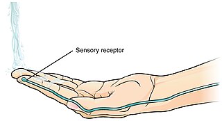
In physiology, a stimulus is a detectable change in the physical or chemical structure of an organism's internal or external environment. The ability of an organism or organ to detect external stimuli, so that an appropriate reaction can be made, is called sensitivity (excitability). Sensory receptors can receive information from outside the body, as in touch receptors found in the skin or light receptors in the eye, as well as from inside the body, as in chemoreceptors and mechanoreceptors. When a stimulus is detected by a sensory receptor, it can elicit a reflex via stimulus transduction. An internal stimulus is often the first component of a homeostatic control system. External stimuli are capable of producing systemic responses throughout the body, as in the fight-or-flight response. In order for a stimulus to be detected with high probability, its level of strength must exceed the absolute threshold; if a signal does reach threshold, the information is transmitted to the central nervous system (CNS), where it is integrated and a decision on how to react is made. Although stimuli commonly cause the body to respond, it is the CNS that finally determines whether a signal causes a reaction or not.

<span class="mw-page-title-main">Free nerve ending</span> Type of nerve fiber carrying sensory signals

A free nerve ending (FNE) or bare nerve ending, is an unspecialized, afferent nerve fiber sending its signal to a sensory neuron. Afferent in this case means bringing information from the body's periphery toward the brain. They function as cutaneous nociceptors and are essentially used by vertebrates to detect noxious stimuli that often result in pain.

<span class="mw-page-title-main">Thermoreceptor</span> Receptive portion of a sensory neuron

A thermoreceptor is a non-specialised sense receptor, or more accurately the receptive portion of a sensory neuron, that codes absolute and relative changes in temperature, primarily within the innocuous range. In the mammalian peripheral nervous system, warmth receptors are thought to be unmyelinated C-fibres, while those responding to cold have both C-fibers and thinly myelinated A delta fibers. The adequate stimulus for a warm receptor is warming, which results in an increase in their action potential discharge rate. Cooling results in a decrease in warm receptor discharge rate. For cold receptors their firing rate increases during cooling and decreases during warming. Some cold receptors also respond with a brief action potential discharge to high temperatures, i.e. typically above 45 °C, and this is known as a paradoxical response to heat. The mechanism responsible for this behavior has not been determined.

<span class="mw-page-title-main">Nociceptor</span> Sensory neuron that detects pain

A nociceptor is a sensory neuron that responds to damaging or potentially damaging stimuli by sending "possible threat" signals to the spinal cord and the brain. The brain creates the sensation of pain to direct attention to the body part, so the threat can be mitigated; this process is called nociception.

<span class="mw-page-title-main">Spinothalamic tract</span> Sensory pathway from the skin to the thalamus

The spinothalamic tract is a part of the anterolateral system or the ventrolateral system, a sensory pathway to the thalamus. From the ventral posterolateral nucleus in the thalamus, sensory information is relayed upward to the somatosensory cortex of the postcentral gyrus.

<span class="mw-page-title-main">Sensory neuron</span> Nerve cell that converts environmental stimuli into corresponding internal stimuli

Sensory neurons, also known as afferent neurons, are neurons in the nervous system, that convert a specific type of stimulus, via their receptors, into action potentials or graded receptor potentials. This process is called sensory transduction. The cell bodies of the sensory neurons are located in the dorsal ganglia of the spinal cord.

<span class="mw-page-title-main">Dorsal root ganglion</span> Cluster of neurons in a dorsal root of a spinal nerve

A dorsal root ganglion is a cluster of neurons in a dorsal root of a spinal nerve. The cell bodies of sensory neurons known as first-order neurons are located in the dorsal root ganglia.

Neuropathic pain is pain caused by a lesion or disease of the somatosensory nervous system. Neuropathic pain may be associated with abnormal sensations called dysesthesia or pain from normally non-painful stimuli (allodynia). It may have continuous and/or episodic (paroxysmal) components. The latter resemble stabbings or electric shocks. Common qualities include burning or coldness, "pins and needles" sensations, numbness and itching.

<span class="mw-page-title-main">Gate control theory</span> Theory about pain and the nervous system

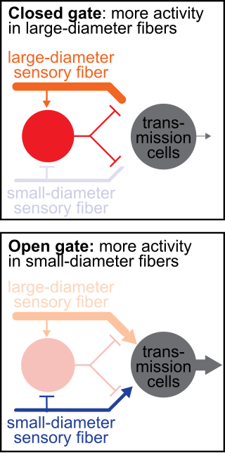
The gate control theory of pain asserts that non-painful input closes the nerve "gates" to painful input, which prevents pain sensation from traveling to the central nervous system.

<span class="mw-page-title-main">Allodynia</span> Feeling of pain from stimuli which do not normally elicit pain

Allodynia is a condition in which pain is caused by a stimulus that does not normally elicit pain. For example, sunburn can cause temporary allodynia, so that usually painless stimuli, such as wearing clothing or running cold or warm water over it, can be very painful. It is different from hyperalgesia, an exaggerated response from a normally painful stimulus. The term comes from Ancient Greek άλλος (állos) 'other', and οδύνη (odúnē) 'pain'.

<span class="mw-page-title-main">Microneurography</span>

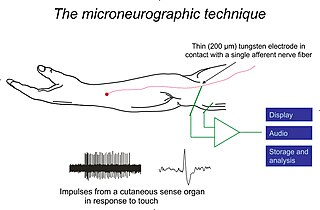
Microneurography is a neurophysiological method employed to visualize and record the traffic of nerve impulses that are conducted in peripheral nerves of waking human subjects. It can also be used in animal recordings. The method has been successfully employed to reveal functional properties of a number of neural systems, e.g. sensory systems related to touch, pain, and muscle sense as well as sympathetic activity controlling the constriction state of blood vessels. To study nerve impulses of an identified nerve, a fine tungsten needle microelectrode is inserted into the nerve and connected to a high input impedance differential amplifier. The exact position of the electrode tip within the nerve is then adjusted in minute steps until the electrode discriminates nerve impulses of interest. A unique feature and a significant strength of the microneurography method is that subjects are fully awake and able to cooperate in tests requiring mental attention, while impulses in a representative nerve fibre or set of nerve fibres are recorded, e.g. when cutaneous sense organs are stimulated or subjects perform voluntary precision movements.

<span class="mw-page-title-main">TRPM8</span> Protein-coding gene in the species Homo sapiens

Transient receptor potential cation channel subfamily M (melastatin) member 8 (TRPM8), also known as the cold and menthol receptor 1 (CMR1), is a protein that in humans is encoded by the TRPM8 gene. The TRPM8 channel is the primary molecular transducer of cold somatosensation in humans. In addition, mints can desensitize a region through the activation of TRPM8 receptors.

<span class="mw-page-title-main">Axon reflex</span>

The axon reflex is the response stimulated by peripheral nerves of the body that travels away from the nerve cell body and branches to stimulate target organs. Reflexes are single reactions that respond to a stimulus making up the building blocks of the overall signaling in the body's nervous system. Neurons are the excitable cells that process and transmit these reflex signals through their axons, dendrites, and cell bodies. Axons directly facilitate intercellular communication projecting from the neuronal cell body to other neurons, local muscle tissue, glands and arterioles. In the axon reflex, signaling starts in the middle of the axon at the stimulation site and transmits signals directly to the effector organ skipping both an integration center and a chemical synapse present in the spinal cord reflex. The impulse is limited to a single bifurcated axon, or a neuron whose axon branches into two divisions and does not cause a general response to surrounding tissue.

Mechanosensation is the transduction of mechanical stimuli into neural signals. Mechanosensation provides the basis for the senses of light touch, hearing, proprioception, and pain. Mechanoreceptors found in the skin, called cutaneous mechanoreceptors, are responsible for the sense of touch. Tiny cells in the inner ear, called hair cells, are responsible for hearing and balance. States of neuropathic pain, such as hyperalgesia and allodynia, are also directly related to mechanosensation. A wide array of elements are involved in the process of mechanosensation, many of which are still not fully understood.

Zucapsaicin (Civanex) is a medication used to treat osteoarthritis of the knee and other neuropathic pain. Zucapsaicin is a member of phenols and a member of methoxybenzenes. It is a modulator of transient receptor potential cation channel subfamily V member 1 (TRPV-1), also known as the vanilloid or capsaicin receptor 1 that reduces pain, and improves articular functions. It is the cis-isomer of capsaicin. Civamide, manufactured by Winston Pharmaceuticals, is produced in formulations for oral, nasal, and topical use.

Group A nerve fibers are one of the three classes of nerve fiber as generally classified by Erlanger and Gasser. The other two classes are the group B nerve fibers, and the group C nerve fibers. Group A are heavily myelinated, group B are moderately myelinated, and group C are unmyelinated.

<span class="mw-page-title-main">Presynaptic inhibition</span>

Presynaptic inhibition is a phenomenon in which an inhibitory neuron provides synaptic input to the axon of another neuron to make it less likely to fire an action potential. Presynaptic inhibition occurs when an inhibitory neurotransmitter, like GABA, acts on GABA receptors on the axon terminal. Or when endocannabinoids act as retrograde messengers by binding to presynaptic CB1 receptors, thereby indirectly modulating GABA and the excitability of dopamine neurons by reducing it and other presynaptic released neurotransmitters. Presynaptic inhibition is ubiquitous among sensory neurons.

References

  1. 1 2 3 4 5 6 7 8 9 10 11 12 13 Purves, Dale; et al. (2004). Neuroscience. Massachusetts: Sinauer Associates, Inc. ISBN   978-0-87893-725-7.
  2. Craig, AD (August 2002). "How do you feel? Interoception: the sense of the physiological condition of the body". Nature Reviews Neuroscience. 3 (8): 655–66. doi:10.1038/nrn894. PMID   12154366. S2CID   17829407.
  3. 1 2 3 4 5 6 7 8 9 10 11 12 13 Murinson, BB; JW Griffin (2004). "C-fiber structure varies with location in peripheral nerve". Journal of Neuropathology and Experimental Neurology. 63 (3): 246–254. doi: 10.1093/jnen/63.3.246 . PMID   15055448.
  4. 1 2 3 Fagan, Tom (2003). "Glial Cells Critical for Peripheral Nervous System Health". News from Harvard Medical, Dental and Public Health Schools.
  5. 1 2 3 4 5 6 7 8 9 10 11 12 13 14 Baron, Ralf (2006). "Mechanisms of Disease: neuropathic pain—a clinical perspective". Nature Clinical Practice Neurology. 2 (2): 95–106. doi:10.1038/ncpneuro0113. ISSN   1745-834X. PMID   16932531. S2CID   12925061.
  6. 1 2 Chung, J.M.; et al. (1979). "Excitation of primate spinothalamic neurons by cutaneous C-fiber volleys". Journal of Neurophysiology. 42 (5): 1354–1369. doi:10.1152/jn.1979.42.5.1354. PMID   114611.
  7. Yam, Mun Fei; Loh, Yean Chun; Tan, Chu Shan; Khadijah Adam, Siti; Abdul Manan, Nizar; Basir, Rusliza (July 24, 2018). "General Pathways of Pain Sensation and the Major Neurotransmitters Involved in Pain Regulation". International Journal of Molecular Sciences. 19 (8): 2164. doi: 10.3390/ijms19082164 . ISSN   1422-0067. PMC   6121522 . PMID   30042373.
  8. 1 2 3 4 5 6 Craig, AD (2003). "Interoception: the sense of the physiological condition of the body". Current Opinion in Neurobiology. 13 (4): 500–505. doi:10.1016/S0959-4388(03)00090-4. PMID   12965300. S2CID   16369323.
  9. 1 2 Loken, L (2009). "Coding of pleasant touch by unmyelinated afferents in humans". Nature Neuroscience. 12 (5): 548–549. doi:10.1038/nn.2312. PMID   19363489. S2CID   4311541.
  10. 1 2 3 Purves, Dale (2011). Neuroscience (5. ed.). Sunderland, Mass.: Sinauer. pp. 211–212. ISBN   978-0-87893-695-3.
  11. 1 2 Davis, JB; et al. (2000). "Vanilloid receptor-1 is essential for inflammatory thermal hyperalgesia". Nature. 405 (6783): 183–7. Bibcode:2000Natur.405..183D. doi:10.1038/35012076. PMID   10821274. S2CID   4337894.
  12. 1 2 3 4 5 6 7 8 9 10 Scholz, Joachim; Clifford Woolf (2002). "Can we conquer pain?". Nature Neuroscience. 5: 1062–1067. doi:10.1038/nn942. PMID   12403987. S2CID   15781811.
  13. Baron, Ralph; Ogawa, Setsuro; Katz, Joel; Nagai, Hitoshi; Kashiwazaki, Miho; Saeki, Shigeru; Suzuki, Hajime (2000). "Peripheral neuropathic pain: from mechanisms to symptoms". Clinical Journal of Pain. 16 (2 Suppl): S12–20. doi:10.1097/00002508-200003000-00003. PMID   10870735.
  14. 1 2 Lai, J; Hunter, John C; Porreca, Frank (2003). "The role of voltage-gated sodium channels in neuropathic pain". Current Opinion in Neurobiology. 13 (3): 291–297. doi:10.1016/S0959-4388(03)00074-6. PMID   12850213. S2CID   24745129.
  15. 1 2 3 4 5 6 Staud, Roland; et al. (2007). "Brain activity related to temporal summation of C-fiber evoked pain". Pain. 129 (1–2) (1-2 ed.): 130–142. doi:10.1016/j.pain.2006.10.010. PMC   1997296 . PMID   17156923.
  16. 1 2 3 4 5 Mano, Tadaaki; et al. (2006). "Microneurography as a tool in clinical neurophysiology to investigate peripheral neural traffic in humans". Clinical Neurophysiology. 117 (11): 2357–2384. doi:10.1016/j.clinph.2006.06.002. PMID   16904937. S2CID   22258173.
  17. 1 2 3 4 5 6 7 Moalem-Taylor, Gila; et al. (2007). "Post-spike excitability indicates changes in membrane potential of isolated c-fibers". Muscle and Nerve. 36 (2): 172–182. doi: 10.1002/mus.20793 . PMID   17487863. S2CID   36126632.
  18. Puccetti, Roland (1977). "The Great C-Fiber Myth: A Critical Note". Philosophy of Science. 44 (2): 303–305. doi:10.1086/288746. ISSN   0031-8248. JSTOR   187355.
  19. "David Lewis | Internet Encyclopedia of Philosophy" . Retrieved April 23, 2024.
  20. Puccetti, Roland (1977). "The Great C-Fiber Myth: A Critical Note". Philosophy of Science. 44 (2): 303–305. doi:10.1086/288746. ISSN   0031-8248. JSTOR   187355.
  21. "Identity Theory | Internet Encyclopedia of Philosophy" . Retrieved April 23, 2024.
  22. Aranyosi, István (April 23, 2024). "The Peripheral Mind: Philosophy of Mind and the Peripheral Nervous System | Return of the C Fibers; or, Philosophers' Lack of Nerve". academic.oup.com. OUP. pp. 34–57. Retrieved April 23, 2024.