Sodium channel

Last updated

Sodium channels are integral membrane proteins that form ion channels, conducting sodium ions (Na+) through a cell's membrane. [1] [2] They belong to the superfamily of cation channels.

Contents

Classification

They are classified into 2 types:

Type of sodium channelSynonymsTrigger
(factor that stimulates the channel)
Voltage-gated sodium channels
  • "VGSCs"
  • "voltage-dependent"
  • "voltage-sensitive"
  • "Nav channel"
change in membrane potential, which is also called change in voltage
ligand-gated sodium channels LGSCsbinding of substances such as ligands to the channel
Leak sodium channelNALCNUngated, always open

Function

In excitable cells such as neurons, myocytes, and certain types of glia, sodium channels are responsible for the rising phase of action potentials. These channels go through three different states called resting, active and inactive states. Even though the resting and inactive states would not allow the ions to flow through the channels the difference exists with respect to their structural conformation.

Selectivity

Sodium channels are highly selective for the transport of ions across cell membranes. The high selectivity with respect to the sodium ion is achieved in many different ways. All involve encapsulation of the sodium ion in a cavity of specific size within a larger molecule. [3]

Voltage-gated sodium channels

Structure

Diagram of a voltage-sensitive sodium channel a-subunit. G - glycosylation, P - phosphorylation, S - ion selectivity, I - inactivation. Positive (+) charges in S4 are important for transmembrane voltage sensing. Sodium-channel.svg
Diagram of a voltage-sensitive sodium channel α-subunit. G – glycosylation, P – phosphorylation, S – ion selectivity, I – inactivation. Positive (+) charges in S4 are important for transmembrane voltage sensing.

Sodium channels consist of large alpha subunits that associate with accessory proteins, such as beta subunits. An alpha subunit forms the core of the channel and is functional on its own. When the alpha subunit protein is expressed by a cell, it is able to form a pore in the cell membrane that conducts Na+ in a voltage-dependent way, even if beta subunits or other known modulating proteins are not expressed. When accessory proteins assemble with α subunits, the resulting complex can display altered voltage dependence and cellular localization.

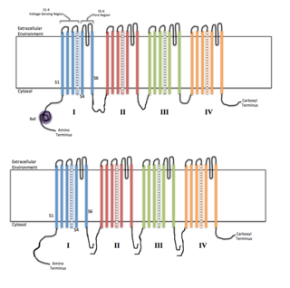
The alpha subunit consists of four repeat domains, labelled I through IV, each containing six membrane-spanning segments, labelled S1 through S6. The highly conserved S4 segment acts as the channel's voltage sensor. The voltage sensitivity of this channel is due to positive amino acids located at every third position. [5] When stimulated by a change in transmembrane voltage, this segment moves toward the extracellular side of the cell membrane, allowing the channel to become permeable to ions. The ions are conducted through the central pore cavity, which consists of two main regions. The more external (i.e., more extracellular) portion of the pore is formed by the "P-loops" (the region between S5 and S6) of the four domains. This region is the most narrow part of the pore and is responsible for its ion selectivity. The inner portion (i.e., more cytoplasmic) of the pore is the pore gate and is formed by the combined S5 and S6 segments of the four domains. The pore domain also features lateral tunnels or fenestrations that run perpendicular to the pore axis. These fenestrations that connect the central cavity to the membrane are proposed to be important for drug accessibility. [6] [7] [8]

In mammalian sodium channels, the region linking domains III and IV is also important for channel function. This DIII-IV linker is responsible for wedging the pore gate shut after channel opening, inactivating it. [9]

Gating

Voltage-gated Na+ channels have three main conformational states: closed, open and inactivated. Forward/back transitions between these states are correspondingly referred to as activation/deactivation (between open and closed, respectively), inactivation/reactivation (between inactivated and open, respectively), and recovery from inactivation/closed-state inactivation (between inactivated and closed, respectively). Closed and inactivated states are ion impermeable.

Before an action potential occurs, the axonal membrane is at its normal resting potential, about −70 mV in most human neurons, and Na+ channels are in their deactivated state, blocked on the extracellular side by their activation gates. In response to an increase of the membrane potential to about −55 mV (in this case, caused by an action potential), the activation gates open, allowing positively charged Na+ ions to flow into the neuron through the channels, and causing the voltage across the neuronal membrane to increase to +30 mV in human neurons. Because the voltage across the membrane is initially negative, as its voltage increases to and past zero (from −70 mV at rest to a maximum of +30 mV), it is said to depolarize. This increase in voltage constitutes the rising phase of an action potential.

Action PotentialMembrane PotentialTarget PotentialGate's Target StateNeuron's Target State
Resting−70 mV−55 mVDeactivated → ActivatedPolarized
Rising−55 mV0 mVActivatedPolarized → Depolarized
Rising0 mV+30 mVActivated → InactivatedDepolarized
Falling+30 mV0 mVInactivatedDepolarized → Repolarized
Falling0 mV−70 mVInactivatedRepolarized
Undershot−70 mV−75 mVInactivated → DeactivatedRepolarized → Hyperpolarized
Rebounding−75 mV−70 mVDeactivatedHyperpolarized → Polarized

At the peak of the action potential, when enough Na+ has entered the neuron and the membrane's potential has become high enough, the Na+ channels inactivate themselves by closing their inactivation gates . The inactivation gate can be thought of as a "plug" tethered to domains III and IV of the channel's intracellular alpha subunit. Closure of the inactivation gate causes Na+ flow through the channel to stop, which in turn causes the membrane potential to stop rising. The closing of the inactivation gate creates a refractory period within each individual Na+ channel. This refractory period eliminates the possibility of an action potential moving in the opposite direction back towards the soma. With its inactivation gate closed, the channel is said to be inactivated. With the Na+ channel no longer contributing to the membrane potential, the potential decreases back to its resting potential as the neuron repolarizes and subsequently hyperpolarizes itself, and this constitutes the falling phase of an action potential. The refractory period of each channel is therefore vital in propagating the action potential unidirectionally down an axon for proper communication between neurons.

When the membrane's voltage becomes low enough, the inactivation gate reopens and the activation gate closes in a process called deinactivation. With the activation gate closed and the inactivation gate open, the Na+ channel is once again in its deactivated state, and is ready to participate in another action potential.

When any kind of ion channel does not inactivate itself, it is said to be persistently (or tonically) active. Some kinds of ion channels are naturally persistently active. However, genetic mutations that cause persistent activity in other channels can cause disease by creating excessive activity of certain kinds of neurons. Mutations that interfere with Na+ channel inactivation can contribute to cardiovascular diseases or epileptic seizures by window currents, which can cause muscle and/or nerve cells to become over-excited.

Modeling the behavior of gates

The temporal behavior of Na+ channels can be modeled by a Markovian scheme or by the Hodgkin–Huxley-type formalism. In the former scheme, each channel occupies a distinct state with differential equations describing transitions between states; in the latter, the channels are treated as a population that are affected by three independent gating variables. Each of these variables can attain a value between 1 (fully permeant to ions) and 0 (fully non-permeant), the product of these variables yielding the percentage of conducting channels. The Hodgkin–Huxley model can be shown to be equivalent to a Markovian model.[ further explanation needed ]

Impermeability to other ions

The pore of sodium channels contains a selectivity filter made of negatively charged amino acid residues, which attract the positive Na+ ion and keep out negatively charged ions such as chloride. The cations flow into a more constricted part of the pore that is 0.3 by 0.5 nm wide, which is just large enough to allow a single Na+ ion with a water molecule associated to pass through. The larger K+ ion cannot fit through this area. Ions of different sizes also cannot interact as well with the negatively charged glutamic acid residues that line the pore. [ citation needed ]

Diversity

Voltage-gated sodium channels normally consist of an alpha subunit that forms the ion conduction pore and one to two beta subunits that have several functions including modulation of channel gating. [10] Expression of the alpha subunit alone is sufficient to produce a functional channel.

Alpha subunits

Figure 1. Likely evolutionary relationship of the nine known human sodium channels. Sodium channel phylogram.png
Figure 1. Likely evolutionary relationship of the nine known human sodium channels.

The family of sodium channels has 9 known members, with amino acid identity >50% in the trans-membrane segments and extracellular loop regions. A standardized nomenclature for sodium channels is currently used and is maintained by the IUPHAR. [11]

The proteins of these channels are named Nav1.1 through Nav1.9. The gene names are referred to as SCN1A through SCN5A, then SCN8A through SCN11A. [11] The "tenth member", Nax, does not act in a voltage-gated way. It has a loosely similar overall structure. Not much is known about its real function, other than that it also associates with beta subunits. [12]

The probable evolutionary relationship between these channels, based on the similarity of their amino acid sequences, is shown in figure 1. The individual sodium channels are distinguished not only by differences in their sequence but also by their kinetics and expression profiles. Some of this data is summarized in table 1, below.

Table 1. Nomenclature and some functions of voltage-gated sodium channel alpha subunits
Protein nameGeneExpression profileAssociated human channelopathies
Nav1.1 SCN1A Central neurons, [peripheral neurons] and cardiac myocytes febrile epilepsy, GEFS+, Dravet syndrome (also known as severe myclonic epilepsy of infancy or SMEI), borderline SMEI (SMEB), West syndrome (also known as infantile spasms), Doose syndrome (also known as myoclonic astatic epilepsy), intractable childhood epilepsy with generalized tonic-clonic seizures (ICEGTC), Panayiotopoulos syndrome, familial hemiplegic migraine (FHM), familial autism, Rasmussens's encephalitis and Lennox-Gastaut syndrome [13]
Nav1.2 SCN2A Central neurons, peripheral neuronsinherited febrile seizures, epilepsy, and autism spectrum disorder
Nav1.3 SCN3A Central neurons, peripheral neurons and cardiac myocytesepilepsy, pain, brain malformations [14] [15]
Nav1.4 SCN4A Skeletal muscle hyperkalemic periodic paralysis, paramyotonia congenita, and potassium-aggravated myotonia
Nav1.5 SCN5A Cardiac myocytes, uninnervated skeletal muscle, central neurons, gastrointestinal smooth muscle cells and Interstitial cells of CajalCardiac: Long QT syndrome Type 3, Brugada syndrome, progressive cardiac conduction disease, familial atrial fibrillation and idiopathic ventricular fibrillation; [16]

Gastrointestinal: Irritable bowel syndrome; [17]

Nav1.6 SCN8A Central neurons, dorsal root ganglia, peripheral neurons, heart, glia cells Epilepsy, [18] ataxia, dystonia, tremor [19]
Nav1.7 SCN9A Dorsal root ganglia, sympathetic neurons, Schwann cells, and neuroendocrine cells erythromelalgia, PEPD, channelopathy-associated insensitivity to pain [14] and recently discovered a disabling form of fibromyalgia (rs6754031 polymorphism) [20]
Nav1.8 SCN10A Dorsal root gangliapain, [14] neuropsychiatric disorders
Nav1.9 SCN11A Dorsal root gangliapain [14]
Nax SCN7A heart, uterus, skeletal muscle, astrocytes, dorsal root ganglion cellsnone known

Beta subunits

Sodium channel beta subunits are type 1 transmembrane glycoproteins with an extracellular N-terminus and a cytoplasmic C-terminus. As members of the Ig superfamily, beta subunits contain a prototypic V-set Ig loop in their extracellular domain. They do not share any homology with their counterparts of calcium and potassium channels. [21] Instead, they are homologous to neural cell adhesion molecules (CAMs) and the large family of L1 CAMs. There are four distinct betas named in order of discovery: SCN1B, SCN2B, SCN3B, SCN4B (table 2). Beta 1 and beta 3 interact with the alpha subunit non-covalently, whereas beta 2 and beta 4 associate with alpha via disulfide bond. [22] Sodium channels are more likely to stay open at the subthreshold membrane potential when interacting with beta toxins, which in turn induces an immediate sensation of pain. [23]

Role of beta subunits as cell adhesion molecules

In addition to regulating channel gating, sodium channel beta subunits also modulate channel expression and form links to the intracellular cytoskeleton via ankyrin and spectrin. [10] [24] [25] Voltage-gated sodium channels also assemble with a variety of other proteins, such as FHF proteins (Fibroblast growth factor Homologous Factor), calmodulin, cytoskeleton or regulatory kinases, [26] [10] [27] [28] [29] which form a complex with sodium channels, influencing its expression and/or function. Several beta subunits interact with one or more extracellular matrix (ECM) molecules. Contactin, also known as F3 or F11, associates with beta 1 as shown via co-immunoprecipitation. [30] Fibronectin-like (FN-like) repeats of Tenascin-C and Tenascin-R bind with beta 2 in contrast to the Epidermal growth factor-like (EGF-like) repeats that repel beta2. [31] A disintegrin and metalloproteinase (ADAM) 10 sheds beta 2's ectodomain possibly inducing neurite outgrowth. [32] Beta 3 and beta 1 bind to neurofascin at Nodes of Ranvier in developing neurons. [33]

Table 2. Nomenclature and some functions of voltage-gated sodium channel beta subunits
Protein nameGene linkAssembles withExpression profileAssociated human channelopathies
Navβ1 SCN1B Nav1.1 to Nav1.7Central Neurons, Peripheral Neurons, skeletal muscle, heart, gliaepilepsy (GEFS+), Brugada syndrome [34]
Navβ2 SCN2B Nav1.1, Nav1.2, Nav1.5 to Nav1.7Central Neurons, peripheral neurons, heart, glia Brugada syndrome [34]
Navβ3 SCN3B Nav1.1 to Nav1.3, Nav1.5central neurons, adrenal gland, kidney, peripheral neurons Brugada syndrome [34]
Navβ4 SCN4B Nav1.1, Nav1.2, Nav1.5heart, skeletal muscle, central and peripheral neuronsnone known

Ligand-gated sodium channels

Ligand-gated sodium channels are activated by binding of a ligand instead of a change in membrane potential.

They are found, e.g. in the neuromuscular junction as nicotinic receptors, where the ligands are acetylcholine molecules. Most channels of this type are permeable to potassium to some degree as well as to sodium.

Role in action potential

Voltage-gated sodium channels play an important role in action potentials. If enough channels open when there is a change in the cell's membrane potential, a small but significant number of Na+ ions will move into the cell down their electrochemical gradient, further depolarizing the cell. Thus, the more Na+ channels localized in a region of a cell's membrane the faster the action potential will propagate and the more excitable that area of the cell will be. This is an example of a positive feedback loop. The ability of these channels to assume a closed-inactivated state causes the refractory period and is critical for the propagation of action potentials down an axon.

Na+ channels both open and close more quickly than K+ channels, producing an influx of positive charge (Na+) toward the beginning of the action potential and an efflux (K+) toward the end.

Ligand-gated sodium channels, on the other hand, create the change in the membrane potential in the first place, in response to the binding of a ligand to it. Leak sodium channels additionally contribute to action potential regulation by modulating the resting potential (and in turn, the excitability) of a cell. [35]

Pharmacologic modulation

Blockers

Activators

The following naturally produced substances persistently activate (open) sodium channels:

Gating modifiers

The following toxins modify the gating of sodium channels:

Sodium leak channel (NALCN)

Sodium leak channels do not show any voltage or ligand gating. Instead, they are always open or "leaking" a small background current to regulate the resting membrane potential of a neuron. [35] In most animals, a single gene encodes the NALCN (sodium leak channel, nonselective) protein. [38]

Structural and functional differences

Despite following the same basic structure as other sodium channels, NALCN is not sensitive to voltage changes. The voltage-sensitive S4 transmembrane domain of NALCN has fewer positively charged amino acids (13 instead of a voltage gated channel's 21) possibly explaining its voltage insensitivity. [35] NALCN is also far less selective for Na+ ions and is permeable to Ca2+ and K+ ions. The EEKE amino acid motif in the pore filter domain of NALCN is similar to both the EEEE motif of voltage-gated calcium channel and the DEKA motif of the voltage-gated sodium channel, possibly explaining its lack of selectivity. [38]

Regulatory pathways and chemicals affecting NALCN function. NALCN activity.png
Regulatory pathways and chemicals affecting NALCN function.

NALCN is not blocked by many common sodium channel blockers, including tetrodotoxin. NALCN is blocked nonspecifically by both Gd3+ and verapamil. [39] Substance P and neurotensin both activate Src family kinases through their respective GPCRs (independent of the coupled G-proteins) which in turn increase the permeability of NALCN through UNC80 activation. [40] Acetylcholine can also increase NALCN activity through M3 muscarinic acetylcholine receptors. [41] Higher levels of extracellular Ca2+ decrease the permeability of NALCN by activating CaSR which inhibits UNC80. [42]

Protein Complex

NALCN complexes with the proteins UNC79, UNC80, and FAM155A. [43] [44] [45] UNC79 appears to be linked to membrane stability of NALCN and linkage with UNC 80. [44] UNC80 mediates chemical modulation of NALCN through multiple pathways. [35] [42] [41] [40] FAM155A helps protein folding in the endoplasmic reticulum, chaperones transport to the axon, and contributes to membrane stability. [45]

Biological function

The resting membrane potential of a neuron is usually -60mV to -80mV, driven primarily by the K+ potential at -90mV. The depolarization from the K+ potential is due primarily to a small Na+ leak current. About 70% of this current is through NALCN. [39] Increasing NALCN permeability lowers the resting membrane potential, bringing it closer to the trigger of an action potential (-55mV), thus increasing the excitability of a neuron.

Role in pathology

Mutations to NALCN lead to severe disruptions to respiratory rhythm in mice [39] and altered circadian locomotion in flies. [46] Mutations to NALCN have also been linked to multiple severe developmental disorders [47] and cervical dystonia. [48] Schizophrenia and bipolar disorder are also linked to mutations to NALCN. [49]

pH modulation

Changes in blood and tissue pH accompany physiological and pathophysiological conditions such as exercise, cardiac ischemia, ischemic stroke, and cocaine ingestion. These conditions are known to trigger the symptoms of electrical diseases in patients carrying sodium channel mutations. Protons cause a diverse set of changes to sodium channel gating, which generally lead to decreases in the amplitude of the transient sodium current and increases in the fraction of non-inactivating channels that pass persistent currents. These effects are shared with disease-causing mutants in neuronal, skeletal muscle, and cardiac tissue and may be compounded in mutants that impart greater proton sensitivity to sodium channels, suggesting a role of protons in triggering acute symptoms of electrical disease. [50]

Molecular mechanisms of proton block

Single channel data from cardiomyocytes have shown that protons can decrease the conductance of individual sodium channels. [51] The sodium channel selectivity filter is composed of a single residue in each of the four pore-loops of the four functional domains. These four residues are known as the DEKA motif. [52] The permeation rate of sodium through the sodium channel is determined by a four carboxylate residues, the EEDD motif, which make up the outer charged ring. [52] The protonation of these carboxylates is one of the main drivers of proton block in sodium channels, although there are other residues that also contribute to pH sensitivity. [53] One such residue is C373 in the cardiac sodium channel which makes it the most pH-sensitive sodium channel among the sodium channels that have been studied to date. [54]

pH modulation of sodium channel gating

As the cardiac sodium channel is the most pH-sensitive sodium channel, most of what is known is based on this channel. Reduction in extracellular pH has been shown to depolarize the voltage-dependence of activation and inactivation to more positive potentials. This indicates that during activities that decrease the blood pH, such as exercising, the probability of channels activating and inactivating is higher more positive membrane potentials, which can lead to potential adverse effects. [55] The sodium channels expressed in skeletal muscle fibers have evolved into relatively pH-insensitive channels. This has been suggested to be a protective mechanism against potential over- or under-excitability in skeletal muscles, as blood pH levels are highly susceptible to change during movement. [56] [57] Recently, a mixed syndrome mutation that causes periodic paralysis and myotonia in the skeletal sodium channel has been shown to impart pH-sensitivity in this channel, making the gating of this channel similar to that of the cardiac subtype. [58]

pH modulation across the subtypes studied thus far

The effects of protonation have been characterized in Nav1.1–Nav1.5. Among these channels, Nav1.1–Nav1.3 and Nav1.5 display depolarized voltage-dependence of activation, while activation in Nav1.4 remains insensitive to acidosis. The voltage-dependence of steady-state fast inactivation is unchanged in Nav1.1–Nav1.4, but steady-state fast inactivation in Nav1.5 is depolarized. Hence, among the sodium channels that have been studied so far, Nav1.4 is the least and Nav1.5 is the most proton-sensitive subtypes. [59]

See also

Related Research Articles

<span class="mw-page-title-main">Ion channel</span> Pore-forming membrane protein

Ion channels are pore-forming membrane proteins that allow ions to pass through the channel pore. Their functions include establishing a resting membrane potential, shaping action potentials and other electrical signals by gating the flow of ions across the cell membrane, controlling the flow of ions across secretory and epithelial cells, and regulating cell volume. Ion channels are present in the membranes of all cells. Ion channels are one of the two classes of ionophoric proteins, the other being ion transporters.

<span class="mw-page-title-main">Membrane potential</span> Type of physical quantity

Membrane potential is the difference in electric potential between the interior and the exterior of a biological cell. That is, there is a difference in the energy required for electric charges to move from the internal to exterior cellular environments and vice versa, as long as there is no acquisition of kinetic energy or the production of radiation. The concentration gradients of the charges directly determine this energy requirement. For the exterior of the cell, typical values of membrane potential, normally given in units of milli volts and denoted as mV, range from –80 mV to –40 mV.

<span class="mw-page-title-main">Potassium channel</span> Ion channel that selectively passes K+

Potassium channels are the most widely distributed type of ion channel found in virtually all organisms. They form potassium-selective pores that span cell membranes. Potassium channels are found in most cell types and control a wide variety of cell functions.

<span class="mw-page-title-main">Voltage-gated ion channel</span> Type of ion channel transmembrane protein

Voltage-gated ion channels are a class of transmembrane proteins that form ion channels that are activated by changes in the electrical membrane potential near the channel. The membrane potential alters the conformation of the channel proteins, regulating their opening and closing. Cell membranes are generally impermeable to ions, thus they must diffuse through the membrane through transmembrane protein channels. They have a crucial role in excitable cells such as neuronal and muscle tissues, allowing a rapid and co-ordinated depolarization in response to triggering voltage change. Found along the axon and at the synapse, voltage-gated ion channels directionally propagate electrical signals. Voltage-gated ion-channels are usually ion-specific, and channels specific to sodium (Na+), potassium (K+), calcium (Ca2+), and chloride (Cl) ions have been identified. The opening and closing of the channels are triggered by changing ion concentration, and hence charge gradient, between the sides of the cell membrane.

Voltage-gated calcium channels (VGCCs), also known as voltage-dependent calcium channels (VDCCs), are a group of voltage-gated ion channels found in the membrane of excitable cells (e.g., muscle, glial cells, neurons, etc.) with a permeability to the calcium ion Ca2+. These channels are slightly permeable to sodium ions, so they are also called Ca2+–Na+ channels, but their permeability to calcium is about 1000-fold greater than to sodium under normal physiological conditions.

<span class="mw-page-title-main">Voltage-gated potassium channel</span> Class of transport proteins

Voltage-gated potassium channels (VGKCs) are transmembrane channels specific for potassium and sensitive to voltage changes in the cell's membrane potential. During action potentials, they play a crucial role in returning the depolarized cell to a resting state.

Na<sub>v</sub>1.4 Protein-coding gene in the species Homo sapiens

Sodium channel protein type 4 subunit alpha is a protein that in humans is encoded by the SCN4A gene.

Na<sub>v</sub>1.7 Protein-coding gene in the species Homo sapiens

Nav1.7 is a sodium ion channel that in humans is encoded by the SCN9A gene. It is usually expressed at high levels in two types of neurons: the nociceptive (pain) neurons at dorsal root ganglion (DRG) and trigeminal ganglion and sympathetic ganglion neurons, which are part of the autonomic (involuntary) nervous system.

Paralytic is a gene in the fruit fly, Drosophila melanogaster, which encodes a voltage gated sodium channel within D. melanogaster neurons. This gene is essential for locomotive activity in the fly. There are 9 different para alleles, composed of a minimum of 26 exons within over 78kb of genomic DNA. The para gene undergoes alternative splicing to produce subtypes of the channel protein. Flies with mutant forms of paralytic are used in fly models of seizures, since seizures can be easily induced in these flies.

T-type calcium channels are low voltage activated calcium channels that become inactivated during cell membrane hyperpolarization but then open to depolarization. The entry of calcium into various cells has many different physiological responses associated with it. Within cardiac muscle cell and smooth muscle cells voltage-gated calcium channel activation initiates contraction directly by allowing the cytosolic concentration to increase. Not only are T-type calcium channels known to be present within cardiac and smooth muscle, but they also are present in many neuronal cells within the central nervous system. Different experimental studies within the 1970s allowed for the distinction of T-type calcium channels from the already well-known L-type calcium channels. The new T-type channels were much different from the L-type calcium channels due to their ability to be activated by more negative membrane potentials, had small single channel conductance, and also were unresponsive to calcium antagonist drugs that were present. These distinct calcium channels are generally located within the brain, peripheral nervous system, heart, smooth muscle, bone, and endocrine system.

Na<sub>v</sub>1.9 Protein-coding gene in the species Homo sapiens

Sodium channel, voltage-gated, type XI, alpha subunit also known as SCN11A or Nav1.9 is a voltage-gated sodium ion channel protein which is encoded by the SCN11A gene on chromosome 3 in humans. Like Nav1.7 and Nav1.8, Nav1.9 plays a role in pain perception. This channel is largely expressed in small-diameter nociceptors of the dorsal root ganglion and trigeminal ganglion neurons, but is also found in intrinsic myenteric neurons.

<span class="mw-page-title-main">Scorpion toxin</span>

Scorpion toxins are proteins found in the venom of scorpions. Their toxic effect may be mammal- or insect-specific and acts by binding with varying degrees of specificity to members of the Voltage-gated ion channel superfamily; specifically, voltage-gated sodium channels, voltage-gated potassium channels, and Transient Receptor Potential (TRP) channels. The result of this action is to activate or inhibit the action of these channels in the nervous and cardiac organ systems. For instance, α-scorpion toxins MeuNaTxα-12 and MeuNaTxα-13 from Mesobuthus eupeus are neurotoxins that target voltage-gated Na+ channels (Navs), inhibiting fast inactivation. In vivo assays of MeuNaTxα-12 and MeuNaTxα-13 effects on mammalian and insect Navs show differential potency. These recombinants exhibit their preferential affinity for mammalian and insect Na+ channels at the α-like toxins' active site, site 3, in order to inactivate the cell membrane depolarization faster[6]. The varying sensitivity of different Navs to MeuNaTxα-12 and MeuNaTxα-13 may be dependent on the substitution of a conserved Valine residue for a Phenylalanine residue at position 1630 of the LD4:S3-S4 subunit or due to various changes in residues in the LD4:S5-S6 subunit of the Navs. Ultimately, these actions can serve the purpose of warding off predators by causing pain or to subdue predators.

<span class="mw-page-title-main">SCN8A</span> Protein-coding gene in the species Homo sapiens

Sodium channel protein type 8 subunit alpha also known as Nav1.6 is a membrane protein encoded by the SCN8A gene. Nav1.6 is one sodium channel isoform and is the primary voltage-gated sodium channel at each node of Ranvier. The channels are highly concentrated in sensory and motor axons in the peripheral nervous system and cluster at the nodes in the central nervous system.

<span class="mw-page-title-main">SCNN1D</span> Protein-coding gene in the species Homo sapiens

The SCNN1D gene encodes for the δ (delta) subunit of the epithelial sodium channel ENaC in vertebrates. ENaC is assembled as a heterotrimer composed of three homologous subunits α, β, and γ or δ, β, and γ. The other ENAC subunits are encoded by SCNN1A, SCNN1B, and SCNN1G.

Na<sub>v</sub>1.8 Protein-coding gene in the species Homo sapiens

Nav1.8 is a sodium ion channel subtype that in humans is encoded by the SCN10A gene.

<span class="mw-page-title-main">Gating (electrophysiology)</span>

In electrophysiology, the term gating refers to the opening (activation) or closing of ion channels. This change in conformation is a response to changes in transmembrane voltage.

A depolarizing prepulse (DPP) is an electrical stimulus that causes the potential difference measured across a neuronal membrane to become more positive or less negative, and precedes another electrical stimulus. DPPs may be of either the voltage or current stimulus variety and have been used to inhibit neural activity, selectively excite neurons, and increase the pain threshold associated with electrocutaneous stimulation.

<span class="mw-page-title-main">Acid-sensing ion channel</span> Class of transport proteins

Acid-sensing ion channels (ASICs) are neuronal voltage-insensitive sodium channels activated by extracellular protons permeable to Na+. ASIC1 also shows low Ca2+ permeability. ASIC proteins are a subfamily of the ENaC/Deg superfamily of ion channels. These genes have splice variants that encode for several isoforms that are marked by a suffix. In mammals, acid-sensing ion channels (ASIC) are encoded by five genes that produce ASIC protein subunits: ASIC1, ASIC2, ASIC3, ASIC4, and ASIC5. Three of these protein subunits assemble to form the ASIC, which can combine into both homotrimeric and heterotrimeric channels typically found in both the central nervous system and peripheral nervous system. However, the most common ASICs are ASIC1a and ASIC1a/2a and ASIC3. ASIC2b is non-functional on its own but modulates channel activity when participating in heteromultimers and ASIC4 has no known function. On a broad scale, ASICs are potential drug targets due to their involvement in pathological states such as retinal damage, seizures, and ischemic brain injury.

<span class="mw-page-title-main">Ball and chain inactivation</span> Model in neuroscience

In neuroscience, ball and chain inactivation is a model to explain the fast inactivation mechanism of voltage-gated ion channels. The process is also called hinged-lid inactivation or N-type inactivation. A voltage-gated ion channel can be in three states: open, closed, or inactivated. The inactivated state is mainly achieved through fast inactivation, by which a channel transitions rapidly from an open to an inactivated state. The model proposes that the inactivated state, which is stable and non-conducting, is caused by the physical blockage of the pore. The blockage is caused by a "ball" of amino acids connected to the main protein by a string of residues on the cytoplasmic side of the membrane. The ball enters the open channel and binds to the hydrophobic inner vestibule within the channel. This blockage causes inactivation of the channel by stopping the flow of ions. This phenomenon has mainly been studied in potassium channels and sodium channels.

References

  1. Jessell TM, Kandel ER, Schwartz JH (2000). Principles of Neural Science (4th ed.). New York: McGraw-Hill. pp.  154–69. ISBN   978-0-8385-7701-1.{{cite book}}: CS1 maint: multiple names: authors list (link)
  2. Bertil Hillel (2001). Ion Channels of Excitable Membranes (3rd ed.). Sunderland, Mass: Sinauer. pp. 73–7. ISBN   978-0-87893-321-1.
  3. Lim C, Dudev T (2016). "Chapter 10. Potassium Versus Sodium Selectivity in Monovalent Ion Channel Selectivity Filters". In Astrid S, Helmut S, Roland KO S (eds.). The Alkali Metal Ions: Their Role in Life. Metal Ions in Life Sciences. Vol. 16. Springer. pp. 325–347. doi:10.1007/978-3-319-21756-7_9. PMID   26860305.
  4. Yu FH, Catterall WA (2003). "Overview of the voltage-gated sodium channel family". Genome Biology. 4 (3): 207. doi: 10.1186/gb-2003-4-3-207 . PMC   153452 . PMID   12620097.
  5. Nicholls, Martin, Fuchs, Brown, Diamond, Weisblat. (2012) "From Neuron to Brain," 5th ed. pg. 86
  6. Hille, B. (1977) Local Anesthetics: Hydrophilic and Hydrophobic Pathways for the Drug-Receptor Reaction. The Journal of General Physiology, 69, 497-515. http://dx.doi.org/10.1085/jgp.69.4.497
  7. Gamal El-Din, Tamer M., et al. "Fenestrations control resting-state block of a voltage-gated sodium channel." Proceedings of the National Academy of Sciences 115.51 (2018): 13111-13116. https://doi.org/10.1073/pnas.1814928115
  8. Tao, Elaine, and Ben Corry. "Characterizing fenestration size in sodium channel subtypes and their accessibility to inhibitors." Biophysical Journal 121.2 (2022): 193-206. https://doi.org/10.1016/j.bpj.2021.12.025
  9. Yan, Zhen, et al. "Structure of the Nav1. 4-β1 complex from electric eel." Cell 170.3 (2017): 470-482. https://doi.org/10.1016/j.cell.2017.06.039
  10. 1 2 3 Isom LL (February 2001). "Sodium channel beta subunits: anything but auxiliary". The Neuroscientist. 7 (1): 42–54. doi:10.1177/107385840100700108. PMID   11486343. S2CID   86422657.
  11. 1 2 Catterall WA, Goldin AL, Waxman SG (December 2005). "International Union of Pharmacology. XLVII. Nomenclature and structure-function relationships of voltage-gated sodium channels". Pharmacological Reviews. 57 (4): 397–409. doi:10.1124/pr.57.4.4. PMID   16382098. S2CID   7332624.
  12. Noland, Cameron L.; Chua, Han Chow; Kschonsak, Marc; Heusser, Stephanie Andrea; Braun, Nina; Chang, Timothy; Tam, Christine; Tang, Jia; Arthur, Christopher P.; Ciferri, Claudio; Pless, Stephan Alexander; Payandeh, Jian (17 March 2022). "Structure-guided unlocking of NaX reveals a non-selective tetrodotoxin-sensitive cation channel". Nature Communications. 13 (1): 1416. doi:10.1038/s41467-022-28984-4. PMC   8931054 . PMID   35301303.
  13. Lossin C. "SCN1A infobase". Archived from the original on 2011-07-21. Retrieved 2009-10-30. compilation of genetic variations in the SCN1A gene that alter the expression or function of Nav1.1
  14. 1 2 3 4 Bennett DL, Clark AJ, Huang J, Waxman SG, Dib-Hajj SD (April 2019). "The Role of Voltage-Gated Sodium Channels in Pain Signaling". Physiological Reviews. 99 (2): 1079–1151. doi: 10.1152/physrev.00052.2017 . PMID   30672368.
  15. Smith RS, Kenny CJ, Ganesh V, Jang A, Borges-Monroy R, Partlow JN, et al. (September 2018). "V1.3) Regulation of Human Cerebral Cortical Folding and Oral Motor Development". Neuron. 99 (5): 905–913.e7. doi: 10.1016/j.neuron.2018.07.052 . PMC   6226006 . PMID   30146301.
  16. Chockalingam P, Wilde A (September 2012). "The multifaceted cardiac sodium channel and its clinical implications". Heart. 98 (17): 1318–24. doi:10.1136/heartjnl-2012-301784. PMID   22875823. S2CID   44433455.
  17. Beyder A, Mazzone A, Strege PR, Tester DJ, Saito YA, Bernard CE, Enders FT, Ek WE, Schmidt PT, Dlugosz A, Lindberg G, Karling P, Ohlsson B, Gazouli M, Nardone G, Cuomo R, Usai-Satta P, Galeazzi F, Neri M, Portincasa P, Bellini M, Barbara G, Camilleri M, Locke GR, Talley NJ, D'Amato M, Ackerman MJ, Farrugia G (June 2014). "Loss-of-function of the voltage-gated sodium channel NaV1.5 (channelopathies) in patients with irritable bowel syndrome". Gastroenterology. 146 (7): 1659–1668. doi:10.1053/j.gastro.2014.02.054. PMC   4096335 . PMID   24613995.
  18. Butler KM, da Silva C, Shafir Y, Weisfeld-Adams JD, Alexander JJ, Hegde M, Escayg A (January 2017). "De novo and inherited SCN8A epilepsy mutations detected by gene panel analysis". Epilepsy Research. 129: 17–25. doi:10.1016/j.eplepsyres.2016.11.002. PMC   5321682 . PMID   27875746.
  19. Meisler MH, Kearney JA (August 2005). "Sodium channel mutations in epilepsy and other neurological disorders". The Journal of Clinical Investigation. 115 (8): 2010–7. doi:10.1172/JCI25466. PMC   1180547 . PMID   16075041.
  20. Vargas-Alarcon G, Alvarez-Leon E, Fragoso JM, Vargas A, Martinez A, Vallejo M, Martinez-Lavin M (February 2012). "A SCN9A gene-encoded dorsal root ganglia sodium channel polymorphism associated with severe fibromyalgia". BMC Musculoskeletal Disorders. 13: 23. doi: 10.1186/1471-2474-13-23 . PMC   3310736 . PMID   22348792.
  21. Catterall WA (April 2000). "From ionic currents to molecular mechanisms: the structure and function of voltage-gated sodium channels". Neuron. 26 (1): 13–25. doi: 10.1016/S0896-6273(00)81133-2 . PMID   10798388. S2CID   17928749.
  22. Isom LL, De Jongh KS, Patton DE, Reber BF, Offord J, Charbonneau H, Walsh K, Goldin AL, Catterall WA (May 1992). "Primary structure and functional expression of the beta 1 subunit of the rat brain sodium channel". Science. 256 (5058): 839–42. Bibcode:1992Sci...256..839I. doi:10.1126/science.1375395. PMID   1375395.
  23. "Blackboard Server Unavailable" (PDF). blackboard.jhu.edu. Retrieved 2020-07-16.
  24. Malhotra JD, Kazen-Gillespie K, Hortsch M, Isom LL (April 2000). "Sodium channel beta subunits mediate homophilic cell adhesion and recruit ankyrin to points of cell-cell contact". The Journal of Biological Chemistry. 275 (15): 11383–8. doi: 10.1074/jbc.275.15.11383 . PMID   10753953.
  25. Malhotra JD, Koopmann MC, Kazen-Gillespie KA, Fettman N, Hortsch M, Isom LL (July 2002). "Structural requirements for interaction of sodium channel beta 1 subunits with ankyrin". The Journal of Biological Chemistry. 277 (29): 26681–8. doi: 10.1074/jbc.M202354200 . PMID   11997395.
  26. Cantrell AR, Catterall WA (June 2001). "Neuromodulation of Na+ channels: an unexpected form of cellular plasticity". Nature Reviews. Neuroscience. 2 (6): 397–407. doi:10.1038/35077553. PMID   11389473. S2CID   22885909.
  27. Shah BS, Rush AM, Liu S, Tyrrell L, Black JA, Dib-Hajj SD, Waxman SG (August 2004). "Contactin associates with sodium channel Nav1.3 in native tissues and increases channel density at the cell surface". The Journal of Neuroscience. 24 (33): 7387–99. doi:10.1523/JNEUROSCI.0322-04.2004. PMC   6729770 . PMID   15317864.
  28. Wittmack EK, Rush AM, Craner MJ, Goldfarb M, Waxman SG, Dib-Hajj SD (July 2004). "Fibroblast growth factor homologous factor 2B: association with Nav1.6 and selective colocalization at nodes of Ranvier of dorsal root axons". The Journal of Neuroscience. 24 (30): 6765–75. doi:10.1523/JNEUROSCI.1628-04.2004. PMC   6729706 . PMID   15282281.
  29. Rush AM, Wittmack EK, Tyrrell L, Black JA, Dib-Hajj SD, Waxman SG (May 2006). "Differential modulation of sodium channel Na(v)1.6 by two members of the fibroblast growth factor homologous factor 2 subfamily". The European Journal of Neuroscience. 23 (10): 2551–62. doi:10.1111/j.1460-9568.2006.04789.x. PMID   16817858. S2CID   21411801.
  30. Kazarinova-Noyes K, Malhotra JD, McEwen DP, Mattei LN, Berglund EO, Ranscht B, Levinson SR, Schachner M, Shrager P, Isom LL, Xiao ZC (October 2001). "Contactin associates with Na+ channels and increases their functional expression". The Journal of Neuroscience. 21 (19): 7517–25. doi:10.1523/JNEUROSCI.21-19-07517.2001. PMC   6762905 . PMID   11567041.
  31. Srinivasan J, Schachner M, Catterall WA (December 1998). "Interaction of voltage-gated sodium channels with the extracellular matrix molecules tenascin-C and tenascin-R". Proceedings of the National Academy of Sciences of the United States of America. 95 (26): 15753–7. Bibcode:1998PNAS...9515753S. doi: 10.1073/pnas.95.26.15753 . PMC   28116 . PMID   9861042.
  32. Kim DY, Ingano LA, Carey BW, Pettingell WH, Kovacs DM (June 2005). "Presenilin/gamma-secretase-mediated cleavage of the voltage-gated sodium channel beta2-subunit regulates cell adhesion and migration". The Journal of Biological Chemistry. 280 (24): 23251–61. doi: 10.1074/jbc.M412938200 . PMID   15833746.
  33. Ratcliffe CF, Westenbroek RE, Curtis R, Catterall WA (July 2001). "Sodium channel beta1 and beta3 subunits associate with neurofascin through their extracellular immunoglobulin-like domain". The Journal of Cell Biology. 154 (2): 427–34. doi:10.1083/jcb.200102086. PMC   2150779 . PMID   11470829.
  34. 1 2 3 Antzelevitch C, Patocskai B (January 2016). "Brugada Syndrome: Clinical, Genetic, Molecular, Cellular, and Ionic Aspects". Current Problems in Cardiology. 41 (1): 7–57. doi:10.1016/j.cpcardiol.2015.06.002. PMC   4737702 . PMID   26671757.
  35. 1 2 3 4 Ren, Dejian (2011-12-22). "Sodium leak channels in neuronal excitability and rhythmic behaviors". Neuron. 72 (6): 899–911. doi:10.1016/j.neuron.2011.12.007. ISSN   1097-4199. PMC   3247702 . PMID   22196327.
  36. Grolleau F, Stankiewicz M, Birinyi-Strachan L, Wang XH, Nicholson GM, Pelhate M, Lapied B (February 2001). "Electrophysiological analysis of the neurotoxic action of a funnel-web spider toxin, delta-atracotoxin-HV1a, on insect voltage-gated Na+ channels". The Journal of Experimental Biology. 204 (Pt 4): 711–21. doi:10.1242/jeb.204.4.711. PMID   11171353.
  37. Possani LD, Becerril B, Delepierre M, Tytgat J (September 1999). "Scorpion toxins specific for Na+-channels". European Journal of Biochemistry. 264 (2): 287–300. doi: 10.1046/j.1432-1327.1999.00625.x . PMID   10491073.
  38. 1 2 Lee, Jung-Ha; Cribbs, Leanne L.; Perez-Reyes, Edward (1999-02-26). "Cloning of a novel four repeat protein related to voltage-gated sodium and calcium channels". FEBS Letters. 445 (2–3): 231–236. doi:10.1016/S0014-5793(99)00082-4. ISSN   0014-5793. PMID   10094463. S2CID   26183219.
  39. 1 2 3 Lu, Boxun; Su, Yanhua; Das, Sudipto; Liu, Jin; Xia, Jingsheng; Ren, Dejian (2007-04-20). "The Neuronal Channel NALCN Contributes Resting Sodium Permeability and Is Required for Normal Respiratory Rhythm". Cell. 129 (2): 371–383. doi: 10.1016/j.cell.2007.02.041 . ISSN   0092-8674. S2CID   17165089.
  40. 1 2 Lu, Boxun; Su, Yanhua; Das, Sudipto; Wang, Haikun; Wang, Yan; Liu, Jin; Ren, Dejian (2009-02-05). "Peptide neurotransmitters activate a cation channel complex of NALCN and UNC-80". Nature. 457 (7230): 741–744. Bibcode:2009Natur.457..741L. doi:10.1038/nature07579. ISSN   1476-4687. PMC   2810458 . PMID   19092807.
  41. 1 2 Swayne, Leigh Anne; Mezghrani, Alexandre; Varrault, Annie; Chemin, Jean; Bertrand, Gyslaine; Dalle, Stephane; Bourinet, Emmanuel; Lory, Philippe; Miller, Richard J.; Nargeot, Joel; Monteil, Arnaud (2009-07-03). "The NALCN ion channel is activated by M3 muscarinic receptors in a pancreatic beta-cell line". EMBO Reports. 10 (8): 873–880. doi:10.1038/embor.2009.125. ISSN   1469-3178. PMC   2710536 . PMID   19575010.
  42. 1 2 Lu, Boxun; Zhang, Qi; Wang, Haikun; Wang, Yan; Nakayama, Manabu; Ren, Dejian (2010-11-04). "Extracellular calcium controls background current and neuronal excitability via an UNC79-UNC80-NALCN cation channel complex". Neuron. 68 (3): 488–499. doi:10.1016/j.neuron.2010.09.014. ISSN   1097-4199. PMC   2987630 . PMID   21040849.
  43. Yeh, Edward; Ng, Sharon; Zhang, Mi; Bouhours, Magali; Wang, Ying; Wang, Min; Hung, Wesley; Aoyagi, Kyota; Melnik-Martinez, Katya; Li, Michelle; Liu, Fang; Schafer, William R.; Zhen, Mei (2008-03-11). "A putative cation channel, NCA-1, and a novel protein, UNC-80, transmit neuronal activity in C. elegans". PLOS Biology. 6 (3): e55. doi: 10.1371/journal.pbio.0060055 . ISSN   1545-7885. PMC   2265767 . PMID   18336069.
  44. 1 2 Humphrey, John A.; Hamming, Kevin S.; Thacker, Colin M.; Scott, Robert L.; Sedensky, Margaret M.; Snutch, Terrance P.; Morgan, Phil G.; Nash, Howard A. (2007-04-03). "A Putative Cation Channel and Its Novel Regulator: Cross-Species Conservation of Effects on General Anesthesia". Current Biology. 17 (7): 624–629. doi: 10.1016/j.cub.2007.02.037 . ISSN   0960-9822. PMID   17350263. S2CID   9395896.
  45. 1 2 Xie, Jiongfang; Ke, Meng; Xu, Lizhen; Lin, Shiyi; Huang, Jin; Zhang, Jiabei; Yang, Fan; Wu, Jianping; Yan, Zhen (2020-11-17). "Structure of the human sodium leak channel NALCN in complex with FAM155A". Nature Communications. 11 (1): 5831. Bibcode:2020NatCo..11.5831X. doi:10.1038/s41467-020-19667-z. ISSN   2041-1723. PMC   7672056 . PMID   33203861.
  46. Lear, Bridget C.; Lin, Jui-Ming; Keath, J. Russel; McGill, Jermaine J.; Raman, Indira M.; Allada, Ravi (2005-12-02). "The Ion Channel Narrow Abdomen Is Critical for Neural Output of the Drosophila Circadian Pacemaker". Neuron. 48 (6): 965–976. doi: 10.1016/j.neuron.2005.10.030 . ISSN   0896-6273. PMID   16364900. S2CID   14910947.
  47. Al-Sayed, Moeenaldeen D.; Al-Zaidan, Hamad; Albakheet, Albandary; Hakami, Hana; Kenana, Rosan; Al-Yafee, Yusra; Al-Dosary, Mazhor; Qari, Alya; Al-Sheddi, Tarfa; Al-Muheiza, Muhammed; Al-Qubbaj, Wafa; Lakmache, Yamina; Al-Hindi, Hindi; Ghaziuddin, Muhammad; Colak, Dilek (2013-10-03). "Mutations in NALCN cause an autosomal-recessive syndrome with severe hypotonia, speech impairment, and cognitive delay". American Journal of Human Genetics. 93 (4): 721–726. doi:10.1016/j.ajhg.2013.08.001. ISSN   1537-6605. PMC   3791267 . PMID   24075186.
  48. Mok, Kin Y.; Schneider, Susanne A.; Trabzuni, Daniah; Stamelou, Maria; Edwards, Mark; Kasperaviciute, Dalia; Pickering-Brown, Stuart; Silverdale, Monty; Hardy, John; Bhatia, Kailash P. (2014-02-01). "Genomewide association study in cervical dystonia demonstrates possible association with sodium leak channel". Movement Disorders: Official Journal of the Movement Disorder Society. 29 (2): 245–251. doi:10.1002/mds.25732. ISSN   1531-8257. PMC   4208301 . PMID   24227479.
  49. Wang, Ke-Sheng; Liu, Xue-Feng; Aragam, Nagesh (2010-12-01). "A genome-wide meta-analysis identifies novel loci associated with schizophrenia and bipolar disorder". Schizophrenia Research. 124 (1): 192–199. doi:10.1016/j.schres.2010.09.002. ISSN   0920-9964. S2CID   21402201.
  50. Peters CH, Ghovanloo MR, Gershome C, Ruben PC (February 2018). "pH Modulation of Voltage-Gated Sodium Channels". Voltage-gated Sodium Channels: Structure, Function and Channelopathies. Handbook of Experimental Pharmacology. Vol. 246. pp. 147–160. doi:10.1007/164_2018_99. ISBN   978-3-319-90283-8. PMID   29460150.
  51. Zhang JF, Siegelbaum SA (December 1991). "Effects of external protons on single cardiac sodium channels from guinea pig ventricular myocytes". The Journal of General Physiology. 98 (6): 1065–83. doi:10.1085/jgp.98.6.1065. PMC   2229074 . PMID   1664454.
  52. 1 2 Sun YM, Favre I, Schild L, Moczydlowski E (December 1997). "On the structural basis for size-selective permeation of organic cations through the voltage-gated sodium channel. Effect of alanine mutations at the DEKA locus on selectivity, inhibition by Ca2+ and H+, and molecular sieving". The Journal of General Physiology. 110 (6): 693–715. doi:10.1085/jgp.110.6.693. PMC   2229404 . PMID   9382897.
  53. Khan A, Romantseva L, Lam A, Lipkind G, Fozzard HA (August 2002). "Role of outer ring carboxylates of the rat skeletal muscle sodium channel pore in proton block". The Journal of Physiology. 543 (Pt 1): 71–84. doi:10.1113/jphysiol.2002.021014. PMC   2290475 . PMID   12181282.
  54. Vilin YY, Peters CH, Ruben PC (2012). "Acidosis differentially modulates inactivation in na(v)1.2, na(v)1.4, and na(v)1.5 channels". Frontiers in Pharmacology. 3: 109. doi: 10.3389/fphar.2012.00109 . PMC   3372088 . PMID   22701426.
  55. Jones DK, Peters CH, Allard CR, Claydon TW, Ruben PC (February 2013). "Proton sensors in the pore domain of the cardiac voltage-gated sodium channel". The Journal of Biological Chemistry. 288 (7): 4782–91. doi: 10.1074/jbc.M112.434266 . PMC   3576083 . PMID   23283979.
  56. Khan A, Kyle JW, Hanck DA, Lipkind GM, Fozzard HA (October 2006). "Isoform-dependent interaction of voltage-gated sodium channels with protons". The Journal of Physiology. 576 (Pt 2): 493–501. doi:10.1113/jphysiol.2006.115659. PMC   1890365 . PMID   16873405.
  57. Hermansen L, Osnes JB (March 1972). "Blood and muscle pH after maximal exercise in man". Journal of Applied Physiology. 32 (3): 304–8. doi:10.1152/jappl.1972.32.3.304. PMID   5010039.
  58. Ghovanloo MR, Abdelsayed M, Peters CH, Ruben PC (April 2018). "A Mixed Periodic Paralysis & Myotonia Mutant, P1158S, Imparts pH-Sensitivity in Skeletal Muscle Voltage-gated Sodium Channels". Scientific Reports. 8 (1): 6304. Bibcode:2018NatSR...8.6304G. doi:10.1038/s41598-018-24719-y. PMC   5908869 . PMID   29674667.
  59. Ghovanloo MR, Peters CH, Ruben PC (October 2018). "Effects of acidosis on neuronal voltage-gated sodium channels: Nav1.1 and Nav1.3". Channels. 12 (1): 367–377. doi:10.1080/19336950.2018.1539611. PMC   6284583 . PMID   30362397.