Secalonic acid

Last updated

Secalonic acids are a group of xanthone derivatives closely related to ergoflavin and ergochrysin A that are collectively called ergochromes and belong to a class of mycotoxins initially isolated as major ergot pigments from the fungi Claviceps purpurea that grows parasitically on rye grasses. [1] [2] From early times and particularly in medieval Europe the consumption of grains containing ergot has repeatedly lead to mass poisonings known as ergotism which was caused by toxic ergot alkaloids and mycotoxins such as the ergochromes, due to contamination of flour by C. purpurea. A cluster of genes responsible for the synthesis of secalonic acids in C. purpurea has been identified. [3] Secalonic acid D the enantiomer of secalonic acid A is a major environmental toxin, isolated from the fungus Penicillium oxalicum , and is a major microbial contaminant of freshly-harvested corn which causes toxicity through contamination of foodstuffs. [1] [2]

Contents

Occurrence

In addition to the occurrence in C. purpurea the secalonic acids A, B, D and ergoflavin have also been isolated from other fungi, and the three secalonic acids have also been found in various lichens. [1] To date at least twenty-two members of the ergochrome family have been isolated and structurally identified, [4] including secalonic acid E (the enantiomer of secalonic acid A) from the fungus Phoma terrestris , secalonic acid F from the fungus Aspergillus aculeatus , and secalonic acid G from the fungus Pyrenochaeta terrestris . [1] In addition the monomeric units of the dimeric secalonic acids, namely hemisecalonic acids B, and E (blennolides A and E) have been isolated from Blennoria sp., an endophytic fungus from Carpobrotus edulis . [4]

Bioactivity

The secalonic family of secondary metabolite mycotoxins exhibit interesting bioactivities. Secalonic acid A has antitumor properties and also reduces colchicines toxicity in rat cortical neurons. [5] In addition, it has been demonstrated that secalonic acid A protects against dopaminergic neuron death in a Parkinson's disease mouse model. [6] Secalonic acid B also has antitumor activity. When tested against B16 murine melanoma it was found to be active in the low micromolar range. [7] It also proved to be an effective antimicrobial agent against the Gram-positive bacteria ( Bacillus megaterium ) and the Gram-negative bacteria ( Escherichia coli ) and was found to be antifungal against ( Microbotryum violaceum ) and antialgal against (Chlorella fusca). [4] Secalonic acid D (SAD) is a toxic and teratogenic metabolite. Teratogenic effects were observed in the development of rats that were exposed to SAD injected during fetal development. [1] SAD exhibited potent cytotoxicity on multidrug resistance (MDR) cells and their parental cells. Investigation of the antitumor activity of SAD showed that it exerted potent cytotoxic activity on SP cells, due to induction of ABCG2 degradation by calpain-1 activation. [8] Ergoflavin showed good anti-inflammatory activity and good anticancer activities including significant inhibition of proliferation particularly in pancreatic, renal, and lung cancer cells, [9] and may be exerting its effects via mechanisms similar to those of secalonic acid D.

Structure

Ergoflavin was first isolated in pure form from Claviceps purpurea (ergot) in 1958. [10] It was shown to be a 2,2’- biaryl linked dimer in 1963 and the structure confirmed that year by single-crystal X-ray analysis. [1] During the following decade the structures of secalonic acids A, B, C, D and ergochrysin A were similarly firmly established, [11] and although there was some early contention whether they were 2,2’-, 4,4’- or even 2,4’-linked [1] it was confirmed that they too were all 2,2’- linked between the biphenyl residues. [12] In all known secalonic acids, the methyl and methoxycarbonyl substituents are found to be trans to each other, and X-ray analysis of the crystal structure of secalonic acid A showed that the 2,2’-biaryl linkage was nonplanar and the angle between the two biphenyl planes was 36.5°. [1]

Isomerisation of 2,2'-, 2,4'-, and 4,4'-linked Secalonic acid A Interconversion of 2,2'-, 2,4'-, and 4,4'-linked Secalonic acid A.svg
Isomerisation of 2,2’-, 2,4’-, and 4,4’-linked Secalonic acid A

The tetrahydroxanthone-containing secalonic acids have been demonstrated to be unstable under basic conditions, and they can easily undergo isomerizations arising from ether linkage replacement. [1] The 2-2’linked secalonic acid A isomerizes in DMSO at room temperature to the 2-4’linked secalonic acid A and 4-4’linked secalonic acid A during 13hr, to reach an equilibrium of 3.2 : 2 : 1. [13] This isomerisation goes faster in the presence of base (DMSO/pyridine).

Synthesis

The common key feature in the synthesis of ergoflavin and the secalonic acids is the biaryl dimerisation of protected iodo-aryl monomers with Cu or Pd. Whalley's synthesis of ergoflavin 3 from hemiergoflavin 1 in 1971 was achieved by a low yield coupling of two protected 2-iodo-hemiegoflavin monomers 2 with copper under the Ullmann reaction conditions, followed by acid deprotection. [11] [12]

Synthesis of Ergoflavin via dimerisation 2-iodo-hemiegoflavin monomers Synthesis of Ergoflavin.svg
Synthesis of Ergoflavin via dimerisation 2-iodo-hemiegoflavin monomers

Similarly more than forty years later Porco's synthesis of the more labile secalonic acid D in 60% yield involved coupling two protected iodo monomers via their stannes with CuCl at room temperature, [14] whereas Tietze achieved a similar synthesis of secalonic acid E by coupling two protected iodo monomers with Pd (OAc)2under Suzuki conditions at 70 °C in 85% yield. [15]

Synthesis of Secalonic Acid D via dimerisation of aryl stannes from 2-iodo-hemisecalonic acid D monomers Synthesis of Secalonic Acid D.svg
Synthesis of Secalonic Acid D via dimerisation of aryl stannes from 2-iodo-hemisecalonic acid D monomers
Synthesis of Secalonic Acid E via dimerisation of 2-iodo-hemisecalonic acid E monomers Synthesis of Secalonic Acid E.svg
Synthesis of Secalonic Acid E via dimerisation of 2-iodo-hemisecalonic acid E monomers

Related Research Articles

<span class="mw-page-title-main">Ergot</span> Group of fungi of the genus Claviceps

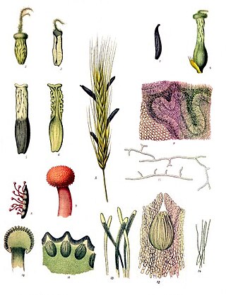
Ergot or ergot fungi refers to a group of fungi of the genus Claviceps.

<span class="mw-page-title-main">Ergine</span> Chemical compound

Ergine, also known as d-lysergic acid amide (LSA) and d-lysergamide, is an ergoline alkaloid that occurs in various species of vines of the Convolvulaceae and some species of fungi. The psychedelic properties in the seeds of ololiuhqui, Hawaiian baby woodrose and morning glories have been linked to ergine and/or isoergine, its epimer, as it is an alkaloid present in the seeds.

A mycotoxin is a toxic secondary metabolite produced by fungi and is capable of causing disease and death in both humans and other animals. The term 'mycotoxin' is usually reserved for the toxic chemical products produced by fungi that readily colonize crops.

<span class="mw-page-title-main">Ergoline</span> Chemical compound

Ergoline is a chemical compound whose structural skeleton is contained in a variety of alkaloids, referred to as ergoline derivatives or ergoline alkaloids. Ergoline alkaloids, one being ergine, were initially characterized in ergot. Some of these are implicated in the condition ergotism, which can take a convulsive form or a gangrenous form. Even so, many ergoline alkaloids have been found to be clinically useful. Annual world production of ergot alkaloids has been estimated at 5,000–8,000 kg of all ergopeptines and 10,000–15,000 kg of lysergic acid, used primarily in the manufacture of semi-synthetic derivatives.

<span class="mw-page-title-main">Ergotamine</span> Chemical compound in the ergot family of alkaloids

Ergotamine, sold under the brand names Cafergot and Ergomar among others, is an ergopeptine and part of the ergot family of alkaloids; it is structurally and biochemically closely related to ergoline. It possesses structural similarity to several neurotransmitters, and has biological activity as a vasoconstrictor.

<span class="mw-page-title-main">Lysergic acid hydroxyethylamide</span> Chemical compound

D-Lysergic acid α-hydroxyethylamide, also known as D-lysergic acid methyl carbinolamide, is a is a Lysergamide and alkaloid of the Ergoline family, it is present in higher concentrations in the parasitic fungi species "Claviceps", mainly the Claviceps Paspali, also in Claviceps Purpurea. This fungi grows in various species in the Convolvulaceae family like the Ipomoea violacea, the Rivea corymbosa (Ololiuhqui), and the Argyreia nervosa. Heavenly Blue Morning Glory and Hawaiian Baby Woodrose especially contain high amounts of LSH, with content varying between species and by how fresh the seeds are. LSH is a psychoactive Ergoline and has effects similar to LSD due to similarity in the structure and is the main psychoactive compound found in Claviceps Paspali and in (fresh) Heavenly Blue Morning Glory Seeds. LSH is unstable and breaks down into LSA quickly, so old seeds often only contains LSA and iso-LSA. When the seeds are fresh, they contain significantly higher amounts of LSH.

<span class="mw-page-title-main">Sclerotium</span> Mycelial mass

A sclerotium, is a compact mass of hardened fungal mycelium containing food reserves. One role of sclerotia is to survive environmental extremes. In some higher fungi such as ergot, sclerotia become detached and remain dormant until favorable growth conditions return. Sclerotia initially were mistaken for individual organisms and described as separate species until Louis René Tulasne proved in 1853 that sclerotia are only a stage in the life cycle of some fungi. Further investigation showed that this stage appears in many fungi belonging to many diverse groups. Sclerotia are important in the understanding of the life cycle and reproduction of fungi, as a food source, as medicine, and in agricultural blight management.

<span class="mw-page-title-main">Indole alkaloid</span> Class of alkaloids

Indole alkaloids are a class of alkaloids containing a structural moiety of indole; many indole alkaloids also include isoprene groups and are thus called terpene indole or secologanin tryptamine alkaloids. Containing more than 4100 known different compounds, it is one of the largest classes of alkaloids. Many of them possess significant physiological activity and some of them are used in medicine. The amino acid tryptophan is the biochemical precursor of indole alkaloids.

<i>Claviceps purpurea</i> Species of fungus

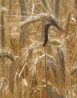
Claviceps purpurea is an ergot fungus that grows on the ears of rye and related cereal and forage plants. Consumption of grains or seeds contaminated with the survival structure of this fungus, the ergot sclerotium, can cause ergotism in humans and other mammals. C. purpurea most commonly affects outcrossing species such as rye, as well as triticale, wheat and barley. It affects oats only rarely.

<span class="mw-page-title-main">Clavicipitaceae</span> Family of fungi

The Clavicipitaceae are a family of fungi within the order Hypocreales. A 2008 estimate placed 43 genera in the family, but a study in 2020 has increased this number to 50.

<span class="mw-page-title-main">Ergocryptine</span> Chemical compound

Ergocryptine is an ergopeptine and one of the ergoline alkaloids. It is isolated from ergot or fermentation broth and it serves as starting material for the production of bromocriptine. Two isomers of ergocryptine exist, α-ergocryptine and β-ergocryptine. The beta differs from the alpha form only in the position of a single methyl group, which is a consequence of the biosynthesis in which the proteinogenic amino acid leucine is replaced by isoleucine. β-Ergocryptine was first identified in 1967 by Albert Hofmann. Ergot from different sources have different ratios of the two isomers.

<span class="mw-page-title-main">Torreyanic acid</span> Group of chemical compounds

Torreyanic acid is a dimeric quinone first isolated and by Lee et al. in 1996 from an endophyte, Pestalotiopsis microspora. This endophyte is likely the cause of the decline of Florida torreya, an endangered species that is related to the taxol-producing Taxus brevifolia. The natural product was found to be cytotoxic against 25 different human cancer cell lines with an average IC50 value of 9.4 µg/mL, ranging from 3.5 (NEC) to 45 (A549) µg/mL. Torreyanic acid was found to be 5-10 times more potent in cell lines sensitive to protein kinase C (PKC) agonists, 12-o-tetradecanoyl phorbol-13-acetate (TPA), and was shown to cause cell death via apoptosis. Torreyanic acid also promoted G1 arrest of G0 synchronized cells at 1-5 µg/mL levels, depending on the cell line. It has been proposed that the eukaryotic translation initiation factor EIF-4a is a potential biochemical target for the natural compound.

<span class="mw-page-title-main">Dideoxyverticillin A</span> Chemical compound

Dideoxyverticillin A, also known as (+)-11,11′-dideoxyverticillin A, is a complex epipolythiodioxopiperazine initially isolated from the marine fungus Penicillium sp. in 1999. It has also been found in the marine fungus Bionectriaceae, and belongs to a class of naturally occurring 2,5-diketopiperazines.

Medicinal fungi are fungi that contain metabolites or can be induced to produce metabolites through biotechnology to develop prescription drugs. Compounds successfully developed into drugs or under research include antibiotics, anti-cancer drugs, cholesterol and ergosterol synthesis inhibitors, psychotropic drugs, immunosuppressants and fungicides.

Penicillium vinaceum is an anamorph species of fungus in the genus Penicillium which produces penicillivinacine, vinaxanthone and citrmycetin.

<span class="mw-page-title-main">Phomoxanthone</span> Class of chemical compounds

The phomoxanthones are a loosely defined class of natural products. The two founding members of this class are phomoxanthone A and phomoxanthone B. Other compounds were later also classified as phomoxanthones, although a unifying nomenclature has not yet been established. The structure of all phomoxanthones is derived from a dimer of two covalently linked tetrahydroxanthones, and they differ mainly in the position of this link as well as in the acetylation status of their hydroxy groups. The phomoxanthones are structurally closely related to other tetrahydroxanthone dimers such as the secalonic acids and the eumitrins. While most phomoxanthones were discovered in fungi of the genus Phomopsis, most notably in the species Phomopsis longicolla, some have also been found in Penicillium sp.

<span class="mw-page-title-main">Phomoxanthone A</span> Chemical compound

The mycotoxin phomoxanthone A, or PXA for short, is a toxic natural product that affects the mitochondria. It is the most toxic and the best studied of the naturally occurring phomoxanthones. PXA has recently been shown to induce rapid, non-canonical mitochondrial fission by causing the mitochondrial matrix to fragment while the outer mitochondrial membrane can remain intact. This process was shown to be independent from the mitochondrial fission and fusion regulators DRP1 and OPA1.

<span class="mw-page-title-main">Phomoxanthone B</span> Chemical compound

The mycotoxin phomoxanthone B, or PXB for short, is a toxic natural product. It is a less toxic isomer of phomoxanthone A and one of the two founding members of the class of phomoxanthone compounds. The phomoxanthones are named after the fungus Phomopsis, from which they were first isolated, and after their xanthonoid structure. Chemically, they are dimers of two tetrahydroxanthones that are covalently linked to each other. PXB itself is a homodimer of two identical diacetylated tetrahydroxanthones. The position of the link between the two tetrahydroxanthones is the only structural difference between PXB and its isomers PXA and dicerandrol C: In PXA, the two xanthonoid monomers are symmetrically linked at C-4,4’, while in PXB, they are asymmetrically linked at C-2,4’, and in dicerandrol C, they are symmetrically linked at C-2,2’.

<span class="mw-page-title-main">Dicerandrol C</span> Chemical compound

Dicerandrol C is a natural product. It is a less toxic isomer of phomoxanthone A (PXA) and phomoxanthone B (PXB), all three of which are members of the class of phomoxanthone compounds. The phomoxanthones are named after the fungus Phomopsis, from which they were first isolated, and after their xanthonoid structure. Chemically, they are dimers of two tetrahydroxanthones that are covalently linked to each other. Dicerandrol C itself is a homodimer of two identical diacetylated tetrahydroxanthones. The position of the link between the two tetrahydroxanthones is the only structural difference between dicerandrol C and its isomers PXA and PXB: In PXA, the two xanthonoid monomers are symmetrically linked at C-4,4’, while in PXB, they are asymmetrically linked at C-2,4’, and in dicerandrol C, they are symmetrically linked at C-2,2’.

<span class="mw-page-title-main">Griseoxanthone C</span> Chemical compound found in some lichens

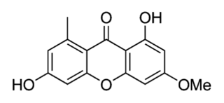
Griseoxanthone C is an organic compound in the structural class of chemicals known as xanthones. Its chemical formula is 1,6-dihydroxy-3-methoxy-8-methylxanthen-9-one, and its molecular formula is C15H12O5. It is found in a plant and some fungi, including a lichen.

References

  1. 1 2 3 4 5 6 7 8 9 Masters KS; Bräse S (May 2012). "Xanthones from fungi, lichens, and bacteria: the natural products and their synthesis". Chemical Reviews. 112 (7): 3717–3776. doi:10.1021/cr100446h. PMID   22617028.
  2. 1 2 Wezeman T, Bräse S, Masters KS (Jan 2015). "Xanthone dimers: a compound family which is both common and privileged". Natural Product Reports. 32 (1): 6–28. doi: 10.1039/c4np00050a . PMID   25226564.
  3. Neubauer, Lisa; Dopstadt, Julian; Humpf, Hans-Ulrich; Tudzynski, Paul (2016). "Identification and characterization of the ergochrome gene cluster in the plant pathogenic fungus Claviceps purpurea". Fungal Biology and Biotechnology. 3: 2. doi: 10.1186/s40694-016-0020-z . ISSN   2054-3085. PMC   5611617 . PMID   28955461.
  4. 1 2 3 Zhang W, Krohn K, Flörke U, Pescitelli G, Di Bari L, Antus S, Kurtán T, Rheinheimer J, Draeger S, Schulz B (May 2008). "New Mono‐and Dimeric Members of the Secalonic Acid Family: Blennolides A–G Isolated from the Fungus Blennoria sp". Chemistry: A European Journal. 14 (16): 4913–4923. doi:10.1002/chem.200800035. PMID   18425741.
  5. Zhai A, Zhang Y, Zhu X, Liang J, Wang X, Lin Y, Chen R (January 2011). "Secalonic acid A reduced colchicine cytotoxicity through suppression of JNK, p38 MAPKs and calcium influx". Neurochemistry International. 58 (1): 85–91. doi:10.1016/j.neuint.2010.10.016. PMID   21073911. S2CID   207121053.
  6. Zhai, Aifeng; Zhu, Xiaonan; Wang, Xuelan; Chen, Ruzhu; Wang, Hai (2013). "Secalonic acid A protects dopaminergic neurons from 1-methyl-4-phenylpyridinium (MPP+)-induced cell death via the mitochondrial apoptotic pathway". European Journal of Pharmacology. 713 (1–3): 58–67. doi:10.1016/j.ejphar.2013.04.029. ISSN   0014-2999. PMID   23665112.
  7. Millot M, Tomasi S, Studzinska E, Rouaud I, Boustie J (November 2009). "Cytotoxic constituents of the lichen Diploicia canescens". Journal of Natural Products. 72 (12): 2177–2180. doi:10.1021/np9003728. PMID   19919064.
  8. Hu, Ya-peng; Tao, Li-yang; Wang, Fang; Zhang, Jian-ye; Liang, Yong-ju; Fu, Li-wu (2013). "Secalonic acid D reduced the percentage of side populations by down-regulating the expression of ABCG2". Biochemical Pharmacology. 85 (11): 1619–1625. doi:10.1016/j.bcp.2013.04.003. ISSN   0006-2952. PMID   23583455.
  9. Deshmukh SK, Mishra PD, Kulkarni-Almeida A, Verekar S, Sahoo MR, Periyasamy G, Goswami H, Khanna A, Balakrishnan A, Vishwakarma R (May 2009). "Anti‐inflammatory and anticancer activity of ergoflavin isolated from an endophytic fungus". Chemistry & Biodiversity. 6 (5): 784–789. doi: 10.1002/cbdv.200800103 . PMID   19479845. S2CID   6353623.
  10. Eglinton G, King FE, Lloyd G, Loder JW, Marshall JR, Robertson A, Whalley WB (1958). "373. The chemistry of fungi. Part XXXV. A preliminary investigation of ergoflavin". Journal of the Chemical Society (Resumed): 1833–1842. doi:10.1039/JR9580001833.
  11. 1 2 Hooper JW, Marlow W, Whalley WB, Borthwick AD, Bowden R (1971). "The chemistry of fungi. Part LXV. The structures of ergochrysin A, isoergochrysin A, and ergoxanthin, and of secalonic acids A, B, C, and D.". Journal of the Chemical Society C: Organic. 21: 3580–3590. doi:10.1039/J39710003580. PMID   5167268.
  12. 1 2 Hooper JW, Marlow W, Whalley WB, Borthwick AD, Bowden R (1971). "The position of the biphenyl linkage in the ergot pigments. A partial synthesis of ergoflavin". Journal of the Chemical Society D: Chemical Communications (2): 111–112. doi:10.1039/C29710000111.
  13. Qin T, Iwata T, Ransom TT, Beutler JA, Porco Jr JA (November 2015). "Syntheses of Dimeric Tetrahydroxanthones with Varied Linkages: Investigation of "Shapeshifting" Properties". Journal of the American Chemical Society. 137 (48): 15225–15233. doi:10.1021/jacs.5b09825. PMC   4863954 . PMID   26544765.
  14. Qin T, Porco JA (March 2014). "Total syntheses of secalonic acids A and D". Angewandte Chemie International Edition. 53 (12): 3107–3110. doi:10.1002/anie.201311260. PMC   4098722 . PMID   24519991.
  15. Ganapathy D, Reiner JR, Löffler LE, Ma L, Gnanaprakasam B, Niepötter B, Koehne I, Tietze LF (November 2015). "Enantioselective Total Synthesis of Secalonic Acid E". Chemistry: A European Journal. 21 (47): 16807–16810. doi:10.1002/chem.201503593. PMID   26447631.