Sordaria fimicola

Last updated

Contents

Sordaria fimicola
Sordaria fimicola perithecium (tan mutant) 40X.png
Sordaria fimicola perithecium
Scientific classification OOjs UI icon edit-ltr.svg
Domain: Eukaryota
Kingdom: Fungi
Division: Ascomycota
Class: Sordariomycetes
Order: Sordariales
Family: Sordariaceae
Genus: Sordaria
Species:
S. fimicola
Binomial name
Sordaria fimicola
(Roberge ex Desm.) Ces. & De Not.
Image taken at 100x Perithecium releasing asci, some free ascospores..jpg
Image taken at 100x

Sordaria fimicola is a species of microscopic fungus. It is commonly found in the feces of herbivores. Sordaria fimicola is often used in introductory biology and mycology labs because it is easy to grow on nutrient agar in dish cultures. The genus Sordaria, closely related to Neurospora and Podospora , is a member of the large class Sordariomycetes, or flask-fungi. The natural habitat of the three species of Sordaria that have been the principal subjects in genetic studies is dung of herbivorous animals. The species S. fimicola is common and worldwide in distribution. The species of Sordaria are similar morphologically, producing black perithecia containing asci with eight dark ascospores in a linear arrangement. These species share a number of characteristics that are advantageous for genetic studies. They all have a short life cycle, usually 7–12 days, and are easily grown in culture. Most species are self-fertile and each strain is isogenic. All kinds of mutants are easily induced and readily obtainable with particular ascospore color mutants. These visual mutants aid in tetrad analysis, especially in analysis of intragenic recombination. [1]

The most common form of S. fimicola is a dark brown. Certain mutants are grey or tan. A common experiment for an introductory biology lab class is to cross one of the mutant types with a wild type and observe the ratio of coloring in the offspring. This experiment illustrates the concepts of genetic inheritance in a haploid organism. The eight ascospores are produced inside an ascus. Sordaria squashes can give us information about crossing over during meiosis. If no crossing over occurs a 4:4 pattern is produced: four black spores, and four tan spores all lined up. If crossing over does occur there is a 2:2:2:2 pattern visible, or a 2:4:2 pattern.

Another common lab use is to observe meiosis and mitosis in the fruit bodies, called perithecia. An interesting feature of S. fimicola is that its fruit body is phototrophic. Thus, as it grows the stalk will bend toward a light source and when the sac bursts, the spores are shot towards the light.

Taxonomy

Research to update Sordariomycete fungal taxonomy is ongoing, and the position of Sordaria fimicola within the taxonomy of the Sordariomycetes is being researched and updated. The sordariomycetes are known as the flask fungi because they are characterized by flask-shaped perithecia and unitunicate asci. [2] Phylogenetic studies of partial 18s ribosomal DNA strands has illuminated the phylogeny of the Sordariomycetes. [3] Morphological features that characterize the Sordariaceae include the differentiation of the hyphal envelope that surrounds the ascogonium into peripheral wall layers and a pseudoparenchymatous centrum. Broad paraphyses composed of delicate, multinucleate cells arise from the cells of the centrum and completely fill the perithecium, crushing the remaining pseudoparenchymatous cells against the perithecial wall. [4] S. fimicola differs from other species of Sordariaceae studied in the aggregation of the ascogenous cells to form a placenta‐like mass in the base of the centrum. Consequently, the asci arise in a cluster rather than in a uniform wall layer. [4]

Fungal morphology

Sordaria fimicola is an ascomycete fungus that grows well on nutrient agar, dung, and in decaying matter in soil. As an ascoymcete it has a sexual teleomorph form and an asexual anamorph form. S. fimicola typically follows the life cycle of an obligate dung fungus; sexual reproduction that is obligate to herbivore dung after passage through the herbivore's gastrointestinal tract. Following meiosis on dung, ascospores are discharged and stick onto plant surfaces where they are thought to remain epiphyllous. [5] [6] S. fimicola grows septate hyphae which at the macro level look like small dark brown fibers forming a fluffy sheet over the substrate. Small black spots will form when sexual reproduction occurs, as these spots are the location of the ascii. Additionally, there are tan and grey mutant strains of S. fimicola. [5] S. fimicola grows best in carbon rich substrates, but it also needs access to nitrogen. Research has shown that S. fimicola grows best with a carbon:nitrogen ratio between 5:1 and 10:1, as S. fimicola grown under these conditions produces markedly more perithecium than in those with higher carbon ratios or higher nitrogen ratios. S. fimicola grows well in a laboratory setting and because of that it is often used in introductory biology laboratories and also as a model organism for research. S. fimicola is also a good tool for teaching meiosis as it quickly produces diploid perithecium which undergoes meiosis to produce ascii with ordered linear tetrads of haploid ascospores. [3]

Ecology

Although S. fimicola was traditionally understood to grow on the dung of herbivorous animals or in decaying plant matter, it has also been discovered that it grows symbiotically with some plants. New research shows that S. fimicola is a facultative dung fungus and S. fimicola has been found to grow among the roots of rye-grass and wheat-grass. [7] In sterilized and unsterilized soil, S. fimicola promoted host growth and prevented mortality. Research shows that S. fimicola in potato dextrose cultures is capable of producing triacontanol and indole-3-carboxaldehyde, both of which have antibacterial properties that may help prevent host disease.[ citation needed ]

S. fimicola has also been shown to inhibit the growth of other species of fungi which are pathogenic to plants including Pestalotiopsis guepinii , Colletotrichum capsici , Curvularia lunata [Cochliobolus lunatus], Alternaria alternata and Fusarium oxysporum . [8]

S. fimicola has also been shown to negatively affect the health of some plants that it can be found growing on. When S. fimicola was first isolated from maize researchers believed that it was most likely an opportunistic pathogen. Healthy maize plants in the laboratory did not grow S. fimicola indicating that healthy maize plants may exclude it. S. fimicola did colonize healthy B. tectorum resulting in reduced fecundity and reduced growth. [7]

S. fimicola is also phototropic and the response is twofold: 1) the number of fruiting bodies produced by the fungus is influenced by light and 2) the direction which the fruiting bodies grow is also influenced. This response is likely mediated by a specific fungal receptor called the S. fimicola white collar-1 photoreceptor (SfWC-1). Other species of phototropic fungi have homologous receptors with similar receptor domains. [9] S. fimicola with a non-functional mutant SfWC-1 receptor had delayed and less-pronounced fruiting-body formation, was defective in phototropism of the perithecial beaks, and lacked the fruiting-body zonation pattern compared with the wild type. [9]

Gene conversion

Each individual meiosis generates four haploid products, and after one further round of mitosis, eight products are formed and all retained as haploid spores within the sac-like ascus (pl. asci). The retention of the products of an individual meiosis in an individual ascus has facilitated certain kinds of genetic analyses, particularly the analysis of the molecular mechanism of genetic recombination. When a wild type (+) strain is mated with a mutant (m) strain, ordinarily each ascus will contain a pattern of four + and four m spores. However, it was found that, with low frequency, some asci had ratios that differed from the expected 4+ : 4m (e.g. 6+: 2m or 2+: 6m or even 5+: 3m or 3+: 5m). In these cases it appeared that the m gene had been converted to the + gene or vice versa. And so the phenomenon was termed "gene conversion." Gene conversion was first detected in S. fimicola in 1951 by Lindsay Olive, and definitively characterized by him in 1959. [10] Olive considered that these gene conversion events resulted from "trans replication, by which a locus is copied more than the normal number of times during replication at meiotic prophase." Since then many studies on the gene conversion phenomenon were carried out with S. fimicola and other organisms, particularly other ascomycetes [see review by Whitehouse [11] (1982)]. Efforts to understand gene conversion at the molecular level have provided important insights into the mechanism and adaptive function of meiotic recombination, which in turn bears on the adaptive function of sexual reproduction.

Images

Related Research Articles

<span class="mw-page-title-main">Ascomycota</span> Division or phylum of fungi

Ascomycota is a phylum of the kingdom Fungi that, together with the Basidiomycota, forms the subkingdom Dikarya. Its members are commonly known as the sac fungi or ascomycetes. It is the largest phylum of Fungi, with over 64,000 species. The defining feature of this fungal group is the "ascus", a microscopic sexual structure in which nonmotile spores, called ascospores, are formed. However, some species of Ascomycota are asexual and thus do not form asci or ascospores. Familiar examples of sac fungi include morels, truffles, brewers' and bakers' yeast, dead man's fingers, and cup fungi. The fungal symbionts in the majority of lichens such as Cladonia belong to the Ascomycota.

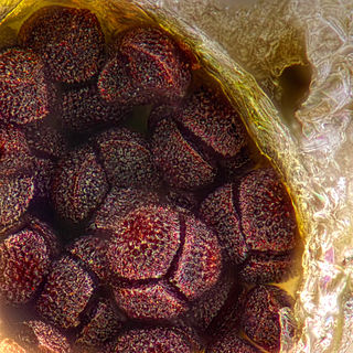
An ascocarp, or ascoma, is the fruiting body (sporocarp) of an ascomycete phylum fungus. It consists of very tightly interwoven hyphae and millions of embedded asci, each of which typically contains four to eight ascospores. Ascocarps are most commonly bowl-shaped (apothecia) but may take on a spherical or flask-like form that has a pore opening to release spores (perithecia) or no opening (cleistothecia).

<span class="mw-page-title-main">Ascus</span> Spore-bearing cell in ascomycete fungi

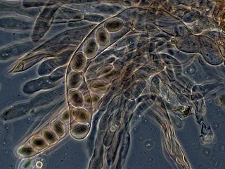
An ascus is the sexual spore-bearing cell produced in ascomycete fungi. Each ascus usually contains eight ascospores, produced by meiosis followed, in most species, by a mitotic cell division. However, asci in some genera or species can occur in numbers of one, two, four, or multiples of four. In a few cases, the ascospores can bud off conidia that may fill the asci with hundreds of conidia, or the ascospores may fragment, e.g. some Cordyceps, also filling the asci with smaller cells. Ascospores are nonmotile, usually single celled, but not infrequently may be coenocytic, and in some cases coenocytic in multiple planes. Mitotic divisions within the developing spores populate each resulting cell in septate ascospores with nuclei. The term ocular chamber, or oculus, refers to the epiplasm that is surrounded by the "bourrelet".

<i>Neurospora crassa</i> Species of ascomycete fungus in the family Sordariaceae

Neurospora crassa is a type of red bread mold of the phylum Ascomycota. The genus name, meaning 'nerve spore' in Greek, refers to the characteristic striations on the spores. The first published account of this fungus was from an infestation of French bakeries in 1843.

Heterothallic species have sexes that reside in different individuals. The term is applied particularly to distinguish heterothallic fungi, which require two compatible partners to produce sexual spores, from homothallic ones, which are capable of sexual reproduction from a single organism.

<span class="mw-page-title-main">Mating in fungi</span> Combination of genetic material between compatible mating types

Fungi are a diverse group of organisms that employ a huge variety of reproductive strategies, ranging from fully asexual to almost exclusively sexual species. Most species can reproduce both sexually and asexually, alternating between haploid and diploid forms. This contrasts with most multicellular eukaryotes such as mammals, where the adults are usually diploid and produce haploid gametes which combine to form the next generation. In fungi, both haploid and diploid forms can reproduce – haploid individuals can undergo asexual reproduction while diploid forms can produce gametes that combine to give rise to the next generation.

<span class="mw-page-title-main">Sordariaceae</span> Family of fungi

The Sordariaceae are a family of perithecial fungi within the Sordariales order.

This is a glossary of some of the terms used in phytopathology.

<span class="mw-page-title-main">Dikarya</span> Subkingdom of fungi

Dikarya is a subkingdom of Fungi that includes the divisions Ascomycota and Basidiomycota, both of which in general produce dikaryons, may be filamentous or unicellular, but are always without flagella. The Dikarya are most of the so-called "higher fungi", but also include many anamorphic species that would have been classified as molds in historical literature. Phylogenetically the two divisions regularly group together. In a 1998 publication, Thomas Cavalier-Smith referred to this group as the Neomycota.

<i>Xylaria polymorpha</i> Species of fungus

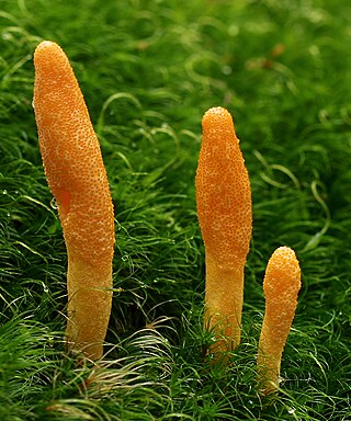
Xylaria polymorpha, commonly known as dead man's fingers, is a cosmopolitan saprobic fungus. It is characterized by its elongated upright, clavate, or strap-like stromata poking up through the ground, much like fingers.

<span class="mw-page-title-main">Tetrad (meiosis)</span> Product of meiosis in spore-producing organisms

The tetrad is the four spores produced after meiosis of a yeast or other Ascomycota, Chlamydomonas or other alga, or a plant. After parent haploids mate, they produce diploids. Under appropriate environmental conditions, diploids sporulate and undergo meiosis. The meiotic products, spores, remain packaged in the parental cell body to produce the tetrad.

Sphaerotheca castagnei is a species of ascomycete fungus in the family Erysiphaceae. A plant pathogen, it causes a form of powdery mildew.

<i>Thelebolus</i> Genus of fungi

Thelebolus is a genus of fungi in the Thelebolaceae family. Often considered related to Pezizales, 18S phylogenies show Thelebolales are a sister group to Pseudeurotiaceae and Leotiales. ITS is an adequate DNA barcode but there are only six variable sites in Thelebolus; β-tubulin is a recommended secondary barcode.

<span class="mw-page-title-main">Cordycipitaceae</span> Family of fungi

The Cordycipitaceae are a family of parasitic fungi in the Ascomycota, class Sordariomycetes and order Hypocreales. The family was first published in 1969 by mycologist Hanns Kreisel, but the naming was invalid according to the code of International Code of Nomenclature for algae, fungi, and plants. It was validly published in 2007.

Arcopilus aureus is a plant and soil fungus in the genus Arcopilus. It was first identified by A. H. Chivers in 1912, who named it Chaetomium aureum. It was later transferred to the genus Arcopilus by Wang and colleagues. The fungus has recently been recognized to have industrial use for the production of the metabolites resveratrol. and sclerotiorin Additionally, A. aureus has high lead tolerance and clearance, suggesting a potential role in environmental biotechnology.

Podospora appendiculata is a coprophilous fungus that is most commonly found in the dung of lagomorphs, such as hares and rabbits, in temperate to warm climates. A member of the division Ascomycota, P. appendiculata is characterized by ovoid, hair-studded perithecia which can bear a distinctive violaceous colouring and peridia which are coriaceous, or leathery, in texture. Podospora appendiculata has been shown to produce three compounds with antimicrobial properties.

Triangularia setosa is a member of the Ascomycota, and of the genus Triangularia. This genus is notable for its widespread appearance on the excrement of herbivores, and is therefore seen as a coprophilous fungus. The fungus itself is characteristically dark in colour and produces sac-like perithecium with a covering of hair. Its dispersion involves the ingestion, passage, and projectile ejection of spores. It has preference for colonizing the dung of lagomorphs, such as hares and rabbits.

Cercophora areolata is a member of the Ascomycota division, and is grouped into the Lasiosphaeriaceae family based on morphology. C. areolata is a coprophilous fungus that has been most recently isolated from porcupine dung. Defining features of C. areolata include: 1) ovoid-conical, glabrous ascomata, 2) black, carbonaceous, areolate peridium and 3) clavate-shaped, single-walled asci. From studies on C. areolata, this fungus produces multiple antifungal compounds, which inhibit other competitor fungi.

Magnaporthe rhizophila is a fungus species in the family Magnaporthaceae. These dark mycelial fungi are common pathogens of cereal and grass roots. Rice blast is one disease known to be caused by M. rhizophila and presents with vascular discoloration in the host organism. The fungus lives best in drier humid conditions, explaining why it is most often found in the soils of Australia, South Africa, and the Southeastern United States.

Protothelenella is a genus of fungi in the family Protothelenellaceae. It contains 11 species, some of which form lichens. Protothelenella species have a crustose thallus with spherical to pear-shaped, dark brown to blackish perithecia. Microscopic characteristics of the genus include bitunicate asci with an amyloid tholus, and ascospores that are colourless and contain multiple internal partitions. Some species grow on acidic substrates including rocks, soil, bryophytes, plant detritus or rotten wood. Other species are lichenicolous (lichen-dwelling), growing on species of Solorina, Peltigera, Pseudocyphellaria, or Cladonia.

References

  1. Alexopoulos CJ, Mims CW, Blackwell M (1996). Introductory Mycology. John Wiley and Sons. p. 361. ISBN   0-471-52229-5.
  2. Hongsanan, Sinang; Maharachchikumbura, Sajeewa S. N.; Hyde, Kevin D.; Samarakoon, Milan C.; Jeewon, Rajesh; Zhao, Qi; Al-Sadi, Abdullah M.; Bahkali, Ali H. (1 May 2017). "An updated phylogeny of Sordariomycetes based on phylogenetic and molecular clock evidence". Fungal Diversity. 84 (1): 25–41. doi:10.1007/s13225-017-0384-2. ISSN   1878-9129. S2CID   5062936.
  3. 1 2 Duong, Lam (2004). "Emarcea castanopsidicola gen. et sp. nov. from Thailand, a new xylariaceous taxon based on morphology and DNA sequences". Studies in Mycology. 50: 253–260.
  4. 1 2 Mai, Shing H. (1977). "Morphological Studies in Sordaria Fimicola and Gelasinospora Longispora". American Journal of Botany. 64 (5): 489–495. doi:10.1002/j.1537-2197.1977.tb11884.x. ISSN   1537-2197.
  5. 1 2 Alexopoulous, C. J. (1996). Introductory Mycology. John Wiley and Sons. p. 361.
  6. "Life Cycle of Sordaria Fimicola". Sciencing. Retrieved 20 May 2021.
  7. 1 2 Newcombe, George; Campbell, Jason; Griffith, David; Baynes, Melissa; Launchbaugh, Karen; Pendleton, Rosemary (3 February 2016). Albrectsen, Benedicte Riber (ed.). "Revisiting the Life Cycle of Dung Fungi, Including Sordaria fimicola". PLOS ONE. 11 (2): e0147425. Bibcode:2016PLoSO..1147425N. doi: 10.1371/journal.pone.0147425 . ISSN   1932-6203. PMC   4739622 . PMID   26839959.
  8. Jeamjitt, O. (2007). "Coprophilous fungi and antagonistic effect of Sordaria fimicola against plant pathogenic fungi in vitro". Plants: 593–600.
  9. 1 2 Krobanan, Kulsumpun; Liang, Syun-Wun; Chiu, Ho-Chen; Shen, Wei-Chiang (12 April 2019). Druzhinina, Irina S. (ed.). "The Blue-Light Photoreceptor Sfwc-1 Gene Regulates the Phototropic Response and Fruiting-Body Development in the Homothallic Ascomycete Sordaria fimicola". Applied and Environmental Microbiology. 85 (12): e02206–18, /aem/85/12/AEM.02206–18.atom. Bibcode:2019ApEnM..85E2206K. doi:10.1128/AEM.02206-18. ISSN   0099-2240. PMC   6544823 . PMID   30979837.
  10. Olive LS (1959). "Aberrant Tetrads in Sordaria Fimicola". Proc. Natl. Acad. Sci. U.S.A. 45 (5): 727–32. Bibcode:1959PNAS...45..727O. doi: 10.1073/pnas.45.5.727 . PMC   222623 . PMID   16590433.
  11. Harold L. K. Whitehouse. 1982. Genetic Recombination. New York: Wiley ISBN   978-0471102052