Streptobacillus moniliformis

Last updated

Streptobacillus moniliformis
Streptobacillus moniliformis.png
Streptobacillus moniliformis
Scientific classification OOjs UI icon edit-ltr.svg
Domain: Bacteria
Phylum: Fusobacteriota
Class: Fusobacteriia
Order: Fusobacteriales
Family: Leptotrichiaceae
Genus: Streptobacillus
Species:
S. moniliformis
Binomial name
Streptobacillus moniliformis
Levaditi et al. 1925 [1]

Streptobacillus moniliformis is a non-motile, Gram-negative rod-shaped bacterium that is a member of the family Leptotrichiaceae. [2] The genome of S. moniliformis is one of two completed sequences of the order Fusobacteriales. [3]

Contents

Etymology

Its genus name comes from the Ancient Greek word στρεπτός : streptós for "curved" or "twisted", and the Latin word bacillus meaning "small rod." The specific name moniliformis means "necklace like". [4] S. moniliformis is microaerophilic, requiring less oxygen than is present in the atmosphere for its growth. [5]

Background

History

S. moniliformis was first isolated from a rat-bitten man in 1914 by German microbiologist H. Schottmüller, who described it as Streptothrix muris ratti. [6] In the United States during the year 1916, S. moniliformis was determined to be the causative source of rat-bite fever. [7]

Microbiology

Some isolates of S. moniliformis have been collected from the upper respiratory tract of domestic and wild rats. [8] Two known variants of S. moniliformis have been identified. The bacillary type is pathogenic. [5] In contrast, the spontaneously occurring L-form, which lacks a cell wall and whose colonies grow in a "fried egg" formation, is non-pathogenic. [5]

Morphology

The bacterium S. moniliformis is a gram-negative pleomorphic rod occurring frequently in chains and tangled filaments with bulbous or Monilia-like swellings. The organism presents phenotypically as being facultatively anaerobic, non-motile, weakly ferments glucose and maltose, is catalase and oxidase-negative, does not reduce nitrate, and exhibits no growth on MacConkey agar. Morphologically, colonies of S. moniliformis are 1–2 mm in size, smooth, convex, non-hemolytic (varied presentation with an α-hemolysis is not uncommon) and gray in color. When cultured in broth, a typical "puff-ball" appearance is seen. [9]

Taxonomy

Streptobacillus moniliformis was previously classified under the family Fusobacteriaceae. [10] It was later regrouped with three other genera: Sebaldella , Sneathia , and Leptotrichia . [2] [3] [11] These four genera were classified under the family Leptotrichiaceae following comparative analyses of the 16S ribosomal RNA gene sequences [12] and 16S-23S rDNA internal transcribed spacer sequences among members of the phylum Fusobacteria. [11] Identification of conserved signature indels unique to Fusobacteria and its primary clades as well as phylogenetic analysis of members of Fusobacteria based on concatenated sequences of 17 conserved proteins further support the distinction between the two families. [11]

S. moniliformis was formerly classified as the only member of the genus Streptobacillus. [2] However, Streptobacillus strains HKU33T and HKU34 were isolated in Hong Kong in September 2014. [2] Streptobacillus HKU33T was found in pus isolated from the abscess of a 38-year-old patient with quinsy and HKU34 from the elbow joint fluid of a 64-year-old patient with septic arthritis. [2] Following analysis of the 16S rRNA gene sequences found in members of Leptotrichiaceae and partial sequences of the recA, groEl, and gyrB genes present in both isolates, the two strains were taxonomically grouped under the novel species Streptobacillus hongkongensis sp. nov. [2]

Genomics

The U.S. Department of Energy's Joint Genome Institute (DOE JGI) sequenced the complete genome of S. moniliformis DSM 12112. [3] It is made up of one circular chromosome of 1,673,280 base pairs as determined from a combination of Sanger and 454 sequencing. [3] The mol% of guanine and cytosine in the DNA is 26.3% with 1,511 protein coding genes out of the 1,566 genes predicted. [3] These low G+C values were previously only seen in members of the order Mycoplasmatales, which includes the genus Mycoplasma , indicating a relationship between Mycoplasma and S. moniliformis. [3] [13] However, 16S rRNA gene analysis showed this relation to be incorrect. [12] S. moniliformis also has a single circular plasmid pSMON01 that is 10,702 base pairs long with 1,511 protein coding genes. [3]

Isolation and identification

The fastidious nature of Streptobacillus moniliformis makes it difficult to culture, with current published recommendations stating it requires media supplemented with 20% serum, ascitic fluid, or whole blood in order to grow. Numerous published reports also state that the organism is inhibited by the polyanionic detergent sodium polyanethole sulfonate (SPS-trade name; Liquoid; Hoffman-La Roche, Inc., Nutley, N.J.), the main anticoagulant in modern commercially available blood culture bottles, used in automated continuous blood culture instruments. [9]

Optimizing the best chances for organism recovery should include the aseptic collection of blood or sterile body fluids into commercial blood culture bottles (documenting the amount of blood drawn from the patient, if applicable) during the acute phase of infection, and collected in duplicate with optimal volumes of inoculation for the isolation and growth of S. moniliformis being 10 ml for adolescent to adult patients. In a pediatric population, 4–5 ml should be extrapolated. Bottles should be incubated minimally for 7 days to ensure the best chance for organism recovery when working with low levels of bacteremia. If not yet detected by day 7, they should be blind sub-cultured to rule out a false-negative blood culture result. Automated continuous blood culturing instrumentation growth and fluorescent detection data should be checked manually during the course of incubation in order to rule out missed false-negative samples. [9]

Once a bottle shows signs of organism growth and detection, a Gram stain should be performed followed by inoculation of blood onto rabbit or sheep blood agar and brain heart infusion broth and incubated aerobically at 35 °C, 35–37 °C, or 5% CO2 enriched (microaerophilic) environment for a minimum of 3–4 days. [9]

While the organism has been reported to be inhibited by specific blood culture additives, specifically SPS in the clinical lab, current research has proposed methods to overcome this limitation with "100% culture, growth success rates". According to Szewc et al., their research into the "reported" fastidious nature of S. moniliformis, and its inhibition by the anticoagulant SPS showed, that when using a specific volume of blood for inoculum, it resulted in 100% recovery and successful growth of this organism and appeared to overcome the inhibitions and limitations that historically have been observed when using SPS for blood culturing and recovering S. moniliformis in a clinical setting. [9]

Relevance

Rat bite fever and Haverhill fever

In the U.S., rat bite fever is primarily caused by transmission of S. moniliformis from the bite of a rat. [14] However, approximately 30% of patients diagnosed with rat bite fever do not recall being scratched or bitten by an infected animal. [8] [15] Transmission of the bacterium is also known to occur via consumption of infected water, close contact with, or handling of rats. [14] [16] Haverhill fever, named after the 1926 outbreak of the disease in Haverhill, Massachusetts, is a form of rat bite fever that can result from ingesting food contaminated with S. moniliformis. [5] In 1986 at a boarding school in the United Kingdom, another outbreak of Haverhill fever was reported. Some 304 people were reported to have been afflicted. [17] Infection was suspected to have resulted from the consumption of either unpasteurized milk or water contaminated with rat feces. [18] Infected individuals described symptoms including a sudden development of vomiting, severe headache, and cold sweats with a high fever. [18] Parker and Hudson first isolated the cause of this outbreak, which they named Haverhilia multiformis. [15] This organism was later matched to S. moniliformis after further research. [19]

Symptoms of rate bite fever include the abrupt onset of fever ranging from 38 °C to 41 °C. [5] Approximately 75% of infected individuals develop a rash in addition to hemorrhaging vesicles. [14] Both the rash and vesicles are usually located on the hands and feet, although the rash has been known to spread to other parts of the body. [10]

The microaerophilic nature of S. moniliformis makes identification difficult. [5] PCR testing is being utilized more for its identification. [19] However, there is still a 13% mortality rate for untreated cases. [16] Immunocompromised individuals, such as HIV-positive individuals, are more at risk of death from this disease. [20] Lab personnel and pet store workers, who work closely with animals on a daily basis, also have an increased risk of infection. [5]

Although S. moniliformis is believed to be part of the commensal bacteria of the respiratory tract of rats, [13] rats have occasionally shown signs of the disease. [5] Antibiotics used to treat infection may cause the formation of the L-form, which persists in the body, although this form is not pathogenic. [5]

Related Research Articles

<i>Yersinia pestis</i> Species of bacteria, cause of plague

Yersinia pestis is a gram-negative, non-motile, coccobacillus bacterium without spores that is related to both Yersinia enterocolitica and Yersinia pseudotuberculosis, the pathogen from which Y. pestis evolved and responsible for the Far East scarlet-like fever. It is a facultative anaerobic organism that can infect humans via the Oriental rat flea. It causes the disease plague, which caused the Plague of Justinian and the Black Death, the deadliest pandemic in recorded history. Plague takes three main forms: pneumonic, septicemic, and bubonic. Yersinia pestis is a parasite of its host, the rat flea, which is also a parasite of rats, hence Y. pestis is a hyperparasite.

<i>Rickettsia</i> Genus of bacteria

Rickettsia is a genus of nonmotile, gram-negative, nonspore-forming, highly pleomorphic bacteria that may occur in the forms of cocci, bacilli, or threads. The genus was named after Howard Taylor Ricketts in honor of his pioneering work on tick-borne spotted fever.

<i>Campylobacter jejuni</i> Species of bacterium

Campylobacter jejuni is a species of pathogenic bacteria, one of the most common causes of food poisoning in Europe and in the US. The vast majority of cases occur as isolated events, not as part of recognized outbreaks. Active surveillance through the Foodborne Diseases Active Surveillance Network (FoodNet) indicates that about 20 cases are diagnosed each year for each 100,000 people in the US, while many more cases are undiagnosed or unreported; the CDC estimates a total of 1.5 million infections every year. The European Food Safety Authority reported 246,571 cases in 2018, and estimated approximately nine million cases of human campylobacteriosis per year in the European Union.

<i>Streptobacillus</i> Genus of bacteria

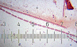
Streptobacillus is a genus of fastidious microaerophilic Gram-negative bacteria, which grow in culture as rods in chains.

Rat-bite fever (RBF) is an acute, febrile human illness caused by bacteria transmitted by rodents, in most cases, which is passed from rodent to human by the rodent's urine or mucous secretions. Alternative names for rat-bite fever include streptobacillary fever, streptobacillosis, spirillary fever, bogger, and epidemic arthritic erythema. It is a rare disease spread by infected rodents and caused by two specific types of bacteria:

  1. Streptobacillus moniliformis, the only reported bacteria that causes RBF in North America
  2. Spirillum minus, common in Asia. Most cases occur in Japan, but specific strains of the disease are present in the United States, Europe, Australia, and Africa.

Sodoku is a bacterial zoonotic disease. It is caused by the Gram-negative rod Spirillum minus. It is a form of rat-bite fever (RBF).

Seoul orthohantavirus (SEOV) is a member of the genus Orthohantavirus of rodent-borne viruses, and is one of the four hantaviruses that are known to cause Hantavirus hemorrhagic fever with renal syndrome (HFRS). It is an Old World hantavirus; a negative sense, single-stranded, tri-segmented RNA virus.

<i>Mycobacterium cosmeticum</i> Species of bacterium

Mycobacterium cosmeticum is a rapidly growing mycobacterium that was first isolated from cosmetic patients and sites performing cosmetic procedures.

Burkholderia gladioli is a species of aerobic gram-negative rod-shaped bacteria that causes disease in both humans and plants. It can also live in symbiosis with plants and fungi and is found in soil, water, the rhizosphere, and in many animals. It was formerly known as Pseudomonas marginata.

<i>Pseudomonas stutzeri</i> Species of bacterium

Pseudomonas stutzeri is a Gram-negative soil bacterium that is motile, has a single polar flagellum, and is classified as bacillus, or rod-shaped. While this bacterium was first isolated from human spinal fluid, it has since been found in many different environments due to its various characteristics and metabolic capabilities. P. stutzeri is an opportunistic pathogen in clinical settings, although infections are rare. Based on 16S rRNA analysis, this bacterium has been placed in the P. stutzeri group, to which it lends its name.

<i>Enterobacter cloacae</i> Species of bacterium

Enterobacter cloacae is a clinically significant Gram-negative, facultatively-anaerobic, rod-shaped bacterium.

Bartonella quintana, originally known as Rochalimaea quintana, and "Rickettsia quintana", is a bacterium transmitted by the human body louse that causes trench fever. This bacterial species caused outbreaks of trench fever affecting 1 million soldiers in Europe during World War I.

<i>Orientia tsutsugamushi</i> Species of bacterium

Orientia tsutsugamushi is a mite-borne bacterium belonging to the family Rickettsiaceae and is responsible for a disease called scrub typhus in humans. It is a natural and an obligate intracellular parasite of mites belonging to the family Trombiculidae. With a genome of only 2.0–2.7 Mb, it has the most repeated DNA sequences among bacterial genomes sequenced so far. The disease, scrub typhus, occurs when infected mite larvae accidentally bite humans. Primarily indicated by undifferentiated febrile illnesses, the infection can be complicated and often fatal.

<span class="mw-page-title-main">Haverhill fever</span> Medical condition

Haverhill fever is a systemic illness caused by the bacterium Streptobacillus moniliformis, an organism common in rats and mice. If untreated, the illness can have a mortality rate of up to 13%. Among the two types of rat-bite fever, Haverhill fever caused by Streptobacillus moniliformis is most common in North America. The other type of infection caused by Spirillum minus is more common in Asia and is also known as Sodoku.

Spirillum minus is an organism associated with rat-bite fever that has never been fully identified and was assigned to the genus Spirillum in 1887 based on morphology, although it is not a validly published name. As Spirillum species generally obligately microaerophiles and are not found in mammals, this organism may be misclassified. Sequencing data should help to resolve this question.

<i>Ehrlichia ewingii</i> Species of bacterium

Ehrlichia ewingii is a species of rickettsiales bacteria. It has recently been associated with human infection, and can be detected via PCR serological testing. The name Ehrlichia ewingii was proposed in 1992.

<i>Streptococcus zooepidemicus</i> Species of bacterium

Streptococcus zooepidemicus is a Lancefield group C streptococcus that was first isolated in 1934 by P. R. Edwards, and named Animal pyogens A. It is a mucosal commensal and opportunistic pathogen that infects several animals and humans, but most commonly isolated from the uterus of mares. It is a subspecies of Streptococcus equi, a contagious upper respiratory tract infection of horses, and shares greater than 98% DNA homology, as well as many of the same virulence factors.

<i>Staphylococcus</i> Genus of Gram-positive bacteria

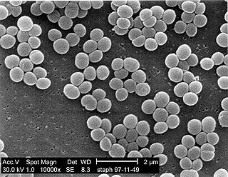
Staphylococcus is a genus of Gram-positive bacteria in the family Staphylococcaceae from the order Bacillales. Under the microscope, they appear spherical (cocci), and form in grape-like clusters. Staphylococcus species are facultative anaerobic organisms.

Dialister pneumosintes is a nonfermentative, anaerobic, gram-negative rod that grows with small, circular, transparent, shiny, smooth colonies on blood agar. D. pneumosintes has been recovered from deep periodontal pockets, but little is known about the relationship between the organism and destructive periodontal disease.

<i>Brevinema andersonii</i> Species of bacterium

Brevinema andersonii, named for John F. Anderson, who first described the organism. This organism is a Gram-negative, microaerophilic, helical shaped, chemoorganotrophic organism from the genus Brevinema. Brevinema andersonii is host associated, strains have been isolated from blood and other tissues of short-tailed shrews and white-footed mice and are infectious for laboratory mice and Syrian hamsters.B. andersonii is readily identified by restriction enzyme analysis, and SDS-PAGE, or fatty acid composition data. Another identifier for B. andersonii is the sheathed periplasmic flagella in the 1-2-1 configuration. While cells are visible by dark-field or phase-contrast microscopy, they cannot be seen when bright-field microscopy is used.

References

  1. Levaditi, C.; Nicolau, S.; Poincloux, P. (1925). "Sur le role etiologique de Streptobacillus moniliformis (nov. spec.) dans l'erytheme polymorphe aigu septicemique". Comptes Rendus Hebdomadaires des Séances de l'Académie des Sciences. Paris. 180: 1188–1190.
  2. 1 2 3 4 5 6 Woo, Patrick (2014). "Streptobacillus hongkongensis sp. nov., Isolated from Patients with Quinsy and Septic Arthritis, and Emended Descriptions of the Genus Streptobacillus and Streptobacillus moniliformis". International Journal of Systematic and Evolutionary Microbiology. 64 (Pt 9): 3034–3039. doi: 10.1099/ijs.0.061242-0 . PMID   24912824.
  3. 1 2 3 4 5 6 7 Nolan, M.; Gronow, S.; Lapidus, A.; Ivanova, N.; Copeland, A.; Lucas, S.; Glavina Del Rio, T.; Chen, F.; Tice, H.; et al. (2009). "Complete genome sequence of Streptobacillus monoiliformis type strain (9901T)". Standards in Genomic Sciences. 1 (3): 300–307. doi:10.4056/sigs.48727. ISSN   1944-3277. PMC   3035246 . PMID   21304670.
  4. Skerman, V.B.D.; McGowan, V.; Sneath, P.H.A. (1989). Approved Lists of Bacterial Names (Amended). Washington, D.C.: ASM Press. ISBN   978-1-55581-014-6. PMID   20806452.
  5. 1 2 3 4 5 6 7 8 9 Elliot, Sean P. (2007). "Rat Bite Fever and Streptobacillus moniliformis". Clinical Microbiology Reviews. 20 (1): 13–22. doi:10.1128/CMR.00016-06. PMC   1797630 . PMID   17223620.
  6. Hagelskjaer, Lena; Sørensen, Inger; Randers, Else (1998). "Streptobacillus moniliformis infection: 2 cases and a literature review". Scandinavian Journal of Infectious Diseases. 30 (3): 309–311. doi:10.1080/00365549850161016. PMID   9790145.
  7. Blake, F.G. (January 1916). "The etiology of ratbite fever". Journal of Experimental Medicine. 23 (1): 39–60. doi:10.1084/jem.23.1.39. PMC   2125348 . PMID   19867970.
  8. 1 2 Koopman, J.P.; Brink Van Den, M.E.; Vennix, P.P.C.A.; Kuypers, W.; Boot, R.; Bakker, R.H. (1991). "Isolation of Streptobacillus moniliformis from the middle ear of rats". Laboratory Animals. 25 (1): 35–39. doi: 10.1258/002367791780808211 . PMID   1826334.
  9. 1 2 3 4 5 Szewc, A.M.; Bell, M.E.; Kelly, A.J.; Humrighouse, B.W.; McQuiston, J.R. (November 2021). "Using the BDFX40 Automated Continuous Blood Culture System to Isolate and Recover Streptobacillus moniliformis in the Presence of 0.05% SPS: A 55-Year, 56-Strain Retrospective Study". Laboratory Medicine. 52 (6): 536–549. doi: 10.1093/labmed/lmab009 . PMID   33693831.
  10. 1 2 Gaastra, Wim; Boot, Ron; Ho, T.K.; Lipman, Len J.A. (2009). "Rat bite fever". Veterinary Medicine. 133 (3): 211–228. doi:10.1016/j.vetmic.2008.09.079. PMID   19008054.
  11. 1 2 3 Gupta, Radhey S.; Sethi, Mohit (August 2014). "Phylogeny and molecular signatures for the phylum Fusobacteria and its distinct subclades". Anaerobe. 28: 182–198. doi:10.1016/j.anaerobe.2014.06.007. PMID   24969840.
  12. 1 2 Staley, James T.; Whitman, William B.; Krieg, Noel R.; Brown, Daniel R.; Hedlund, Brian P.; Paster, Bruce J.; Ward, Naomi L.; Ludwig, Wolfgang (24 November 2010). Bergey's Manual of Systematic Bacteriology Volume 4 (2nd ed.). Springer. p. 789. ISBN   978-0-387-95042-6.
  13. 1 2 Wullenweber, Michael (1995). "Streptobacillus moniliformis – a zoonotic pathogen. Taxonomic considerations, host species, diagnosis, therapy, geographical distribution". Laboratory Animals. 29 (1): 1–15. doi: 10.1258/002367795780740375 . PMID   7707673.
  14. 1 2 3 Cunningham, B.B.; Paller, A.S.; Katz, B.Z. (1998). "Rat bite fever in a pet lover". Journal of the American Academy of Dermatology. 38 (2 Pt 2): 330–332. doi:10.1016/s0190-9622(98)70576-6. PMID   9486709.
  15. 1 2 Parker, R.H. (1989). "Rat-bite fever". In Hoeprich, P.D.; Jordan, M.C. (eds.). Infectious diseases: a modern treatise of infectious processes (4th ed.). Philadelphia: Lippincott. pp. 1310–1312. ISBN   0397508514.
  16. 1 2 Glasman, Peter James; Thuraisingam, Adrian (2009). "Rat bite fever: a misnomer?". BMJ Case Reports. 25 (2): 268–272. doi:10.1136/bcr.04.2009.1795. PMC   265881 . PMID   3029161.
  17. Pilsworth, R. (1983). "Haverhill fever". Lancet. 2 (8345): 336–337. doi:10.1016/s0140-6736(83)90308-2. PMID   6135846. S2CID   40022613.
  18. 1 2 Shanson, D.C.; Midgley, J.; Gazzard, B.G.; Dixey, J. (1983). "Streptobacillus moniliformis isolated from blood in four cases of Haverhill fever – first outbreak in Britain". Lancet. 2 (8341): 92–94. doi:10.1016/S0140-6736(83)90072-7. PMID   6134972. S2CID   38354599.
  19. 1 2 Boot, R.; Bakker, R.H.; Thuis, S.H.; Veenema, J.L.; DeHoo, H. (1993). "An enzyme-linked immune sorbent assay (ELISA) for monitoring rodent colonies for S. moniliformis antibodies". Laboratory Animals. 27 (4): 350–357. doi: 10.1258/002367793780745516 . PMID   8277708.
  20. Rordorf, T.; Zuger, C.; Zbinden, R. (2000). "Streptobacillus moniliformis endocarditis in an HIV-positive patient". Infection. 28 (6): 393–394. doi:10.1007/s150100070012. PMID   11139161. S2CID   41626361.