It is important to note that the TAZ gene was frequently confused with a protein called TAZ (transcriptional coactivator with PDZ-binding motif, a 50kDA protein). which is a part of the Hippo pathway and entirely unrelated to the gene of interest. The Hippo pathway TAZ protein has an official gene symbol of WWTR1.
Structure
The TAFAZZIN gene is located on the q arm of chromosome X at position 28 and it spans 10,208 base pairs.[5] The TAFAZZIN gene produces a 21.3kDa protein composed of 184 amino acids.[13][14] The structure of the encoded protein has been found to differ at their N terminus and the central region, which are two functionally notable regions. A 30 residue hydrophobic stretch at the N terminus may function as a membrane anchor, which does not exist in the shortest forms of tafazzins. The second region is a variable exposed loop located between amino acids 124 and 195 in the central region. This hydrophilic region is known to interact with other proteins. TAZ has no known resemblance to other proteins.[15] The half-life of tafazzin is just 3–6 hours, considerably shorter than most mitochondrial proteins, which may explain research difficulties in studying its structure.[16]
The putative phospholipid-binding site, which is the active site of Tafazzin, is a 57 amino acid cleft with two open ends and positively charged residues.[17] In addition, tafazzin localizes to the membrane leaflets facing the intermembrane space (IMS), which is crucial for remodeling.[18][19] Tafazzin differs from phospholipases in that it contains a conserved histidineresidue, His-77, as part of the conserved HX4D motif seen in acyltransferases. This motif is responsible for facilitating the Asp-His dyad mechanism seen in many serine proteases.[20] Many unique forms of tafazzin have been identified, with lengths from 129 to 292 amino acids.[15] Tafazzin has at least four different isoforms. It has a molecular weight around 35kDa but may also appear in lower molecular weights due to species differences in isoform expression. Seven functional classes of TAFAZZIN mutations have been classified based on the pathogenic loss-of-function mechanisms of each mutation.[21]
The TAFAZZIN gene contains two peptides independent of its active site for directing the protein to the mitochondria, forming residues 84–95 in exon 3 and residues 185–200 in exon 7/8 targets.[22] Tafazzin localizes with peripheral association to membrane leaflets between the inner mitochondrial membrane (IMM) and outer mitochondrial membrane (OMM), facing the intermembrane space (IMS).[18][19] Tafazzin's characteristic interfacial anchoring is achieved by its hydrophobic sequence from residues 215–232.[23] Finally, the translocase of the outer membrane (TOM) and the translocase of the inner membrane (TIM) mediates tafazzin's movement and insertion into the OMM and anchoring to IMM.[23]
Function
The TAFAZZIN gene provides instructions for producing a protein called tafazzin, which is localized to mitochondria, the energy-producing centers of cells. Tafazzin transacylase activity is responsible for cardiolipin remodeling, critical to maintaining mitochondrial inner membrane structure and function. It also has unique acyl specificity and membrane curvature sensing capabilities.[24]
Transacylase (remodeling)
After its synthesis, cardiolipin cannot exert its proper functions until it is actively remodeled. Tafazzin, an acyl-specific transferase, catalyzes the acyl transfer reaction between phospholipids and lysophospholipids in a CoA-independent manner. The remodeling process of cardiolipin involves reaching a final acyl composition that is primarily linoleoyl residues.[7][25]TAZ interacts with an immature cardiolipin by adding the fatty acid linoleic acid, which catalyzes the remodeling of the cardiolipin. The remodeling is achieved by transacylation or the deacylation-reacylation cycle. The deacylation-reacylation cycle, also known as the Lands cycle, begins with deacylation mediated by phospholipase Cld1 to form monolysocardiolipin (MLCL).[26] MLCL is reacylated by tafazzin in a single-step reaction which transfers a linoleic acid group from phosphatidylcholine (PC), completing the CL deacylation-reacylation cycle.[7][27] In contrast, transacylation involves the transfer of a linoleic acid group from phosphatidylcholine (PC) to MLCL. Such enzymatic activity forms lyso-PC and CL, and enriches the specific acyl chain of cardiolipin. The process has been shown to be specific for linoleoyl-containing PC. Such remodeling processes converts cardiolipin into a mature composition that contains a predominance of tetralinoleoyl moieties. CL remodeling in mammals requires additional enzymes, such as monolysocardiolipin acyltransferase (MLCLAT), acyl-CoA:lysocardiolipin acyltransferase (ALCAT), and phospholipase.[28] The process enables the proper function of cardiolipin.[8][9][29]
Acyl specificity and sensing curvature
Tafazzin in CL remodeling has been shown to have a clear preference for linoleoyl-containing PC in forming mature CL.[30] This specificity leads a mature composition of CL that contains a predominance of tetralinoleoyl moieties, which leads to the enrichment of tetralinoleoyl-cardiolipin (CL4).[29] The preference for lineoyl groups has been reported to be ten times greater than that of oleoyl groups and twenty times greater than that of arachidonoyl groups.[7] Conflicting explanations for this preference have included causation from energy minimization with influences by the surrounding microenvironment, known as the thermodynamic remodeling hypothesis,[31] or the inherent enzymatic preference of tafazzin for specific acyl residues.[19]
Tafazzin and cardiolipin in mitochondrial structure and function
Cardiolipin is a complex glycerophospholipid which contains four acyl groups linked to three glycerol moietie localized in the mitochondrial inner membrane. These acyl groups include oleic acid and linoleic acid. Due to this composition, cardiolipin exhibits a conical structure, which allows for membrane curvature called cristae. Further, CL plays important roles in oxidative phosphorylation by stabilizing the chain complexes with its linkages between acyl chains,[29] binding to the c-rings of ATP synthase for proper function,[32] maintaining respiratory chain supercomplex formation with proteins localized in the inner mitochondrial matrix including ATP/ADP translocase, pyruvate carrier, carnitine carrier, and all of the respiratory chain complexes (I, III, IV, V).[33][34][35] Cardiolipin also facilitates proton trapping in the intermembrane space to aid ATP synthase in channeling protons into the mitochondrial matrix. Properly formed CL is critical in maintaining mitochondrial shape, energy production, and protein transport within cells,[9] and remodeling by tafazzin aids in removing and replacing acyl chains damaged by oxidative stress.[36] During apoptosis and similar processes, CL is known to act as a platform for proteins and other machinery involved with its interactions with members of the Bcl-2 family, caspases, Bid, Bax, and Bak.[37]
Barth syndrome (BTHS) is an X-linked disease caused by mutations in the TAFAZZIN gene.[38] More than 160 mutations in the TAFAZZIN gene have been linked to this disease. It is a rare disease, found in 1 out of every 300,000 to 400,000 live births, though it is widely known that the disease is underdiagnosed.[39][40] Although BTHS occurs almost exclusively in males, there has been one identified case of BTHS in a female patient.[41] Tafazzin is responsible for remodeling of a phospholipid cardiolipin (CL),[42] the signature lipid of the mitochondrial inner membrane. TAFAZZIN gene mutations that cause Barth syndrome result in the production of tafazzin proteins with little or no function. As a result, linoleic acid is not added to cardiolipin, which disrupts normal mitochondrial shape and function, including energy production and protein transport. Barth syndrome patients exhibit defects in cardiolipin metabolism, including aberrant cardiolipin fatty acyl composition, accumulation of monolysocardiolipin (MLCL), and reduced total cardiolipin levels.[43][44] This may lead to acute metabolic decompensation and sudden death. Tissues with high energy demands, such as the heart and other muscles, are most susceptible to cell death due to reduced energy production in mitochondria and protein transport.[citation needed]
Additionally, affected white blood cells have abnormally shaped mitochondria, which could impair their ability to grow (proliferate) and mature (differentiate), leading to a weakened immune system and recurrent infections. Dysfunctional mitochondria likely lead to other signs and symptoms of Barth syndrome.[9]
There is no known cure for BTHS, and treatment of BTHS is convoluted and delayed due to the disease's varying phenotypes and its sheer complexity.[40] Thus, many treatments focus on cardiovascular and metabolic disorders, rather than treating the symptom itself. Elamipretide, an agent which protects CL from oxidative damage to maintain mitochondrial cristae and oxidative phosphorylation, is currently being tested in clinical trials.[51][52] Further, dietary fatty acids have been used to enhance bioenergetics and cardiac function in BTHS.[53] However, severe manifestations of the symptoms in BTHS patients require heart transplantation. Statistics show 9 out of 73 (12%) surviving patients who have undergone cardiac transplantation at the last update.[54][47] Heart transplantation in BTHS patients has generally been successful.[47]
Cardiovascular pathology
Cardiomyopathy is a prominent feature of Barth syndrome. The change in acyl chain composition and lipid peroxidation caused by defective tafazzin can cause defective sarcomeric action, which may lead to an insufficient power stroke, severely weakened tissue, enlarged left ventricle, partial or incomplete contraction, and decreased ejection volume. Such consequences contribute to the cardiomyopathic phenotypes of Barth syndrome, marked by a weakened heart and diminished contractility.[35][55] Alternatively, reactive oxygen species (ROS) has been suggested as the primary cause of cardiovascular impairments in BTHS.[56]
Cardiomyopathy in BTHS is exhibited at varying levels. A cohort study of BTHS patients showed 41.5% of all diagnosed cardiomyopathies in the range from birth to one month of age, and 95% exhibited a history of cardiomyopathy.[54] Furthermore, there have been cases with mild or late-onset cardiomyopathies, such as two infantile patients without cardiomyopathic phenotypes at the time of diagnosis.[57] Cardiomyopathy in Barth syndrome is primarily exhibited in multiple forms, including dilated cardiomyopathy (DCM), left ventricular condition in which the heart becomes weakened and enlarged, and therefore cannot pump blood efficiently. The resulting decrease in blood flow can lead to swelling in the legs and abdomen, fluid in the lungs, and an increased risk of blood clots.[9] Some mutations in the TAFAZZIN gene cause dilated cardiomyopathy without the other features of Barth syndrome. LVNC is a condition in which the left ventricle, characterized by a spongy structure on the ventricular wall, exhibits prominent trabeculations and deep intertrabecular recesses.[58] INVM occurs when the lower left chamber of the heart (left ventricle) does not develop correctly. In INVM, the heart muscle is weakened and cannot pump blood efficiently, frequently causing heart failure.[59][35] Abnormal heart rhythms (arrythmias) can also occur.[citation needed]
Musculoskeletal pathology
Musculoskeletalpathology is exhibited in varying forms in BTHS patients. A common phenotype is both generalized and local weakness. Weakness is exhibited as overt muscle weakness and increased exertional fatigue due to skeletalmyopathy. It worsens when present with the cardiovascular symptoms of Barth syndrome.[60] Additional symptoms of musculoskeletal pathology include hypotonia, delayed motor development, short stature, and facial dysmorphia in varying degrees.[61] Furthermore, it has been shown that a downshift in weight, length, and height relative to the normal population is exhibited in BTHS patients.[citation needed]
Treatment for developmental delays have included cornstarch supplementation as an alternative source of glucose.[47] Metabolic deficiencies have been treated by oral arginine and carnitine supplementation, which has been shown to ameliorate cardiac function and muscle weakness in some patients.[62][63][64] However, no formal assessment of the utility of carnitine and arginine supplementation has been published, and its uses have only been effective in patients with specific deficiencies.[62][64]
Neurological pathology
Cognitive impairments are common in BTHS patients in varying degrees. While a higher incidence of cognitive impairment[65] and mild learning and speech difficulties[54] are often manifested, many BTHS patients have also displayed normal cognitive abilities.[61] This shows the limited neurologic involvement in BTHS, despite tafazzin's crucial roles in brain mitochondrial respiration and normal cognitive function.[66] One study has shown that the brain has a distinct CL composition, with more diverse and less tetralinoleoyl-dependent CL. This composition diminishes the need for CL remodeling, resulting in a less tafazzin-dependent composition.[29] Another study found that the brain has a higher concentration of saturated acyl chains.[67] Finally, the brain has a higher ROS scavenging capability, which allows the circumvention of the harmful effects of ROS.[66] These findings explain the neurological phenotypes in BTHS patients.[68]
Metabolic disorders
Metabolic disorders in BTHS are exhibited in the form of 3-methylglutaconic aciduria (3-MGA), a condition characterized by increased levels of organic acids in urine, including 3‐methylglutaconic acid, 3‐methylglutaric acid, and 2‐ethyl-hydracrylic acid.[69] While 3-MGA is largely excreted in BTHS patients, some patients have been found to have normal levels of organic acids in urine.[70] Treatment of 3-MGA and metabolic deficiencies have included riboflavin or coenzyme Q10, which have shown significant improvement in patients.[69]
Hematologic pathology
The major hematologic pathology for BTHS patients is neutropenia, a condition characterized by a decline in total number of neutrophils in circulation with an increase in monocytes and eosinophils and no fluctuations in lymphocytes.[47] Presentation of neutropenia varies from mild to severe, cyclical to non-cyclical, and intermittent to chronic.[71] An absolute neutrophil count of <500/μL, defined as severe chronic neutropenia (SCN), is the most deleterious form.[72]
Cancer
TAFAZZIN has been found to be highly expressed in gastric cancer cells resistant to cisplatin. This resistance was identified to be due to the acquired ability of the cancer cells to undergo epithelial-mesenchymal transition (EMT). The findings that TAFAZZIN is involved in inducing EMT as well as its high levels in these cancer cells may point to its involvement in gastric cancer.[10][11]TAFAZZIN overexpression has been linked to rectal cancer,[73] prostate cancer,[74] thyroid neoplasm,[75] and cervical cancer.[12] In a study of 140 Swedish rectal cancer patients, TAFAZZIN overexpression was associated with an increase in the expression of oncogenes (FXYD-3 and Livin). It was also found to enhance cell anti-apoptosis response and abnormal cell growth and was even found to be an indicator of rectal cancer's stage, type, and progression.[73] Additionally, the levels of TAFAZZIN were connected to the radiotherapy response of the patients, potentially offering insight into cancer recurrence in patients.[73] A potential link between PI3K and TAFAZZIN indicates a possible association between PI3K signaling and TAFAZZIN as both were highly elevated in PTEN mutant cancer cells.[10] In prostate cancer, CL, which is remodeled by tafazzin, was shown to have high palmitoleic acid content, which was found to have the ability to stimulate prostate cancer cell proliferation and reduce the rate of apoptosis.[74] In thyroid neoplasm, TAFAZZIN allows follicular adenomas to be distinguished from follicular carcinomas,[75] while in cervical cancer tafazzin levels increased from normal tissue, to squamous intraepithelial lesions, to squamous cervical carcinoma. Based on studies of cervical cancer progression, it is believed that TAFAZZIN may induce cancer by inhibiting apoptosis and promoting cancer cell growth, viability, and tumorigenesis.[12]
The protein was discovered in 1996 by Italian scientists Silvia Bione et al..[15] Owing to the complex procedure required for the identification of tafazzin, the protein was named after Tafazzi, an Italian comedy character played by Giacomo Poretti who enthusiastically beats his groin with a plastic bottle.[77]
Barth syndrome (BTHS) is a rare but serious X-linked genetic disorder, caused by changes in phospholipid structure and metabolism. It may affect multiple body systems, and is potentially fatal. The syndrome is diagnosed almost exclusively in males.
Succinate dehydrogenase [ubiquinone] cytochrome b small subunit, mitochondrial (CybS), also known as succinate dehydrogenase complex subunit D (SDHD), is a protein that in humans is encoded by the SDHD gene. Names previously used for SDHD were PGL and PGL1. Succinate dehydrogenase is an important enzyme in both the citric acid cycle and the electron transport chain. Hereditary PGL-PCC syndrome is caused by a parental imprint of the SDHD gene. Screening can begin by 6 years of age.
Cardiolipin is an important component of the inner mitochondrial membrane, where it constitutes about 20% of the total lipid composition. It can also be found in the membranes of most bacteria. The name "cardiolipin" is derived from the fact that it was first found in animal hearts. It was first isolated from the beef heart in the early 1940s by Mary C. Pangborn. In mammalian cells, but also in plant cells, cardiolipin (CL) is found almost exclusively in the inner mitochondrial membrane, where it is essential for the optimal function of numerous enzymes that are involved in mitochondrial energy metabolism.
Very long-chain specific acyl-CoA dehydrogenase, mitochondrial (VLCAD) is an enzyme that in humans is encoded by the ACADVL gene.
Phosphatase and tensin homolog (PTEN) is a phosphatase in humans and is encoded by the PTEN gene. Mutations of this gene are a step in the development of many cancers, specifically glioblastoma, lung cancer, breast cancer, and prostate cancer. Genes corresponding to PTEN (orthologs) have been identified in most mammals for which complete genome data are available.
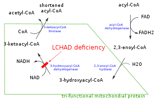
Mitochondrial trifunctional protein (MTP) is a protein attached to the inner mitochondrial membrane which catalyzes three out of the four steps in beta oxidation. MTP is a hetero-octamer composed of four alpha and four beta subunits:
Succinate dehydrogenase complex, subunit A, flavoprotein variant is a protein that in humans is encoded by the SDHA gene. This gene encodes a major catalytic subunit of succinate-ubiquinone oxidoreductase, a complex of the mitochondrial respiratory chain. The complex is composed of four nuclear-encoded subunits and is localized in the mitochondrial inner membrane. SDHA contains the FAD binding site where succinate is deprotonated and converted to fumarate. Mutations in this gene have been associated with a form of mitochondrial respiratory chain deficiency known as Leigh Syndrome. A pseudogene has been identified on chromosome 3q29. Alternatively spliced transcript variants encoding different isoforms have been found for this gene.
Trifunctional enzyme subunit alpha, mitochondrial also known as hydroxyacyl-CoA dehydrogenase/3-ketoacyl-CoA thiolase/enoyl-CoA hydratase, alpha subunit is a protein that in humans is encoded by the HADHA gene. Mutations in HADHA have been associated with trifunctional protein deficiency or long-chain 3-hydroxyacyl-coenzyme A dehydrogenase deficiency.
Trifunctional enzyme subunit beta, mitochondrial (TP-beta) also known as 3-ketoacyl-CoA thiolase, acetyl-CoA acyltransferase, or beta-ketothiolase is an enzyme that in humans is encoded by the HADHB gene.
Mitochondrial import inner membrane translocase subunit Tim8 A, also known as deafness-dystonia peptide or protein is an enzyme that in humans is encoded by the TIMM8A gene. This translocase has similarity to yeast mitochondrial proteins that are involved in the import of metabolite transporters from the cytoplasm into the mitochondrial inner membrane. The gene is mutated in deafness-dystonia syndrome and it is postulated that MTS/DFN-1 is a mitochondrial disease caused by a defective mitochondrial protein import system.
The human gene AGK encodes the enzyme mitochondrial acylglycerol kinase.
Mitochondrial import inner membrane translocase subunit TIM14 is an enzyme that in humans is encoded by the DNAJC19 gene on chromosome 3. TIM14 belongs to the DnaJ family, which has been involved in Hsp40/Hsp70 chaperone systems. As a mitochondrial chaperone, TIM14 functions as part of the TIM23 complex import motor to facilitate the import of nuclear-encoded proteins into the mitochondria. TIM14 also complexes with prohibitin complexes to regulate mitochondrial morphogenesis, and has been implicated in dilated cardiomyopathy with ataxia.
Acyl-CoA dehydrogenase family member 9, mitochondrial is an enzyme that in humans is encoded by the ACAD9 gene. Mitochondrial Complex I Deficiency with varying clinical manifestations has been associated with mutations in ACAD9.
Tricarboxylate transport protein, mitochondrial, also known as tricarboxylate carrier protein and citrate transport protein (CTP), is a protein that in humans is encoded by the SLC25A1 gene. SLC25A1 belongs to the mitochondrial carrier gene family SLC25. High levels of the tricarboxylate transport protein are found in the liver, pancreas and kidney. Lower or no levels are present in the brain, heart, skeletal muscle, placenta and lung.
Cytochrome c oxidase assembly protein COX15 homolog (COX15), also known as heme A synthase, is a protein that in humans is encoded by the COX15 gene. This protein localizes to the inner mitochondrial membrane and involved in heme A biosynthesis. COX15 is also part of a three-component mono-oxygenase that catalyses the hydroxylation of the methyl group at position eight of the protoheme molecule. Mutations in this gene has been reported in patients with hypertrophic cardiomyopathy as well as Leigh syndrome, and characterized by delayed onset of symptoms, hypotonia, feeding difficulties, failure to thrive, motor regression, and brain stem signs.
Serine active site-containing protein 1, or Protein SERAC1 is a protein in humans that is encoded by the SERAC1 gene. The protein encoded by this gene is a phosphatidylglycerol remodeling protein found at the interface of mitochondria and endoplasmic reticula, where it mediates phospholipid exchange. The encoded protein plays a major role in mitochondrial function and intracellular cholesterol trafficking. Defects in this gene are a cause of 3-methylglutaconic aciduria with deafness, encephalopathy, and Leigh-like syndrome (MEGDEL). Two transcript variants, one protein-coding and the other non-protein coding, have been found for this gene.
Monolysocardiolipin acyltransferase is a mitochondrial acyltransferase that facilitates the remodeling of monolysocardiolipin (MLCL) into cardiolipin.
Acyl-CoA:lysocardiolipin acyltransferase-1 (ALCAT1) is a polyglycerophospholipid acyltransferase of the endoplasmic reticulum which is primarily known for catalyzing the acylation of monolysocardiolipin back into cardiolipin, although it does catalyze the acylation of other polyglycerophospholipids.
NADH:ubiquinone oxidoreductase complex assembly factor 4, (NDUFAF4) also known as Hormone-regulated proliferation-associated protein of 20 kDa, (HRPAP20) or C6orf66 is a protein that in humans is encoded by the NDUFAF4 gene. NDUFAF4 is a mitochondrial assembly protein involved in the assembly of NADH dehydrogenase (ubiquinone) also known as complex I, which is located in the mitochondrial inner membrane and is the largest of the five complexes of the electron transport chain. Mutations in this gene have been associated with complex I deficiency and infantile mitochondrial encephalomyopathy. Elevations in HRPAP20 have also been implicated in breast cancer.
Transmembrane protein 70 is a protein that in humans is encoded by the TMEM70 gene. It is a transmembrane protein located in the mitochondrial inner membrane involved in the assembly of the F1 and Fo structural subunits of ATP synthase. Mutations in this gene have been associated with neonatal mitochondrial encephalo-cardiomyopathy due to ATP synthase deficiency, causing a wide variety of symptoms including 3-methylglutaconic aciduria, lactic acidosis, mitochondrial myopathy, and cardiomyopathy.
↑ Tang Y, Xia H, Li D (2018). "Membrane Phospholipid Biosynthesis in Bacteria". In Cao Y (ed.). Advances in Membrane Proteins. Springer Singapore. pp.77–119. doi:10.1007/978-981-13-0532-0_4. ISBN9789811305313.
↑ Schlame M (March 2013). "Cardiolipin remodeling and the function of tafazzin". Biochimica et Biophysica Acta (BBA) - Molecular and Cell Biology of Lipids. 1831 (3): 582–8. doi:10.1016/j.bbalip.2012.11.007. PMID23200781.
1 2 3 Barth PG, Valianpour F, Bowen VM, Lam J, Duran M, Vaz FM, Wanders RJ (May 2004). "X-linked cardioskeletal myopathy and neutropenia (Barth syndrome): an update". American Journal of Medical Genetics. Part A. 126A (4): 349–54. doi:10.1002/ajmg.a.20660. PMID15098233. S2CID25566280.
↑ Barth PG, Scholte HR, Berden JA, Van der Klei-Van Moorsel JM, Luyt-Houwen IE, Van 't Veer-Korthof ET, etal. (December 1983). "An X-linked mitochondrial disease affecting cardiac muscle, skeletal muscle and neutrophil leucocytes". Journal of the Neurological Sciences. 62 (1–3): 327–55. doi:10.1016/0022-510x(83)90209-5. PMID6142097. S2CID22790290.
↑ Aprikyan AA, Khuchua Z (May 2013). "Advances in the understanding of Barth syndrome". British Journal of Haematology. 161 (3): 330–8. doi:10.1111/bjh.12271. PMID23432031. S2CID2324502.
↑ Yen TY, Hwu WL, Chien YH, Wu MH, Lin MT, Tsao LY, etal. (August 2008). "Acute metabolic decompensation and sudden death in Barth syndrome: report of a family and a literature review". European Journal of Pediatrics. 167 (8): 941–4. doi:10.1007/s00431-007-0592-y. PMID17846786. S2CID23471712.
↑ Clinical trial number NCT02976038 for "Open-Label Extension Trial to Characterize the Long-term Safety and Tolerability of Elamipretide in Subjects With Genetically Confirmed Primary Mitochondrial Myopathy (PMM)" at ClinicalTrials.gov
↑ Monteiro JP, Oliveira PJ, Jurado AS (October 2013). "Mitochondrial membrane lipid remodeling in pathophysiology: a new target for diet and therapeutic interventions". Progress in Lipid Research. 52 (4): 513–28. doi:10.1016/j.plipres.2013.06.002. hdl:10316/25581. PMID23827885.
1 2 3 Roberts AE, Nixon C, Steward CG, Gauvreau K, Maisenbacher M, Fletcher M, etal. (November 2012). "The Barth Syndrome Registry: distinguishing disease characteristics and growth data from a longitudinal study". American Journal of Medical Genetics. Part A. 158A (11): 2726–32. doi:10.1002/ajmg.a.35609. PMID23045169. S2CID29937658.
↑ Woiewodski L, Ezon D, Cooper J, Feingold B (April 2017). "Barth Syndrome with Late-Onset Cardiomyopathy: A Missed Opportunity for Diagnosis". The Journal of Pediatrics. 183: 196–198. doi:10.1016/j.jpeds.2016.12.070. PMID28108107.
↑ Bleyl SB, Mumford BR, Brown-Harrison MC, Pagotto LT, Carey JC, Pysher TJ, etal. (October 1997). "Xq28-linked noncompaction of the left ventricular myocardium: prenatal diagnosis and pathologic analysis of affected individuals". American Journal of Medical Genetics. 72 (3): 257–65. doi:10.1002/(SICI)1096-8628(19971031)72:3<257::AID-AJMG2>3.0.CO;2-O. PMID9332651.
↑ Spencer CT, Byrne BJ, Bryant RM, Margossian R, Maisenbacher M, Breitenger P, etal. (November 2011). "Impaired cardiac reserve and severely diminished skeletal muscle O₂ utilization mediate exercise intolerance in Barth syndrome". American Journal of Physiology. Heart and Circulatory Physiology. 301 (5): H2122-9. doi:10.1152/ajpheart.00479.2010. PMID21873497.
1 2 Adès LC, Gedeon AK, Wilson MJ, Latham M, Partington MW, Mulley JC, etal. (February 1993). "Barth syndrome: clinical features and confirmation of gene localisation to distal Xq28". American Journal of Medical Genetics. 45 (3): 327–34. doi:10.1002/ajmg.1320450309. PMID8434619.
1 2 Ferreira C, Thompson R, Vernon H (1993). Adam MP, Ardinger HH, Pagon RA, Wallace SE, Bean LJ, Stephens K, Amemiya A (eds.). Barth Syndrome. University of Washington, Seattle. PMID25299040.{{cite book}}: |work= ignored (help)
1 2 Sapandowski A, Stope M, Evert K, Evert M, Zimmermann U, Peter D, etal. (December 2015). "Cardiolipin composition correlates with prostate cancer cell proliferation". Molecular and Cellular Biochemistry. 410 (1–2): 175–85. doi:10.1007/s11010-015-2549-1. PMID26314254. S2CID10664158.
Takeda A, Sudo A, Yamada M, Yamazawa H, Izumi G, Nishino I, Ariga T (November 2011). "Barth syndrome diagnosed in the subclinical stage of heart failure based on the presence of lipid storage myopathy and isolated noncompaction of the ventricular myocardium". European Journal of Pediatrics. 170 (11): 1481–4. doi:10.1007/s00431-011-1576-5. PMID21932011. S2CID23956305.
Bachou T, Giannakopoulos A, Trapali C, Vazeou A, Kattamis A (2009). "A novel mutation in the G4.5 (TAZ) gene in a Greek patient with Barth syndrome". Blood Cells, Molecules & Diseases. 42 (3): 262–4. doi:10.1016/j.bcmd.2008.11.004. PMID19261493.
Gonzalez IL (May 2005). "Barth syndrome: TAZ gene mutations, mRNAs, and evolution". American Journal of Medical Genetics Part A. 134 (4): 409–14. doi:10.1002/ajmg.a.30661. PMID15793838. S2CID119636.
Stelzl U, Worm U, Lalowski M, Haenig C, Brembeck FH, Goehler H, Stroedicke M, Zenkner M, Schoenherr A, Koeppen S, Timm J, Mintzlaff S, Abraham C, Bock N, Kietzmann S, Goedde A, Toksöz E, Droege A, Krobitsch S, Korn B, Birchmeier W, Lehrach H, Wanker EE (September 2005). "A human protein-protein interaction network: a resource for annotating the proteome". Cell. 122 (6): 957–68. doi:10.1016/j.cell.2005.08.029. hdl:11858/00-001M-0000-0010-8592-0. PMID16169070. S2CID8235923.
Mehrle A, Rosenfelder H, Schupp I, del Val C, Arlt D, Hahne F, Bechtel S, Simpson J, Hofmann O, Hide W, Glatting KH, Huber W, Pepperkok R, Poustka A, Wiemann S (January 2006). "The LIFEdb database in 2006". Nucleic Acids Research. 34 (Database issue): D415–8. doi:10.1093/nar/gkj139. PMC1347501. PMID16381901.
McKenzie M, Lazarou M, Thorburn DR, Ryan MT (August 2006). "Mitochondrial respiratory chain supercomplexes are destabilized in Barth Syndrome patients". Journal of Molecular Biology. 361 (3): 462–9. CiteSeerX10.1.1.314.3366. doi:10.1016/j.jmb.2006.06.057. PMID16857210.
Lu B, Kelher MR, Lee DP, Lewin TM, Coleman RA, Choy PC, Hatch GM (October 2004). "Complex expression pattern of the Barth syndrome gene product tafazzin in human cell lines and murine tissues". Biochemistry and Cell Biology. 82 (5): 569–76. doi:10.1139/o04-055. PMID15499385.
This page is based on this Wikipedia article Text is available under the CC BY-SA 4.0 license; additional terms may apply. Images, videos and audio are available under their respective licenses.