WWTR1

Last updated
WWTR1
Identifiers
Aliases WWTR1 , TAZ, WW domain containing transcription regulator 1
External IDs OMIM: 607392 MGI: 1917649 HomoloGene: 9159 GeneCards: WWTR1
Orthologs
SpeciesHumanMouse
Entrez
Ensembl
UniProt
RefSeq (mRNA)

NM_001168278
NM_001168280
NM_015472
NM_001348362

NM_001168281
NM_133784

RefSeq (protein)

NP_001161750
NP_001161752
NP_056287
NP_001335291

NP_001161753
NP_598545

Location (UCSC) Chr 3: 149.52 – 149.74 Mb Chr 3: 57.36 – 57.48 Mb
PubMed search [3] [4]
Wikidata
View/Edit Human View/Edit Mouse

WW domain-containing transcription regulator protein 1 (WWTR1 [5] ), also known as Transcriptional coactivator with PDZ-binding motif (TAZ), is a protein that in humans is encoded by the WWTR1 gene. WWTR1 acts as a transcriptional coregulator and has no effect on transcription alone. [5] When in complex with transcription factor binding partners, WWTR1 helps promote gene expression in pathways associated with development, cell growth and survival, and inhibiting apoptosis. [6] Aberrant WWTR1 function has been implicated for its role in driving cancers. [7] [8] [9] WWTR1 is often referred to as TAZ due to its initial characterization with the name TAZ. However, WWTR1 (TAZ) is not to be confused with the protein tafazzin, which originally held the official gene symbol TAZ, and is now TAFAZZIN.

Contents

Structure

Protein structure of WWTR1 as predicted by AlphaFold. Prredicted WWTR1 Structure - AlphaFold.png
Protein structure of WWTR1 as predicted by AlphaFold.
Differences in binding domains present on transcriptional coregulators, YAP and TAZ. YAP vs TAZ Binding Domains.png
Differences in binding domains present on transcriptional coregulators, YAP and TAZ.

WWTR1 contains a proline rich region, TEAD binding motif, WW domain, coiled coil region, and a transactivation domain (TAD) containing the PDZ domain-binding motif. WWTR1 (TAZ) lacks a DNA binding domain so it can not directly drive transcription. WWTR1 exhibits conserved structural homology with another transcriptional coregulator, yes-associated protein 1 (YAP). [5] Both YAP and TAZ are able to form homodimers and heterodimers with each other through interactions at the coil coil domain. [12] YAP and TAZ cooperate with transcription factors to promote tissue formation. WWTR1 (TAZ) interacts with a variety of transcriptional partners, including the four TEA domain family members (TEAD1 /2/ 3/ 4) through the TEAD-binding motif and several other factors containing the PPXY motif, which consists of a Proline-Proline-X (any amino acid)-Tyrosine sequence. Examples of such partners include Runx/PEBP2, AP2, C/EBP, c-Jun, Krox-20, Krox-24, MEF2B, NF-E2, Oct-4 and p73, which interact with WWTR1 via the WW domain. [6] The transactivation domain at the C-terminal end (amino acids 165–395) was shown to be important in producing transcriptional effects. [6]

Function

WWTR1 (TAZ) and the structurally similar protein, YAP, act as transcriptional coactivators and are regulated by Hippo pathway activation. YAP and TAZ - Biochemical Regulation Diagram.png
WWTR1 (TAZ) and the structurally similar protein, YAP, act as transcriptional coactivators and are regulated by Hippo pathway activation.

WWTR1 (TAZ) plays an important role in embryogenesis and development, [13] [14] which include regulation of organ size, [15] [16] [17] stem cell renewal, [18] tissue regeneration, [19] [18] osteogenesis, [20] and angiogenesis. [21] These functions are effected through coactivation of transcription factors that promote cell growth, migration, and differentiation, [15] [16] [17] such as the four members of the TEAD transcription factor family, Paired box gene 3 (PAX3), and Runt related transcription factors (RUNX1/) 2). [11] The proliferative functions of WWTR1 (TAZ) and its paralog, YAP, are restricted by the Hippo signaling pathway. [22] [23] [24] This suppressive pathway consists of a kinase signaling cascade, the core of which is made up of the serine-threonine kinases, STK3/MST2 and STK4/MST1, which when active and complexed with the regulatory protein, SAV1, will phosphorylate and activate the LATS1 /2 kinases, which in complex with the regulatory protein, MOB1, phosphorylate and downstream inactivate YAP/TAZ. [14] [15] [25] In this way, Hippo activation arrests cell growth by decreasing proliferative gene expression, leading to decreased cell death by ferroptosis [26] [27] and increased cell death by apoptosis. [14] [15]

Functional redundancy with YAP

Similarities

WWTR1 (TAZ) has a similar structural sequence and binding motifs to yes-associated protein 1 (YAP). [11] YAP and TAZ are often considered functionally redundant in existing literature. [11] Both play roles in organ size development as well as cell migration, wound healing, angiogenesis, and metabolism, particularly in lipogenesis. [11] [28] Inactivation of YAP and TAZ occurs through phosphorylation by kinases in the Hippo pathway, namely LATS1 and LATS2. [11] This recruits the binding of the regulatory protein, 14-3-3, which prevents YAP/TAZ from localizing to the nucleus and marks it for ubiquitination, which allows it to be recognized for subsequent degradation by proteasomes. [11]

Differences

TAZ is able to form both heterodimers and heterotetramers with TEADs to initiate transcription (TAZ-TEAD and TAZ-TEAD-TAZ-TEAD), while YAP is only able to form YAP-TEAD heterodimers. [11] These differences impart unique functions to TAZ, such as in the regulation of adipocyte differentiation through interactions with the peroxisome proliferator-activated receptor (PPARγ), as well as osteogenesis through transcriptional coactivation of bone-specific transcription factors, such as RUNX2 (also known as Cbfa1.) [11] Additionally, TAZ independently interacts with Nuclear factor of activated T-cells 5 (NFATC5) in order to repress transcription in renal cells that are undergoing osmotic stress. [11] Both YAP and TAZ associate with Mothers against decapentaplegic family transcription factors (SMAD) complexes to promote TGF-beta signaling and drive differentiation and development, but upregulation of only TAZ occurs upon transduction of this cascade. [11] TAZ is only able to complex with SMAD2, SMAD3, or SMAD4 to promote nuclear shuttling and transcription, but YAP can also interact with SMAD1 and SMAD7 in addition. [11] In vivo murine studies have demonstrated that animals lacking functional TAZ are more viable than animals lacking YAP expression. [11] In contrast, silencing of YAP contributed to a more dramatic effect on cell expansion, glucose uptake, and cell cycle arrest than TAZ. [11] When assayed in non-small-cell lung cancer (NSCLC) cell lines, WWTR1 maintained the extracellular matrix (ECM) organization and adhesion, and controlled migration more than YAP, which more closely regulated cell division and cell cycle progression genes. [11]

Protein interactions

Binding of WWTR1 (TAZ) to transcription factors, such as TEAD, activates proliferative transcription. Structure of TAZ-TEAD complex (Mouse) - Protein Data Bank.png
Binding of WWTR1 (TAZ) to transcription factors, such as TEAD, activates proliferative transcription.
WWTR1 (TAZ) function is inhibited when in complex with 14-3-3 binding protein. 14-3-3 sigma in complex with TAZ pS89 peptide - Protein Data Bank.png
WWTR1 (TAZ) function is inhibited when in complex with 14-3-3 binding protein.
Protein Interaction PartnerFunctional Effects
AMOT, AngiomotinBinding sequesters YAP/TAZ in the cytoplasm, inhibiting their function [12]
AP-1, Activator protein 1 Promoting trancsription [11]
ASPP2, Apoptosis-stimulating protein of p53Promotes dephosphorylation and stabilization of WWTR1 (TAZ) [12]
β-catenin Recruits a destruction complex that inactivates YAP/TAZ [12]
LATS1/LATS2, Large tumor suppressor kinasesPhosphorylation of WWTR1, marking it for ubiquination [11]
NFATC5, Nuclear factor of activated T-cells 5Represses transcription in renal cells undergoing osmotic stress [11]
PF, ParafibrominStimulates WWTR1 (TAZ) function [12]
PAX3, Paired box gene 3Promoting transcription [11]
PAX8 (Paired box gene 8) and NKX2-4 (NK homeobox) [25] Coactivation of transcription factors involved in thyroid regulation [25]
PRRG4, Proline Rich And Gla Domain 4 [25] Suppressing transcription [11]
RUNX1 /2, Runt related transcription factorsPromotes transcription [11]
SMAD2 /3 /4, Mothers against decapentaplegic family transcription factorsNuclear shuttling; promoting transcription [11]
STAT1, Signal transducer and activator of transcriptionInhibiting STAT1/2 dimerization in metabolism [11]
TEAD1, TEA domain family member [25] Transcriptional activation [11]
TEAD2, TEA domain family member, [25]
TEAD3, TEA domain family member, [25]
TEAD4, TEA domain family member [25]
YAP1, Yes-associated protein 1Dimerization dependent transcriptional regulation [11]
YWHAE (14-3-3), Tyrosine 3-Monooxygenase/Tryptophan 5-Monooxygenase Activation Protein Epsilon [25] Restricts WWTR1 translocation to the nucleus [5]
ZO-2, Tight junction protein 2Localizes YAP/TAZ to the nucleus for increased activity [12]

Clinical significance

Roles in disease

WWTR1 has been implicated in many inflammatory diseases, including cancers.

DiseaseClinical Significance of WWTR1
Cancer Associated with metastasis and poor survival prognosis across many cancer types [9]
Steatohepatitis Overexpression of WWTR1 progresses simple steatosis to steatohepatitis by promoting fibrosis [29]
Atherosclerosis Drives excessive endothelial cell proliferation and inflammation [30]
Sjogren Syndrome Decreased WWTR1 localization to the nucleus results in lack of functional salivary/lacrimal gland development [14]
Hypertension Activation of YAP/TAZ promotes glutamine metabolism and increases pulmonary blood pressure [31]
Psoriasis YAP/TAZ activation drive pathologic angiogenesis and inflammation associated with chronic skin disorders [32]
Atopic Dermatitis
Rosacea
Chronic Urticaria

Cancers

WWTR1 (TAZ) is implicated a wide variety of cancers including melanoma, head and neck squamous cell carcinoma, breast cancer, non-small cell lung cancer, and others due to its high gene and histological expression, as well as correlation with increased metastasis and poorer survival in animal studies and patient data. [9] Along with the structurally similar co-regulator YAP, many studies have described their role in promoting oncogenesis, altering neoplastic metabolism, and generating resistance to therapeutic intervention. [8] [9] [33] [34] In particular, TAZ overexpression conferred resistance to cisplatin chemotherapy as well as immunotherapy treatment with a PD-1 antibody. [33]

Medium intensity WWTR1 protein expression in melanoma samples from Protein Atlas. Protein Atlas WWTR1 Protein Expression Medium Intensity - CAB017483 - Patient 5427 and 4940.png
Medium intensity WWTR1 protein expression in melanoma samples from Protein Atlas.
WWTR1 protein is expressed in moderate to high levels across a diverse array of cancer types and is associated with poor clinical outcomes. Protein Atlas WWTR1 Protein Expression Intensity by Cancer - CAB017483.png
WWTR1 protein is expressed in moderate to high levels across a diverse array of cancer types and is associated with poor clinical outcomes.
WWTR1 Protein Expression By Cancer Type (from Protein Atlas)
Cancer Type# Samples with Medium/High ExpressionTotal # Patient Samples% Patient Samples with Medium/High Expression
Glioma1111100%
Thyroid44100%
Lung1212100%
Colorectal1111100%
Head and Neck44100%
Liver1212100%
Carcinoid44100%
Pancreatic99100%
Urothelial99100%
Prostate1010100%
Testis1111100%
Breast1212100%
Cervical99100%
Endometrial1010100%
Ovarian1111100%
Melanoma1212100%
Skin1111100%
Stomach101190.1%
Renal101190.1%
Lymphoma91275%

As a drug target

YAP and TAZ function have been targeted in several therapeutic methods in the treatment of cancers.

The Hippo signaling agonist, C19, increases the phosphorylation of MST1/2 and LATS1/2, resulting in more downstream inactivation of YAP/TAZ. Modulating extracellular matrix stiffness and tension using thiazovivin, cucurbitacin I, dasatinib, fluvastatin, and pazopanib, exhibited positive results in breast cancer cell lines by preventing YAP/WWTR1 translocation to the nucleus. [35] Endogenous hormonal factors that are synthesized for normal physiological functions such as epinephrine and glucagon have also been demonstrated to have similar inhibitory effects on YAP/TAZ function by promoting Hippo pathway activation. [35] The class of cholesterol inhibitors, statins, was shown to inhibit the Rho family of GTP-ases (Rho-GTPase), which are enzymes that signal for upstream inhibition of the Hippo pathway, and exhibited similar effects in attenuating growth of breast cancer and human lung adenocarcinoma cells. [35] Statins inhibit 3-hydroxy-3-methyl-glutaryl-coenzyme A reductase (HMG-CoA reductase), which is the precursor to mevalonate in the mevalonate pathway that synthesizes the lipid building blocks that form cholesterols and the lipid chains responsible for anchoring Rho-GTPases to the cell membrane. [9] The Rho-GTPase, Ras Family Homolog A (RhoA), is activated by prenlylation (the posttranslational modification through addition of hydrophobic groups), and is responsible in part for modulating cytoskeletal elements that reduce Hippo pathway activity. [9] By targeting Rho kinases with thiazovivin, or lipid synthesis through the mevalonate pathway, with statins, RhoA is inhibited and increased Hippo kinase activity may limit proliferation driven by YAP/TAZ. [9] [35] Tyrosine kinases signal in proliferative pathways, some which promote YAP/TAZ function, such as Src family kinases and includes the Yes tyrosine kinase, which is associated with YAP function. Targeting tyrosine kinases with inhibitors such as dasatinib and pazopanib has shown some effect in cancers. [9]

Inhibition of YAP/TAZ function by targeting their interactions with their transcriptional partners in the TEAD family has also been studied. [35] [36] This includes the use of verteporfin, which was investigated in the treatment of skin cancers, particularly melanoma, although it was not taken beyond preclinical studies. [35]

Drug/Molecule NameDrug ClassMechanism of Action
C19Hippo kinase agonistIncreases phosphorylation by MST1/2 and LATS1/2 to inactivate YAP/TAZ and decrease cell proliferation [35]
Dihydrexidine Dopamine agonistIncreases LATS1/2 activity; Decreases YAP/TAZ function and cell proliferation [35]
Epinephrine Hormonal factorIncreases LATS1/2 activity; Decreases YAP/TAZ function and cell proliferation [35]
Glucagon Hormonal factor
Thiazovivin Rho kinase inhibitorInhibits Rho-GTPase; Increases LATS1/2 activity; Decreases YAP/TAZ function and cell proliferation [9] [35]
Cucurbitacin I JAK/STAT3 inhibitor
Dasatinib Tyrosine kinase inhibitor
Fluvastatin Statin
Pazopanib Tyrosine kinase inhibitor
Verteporfin TEAD inhibitorInhibits the binding of YAP/TAZ to TEAD family transcription factors; Decreases proliferative transcription and cell proliferation [35]

Related Research Articles

<span class="mw-page-title-main">Paracrine signaling</span> Form of localized cell signaling

In cellular biology, paracrine signaling is a form of cell signaling, a type of cellular communication in which a cell produces a signal to induce changes in nearby cells, altering the behaviour of those cells. Signaling molecules known as paracrine factors diffuse over a relatively short distance, as opposed to cell signaling by endocrine factors, hormones which travel considerably longer distances via the circulatory system; juxtacrine interactions; and autocrine signaling. Cells that produce paracrine factors secrete them into the immediate extracellular environment. Factors then travel to nearby cells in which the gradient of factor received determines the outcome. However, the exact distance that paracrine factors can travel is not certain.

The JAK-STAT signaling pathway is a chain of interactions between proteins in a cell, and is involved in processes such as immunity, cell division, cell death, and tumour formation. The pathway communicates information from chemical signals outside of a cell to the cell nucleus, resulting in the activation of genes through the process of transcription. There are three key parts of JAK-STAT signalling: Janus kinases (JAKs), signal transducer and activator of transcription proteins (STATs), and receptors. Disrupted JAK-STAT signalling may lead to a variety of diseases, such as skin conditions, cancers, and disorders affecting the immune system.

The Wnt signaling pathways are a group of signal transduction pathways which begin with proteins that pass signals into a cell through cell surface receptors. The name Wnt is a portmanteau created from the names Wingless and Int-1. Wnt signaling pathways use either nearby cell-cell communication (paracrine) or same-cell communication (autocrine). They are highly evolutionarily conserved in animals, which means they are similar across animal species from fruit flies to humans.

<span class="mw-page-title-main">Angiomotin</span> Protein-coding gene in the species Homo sapiens

Angiomotin (AMOT) is a protein that in humans is encoded by the AMOT gene. It belongs to the motin family of angiostatin binding proteins, which includes angiomotin, angiomotin-like 1 (AMOTL1) and angiomotin-like 2 (AMOTL2) characterized by coiled-coil domains at N-terminus and consensus PDZ-binding domain at the C-terminus. Angiomotin is expressed predominantly in endothelial cells of capillaries as well as angiogenic tissues such as placenta and solid tumor.

<span class="mw-page-title-main">Mothers against decapentaplegic homolog 3</span> Protein-coding gene in humans

Mothers against decapentaplegic homolog 3 also known as SMAD family member 3 or SMAD3 is a protein that in humans is encoded by the SMAD3 gene.

In cell biology, contact inhibition refers to two different but closely related phenomena: contact inhibition of locomotion (CIL) and contact inhibition of proliferation (CIP). CIL refers to the avoidance behavior exhibited by fibroblast-like cells when in contact with one another. In most cases, when two cells contact each other, they attempt to alter their locomotion in a different direction to avoid future collision. When collision is unavoidable, a different phenomenon occurs whereby growth of the cells of the culture itself eventually stops in a cell-density dependent manner.

<span class="mw-page-title-main">CDC42</span> Protein-coding gene in the species Homo sapiens

Cell division control protein 42 homolog is a protein that in humans is encoded by the CDC42 gene. Cdc42 is involved in regulation of the cell cycle. It was originally identified in S. cerevisiae (yeast) as a mediator of cell division, and is now known to influence a variety of signaling events and cellular processes in a variety of organisms from yeast to mammals.

<span class="mw-page-title-main">TEAD2</span> Protein-coding gene in the species Homo sapiens

TEAD2, together with TEAD1, defines a novel family of transcription factors, the TEAD family, highly conserved through evolution. TEAD proteins were notably found in Drosophila (Scalloped), C. elegans, S. cerevisiae and A. nidulans. TEAD2 has been less studied than TEAD1 but a few studies revealed its role during development.

<span class="mw-page-title-main">Transforming protein RhoA</span> Protein and coding gene in humans

Transforming protein RhoA, also known as Ras homolog family member A (RhoA), is a small GTPase protein in the Rho family of GTPases that in humans is encoded by the RHOA gene. While the effects of RhoA activity are not all well known, it is primarily associated with cytoskeleton regulation, mostly actin stress fibers formation and actomyosin contractility. It acts upon several effectors. Among them, ROCK1 and DIAPH1 are the best described. RhoA, and the other Rho GTPases, are part of a larger family of related proteins known as the Ras superfamily, a family of proteins involved in the regulation and timing of cell division. RhoA is one of the oldest Rho GTPases, with homologues present in the genomes since 1.5 billion years. As a consequence, RhoA is somehow involved in many cellular processes which emerged throughout evolution. RhoA specifically is regarded as a prominent regulatory factor in other functions such as the regulation of cytoskeletal dynamics, transcription, cell cycle progression and cell transformation.

<span class="mw-page-title-main">PAK1</span> Mammalian protein found in Homo sapiens

Serine/threonine-protein kinase PAK 1 is an enzyme that in humans is encoded by the PAK1 gene.

<span class="mw-page-title-main">ROCK1</span> Protein

ROCK1 is a protein serine/threonine kinase also known as rho-associated, coiled-coil-containing protein kinase 1. Other common names are ROKβ and P160ROCK. ROCK1 is a major downstream effector of the small GTPase RhoA and is a regulator of the actomyosin cytoskeleton which promotes contractile force generation. ROCK1 plays a role in cancer and in particular cell motility, metastasis, and angiogenesis.

<span class="mw-page-title-main">DNA damage-inducible transcript 3</span> Human protein and coding gene

DNA damage-inducible transcript 3, also known as C/EBP homologous protein (CHOP), is a pro-apoptotic transcription factor that is encoded by the DDIT3 gene. It is a member of the CCAAT/enhancer-binding protein (C/EBP) family of DNA-binding transcription factors. The protein functions as a dominant-negative inhibitor by forming heterodimers with other C/EBP members, preventing their DNA binding activity. The protein is implicated in adipogenesis and erythropoiesis and has an important role in the cell's stress response.

<span class="mw-page-title-main">TANK-binding kinase 1</span> Protein-coding gene in the species Homo sapiens

TBK1 is an enzyme with kinase activity. Specifically, it is a serine / threonine protein kinase. It is encoded by the TBK1 gene in humans. This kinase is mainly known for its role in innate immunity antiviral response. However, TBK1 also regulates cell proliferation, apoptosis, autophagy, and anti-tumor immunity. Insufficient regulation of TBK1 activity leads to autoimmune, neurodegenerative diseases or tumorigenesis.

<span class="mw-page-title-main">YAP1</span> Protein-coding gene in the species Homo sapiens

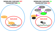
YAP1, also known as YAP or YAP65, is a protein that acts as a transcription coregulator that promotes transcription of genes involved in cellular proliferation and suppressing apoptotic genes. YAP1 is a component in the hippo signaling pathway which regulates organ size, regeneration, and tumorigenesis. YAP1 was first identified by virtue of its ability to associate with the SH3 domain of Yes and Src protein tyrosine kinases. YAP1 is a potent oncogene, which is amplified in various human cancers.

<span class="mw-page-title-main">TEAD1</span> Protein-coding gene in the species Homo sapiens

Transcriptional enhancer factor TEF-1 also known as TEA domain family member 1 (TEAD1) and transcription factor 13 (TCF-13) is a protein that in humans is encoded by the TEAD1 gene. TEAD1 was the first member of the TEAD family of transcription factors to be identified.

<span class="mw-page-title-main">MAP4K2</span> Protein-coding gene in humans

Mitogen-activated protein kinase kinase kinase kinase 2 is an enzyme that in humans is encoded by the MAP4K2 gene.

The Akt signaling pathway or PI3K-Akt signaling pathway is a signal transduction pathway that promotes survival and growth in response to extracellular signals. Key proteins involved are PI3K and Akt.

<span class="mw-page-title-main">Epithelioid hemangioendothelioma</span> Medical condition

Epithelioid hemangioendothelioma (EHE) is a rare tumor, first characterized by Sharon Weiss and Franz Enzinger in 1982 that both clinically and histologically is intermediate between angiosarcoma and hemangioma. However, a distinct, disease-defining genetic alteration recently described for EHE indicates that it is an entirely separate entity from both angiosarcoma and hemangioma.

<span class="mw-page-title-main">Hippo signaling pathway</span> Signaling pathway that controls organ size

The Hippo signaling pathway, also known as the Salvador-Warts-Hippo (SWH) pathway, is a signaling pathway that controls organ size in animals through the regulation of cell proliferation and apoptosis. The pathway takes its name from one of its key signaling components—the protein kinase Hippo (Hpo). Mutations in this gene lead to tissue overgrowth, or a "hippopotamus"-like phenotype.

Barry James Thompson is an Australian and British developmental biologist and cancer biologist. Thompson is known for identifying genes, proteins and mechanisms involved in epithelial polarity, morphogenesis and cell signaling via the Wnt and Hippo signaling pathways, which have key roles in human cancer.

References

  1. 1 2 3 GRCh38: Ensembl release 89: ENSG00000018408 Ensembl, May 2017
  2. 1 2 3 GRCm38: Ensembl release 89: ENSMUSG00000027803 Ensembl, May 2017
  3. "Human PubMed Reference:". National Center for Biotechnology Information, U.S. National Library of Medicine.
  4. "Mouse PubMed Reference:". National Center for Biotechnology Information, U.S. National Library of Medicine.
  5. 1 2 3 4 Kanai F, Marignani PA, Sarbassova D, Yagi R, Hall RA, Donowitz M, et al. (December 2000). "TAZ: a novel transcriptional co-activator regulated by interactions with 14-3-3 and PDZ domain proteins". The EMBO Journal. 19 (24): 6778–6791. doi:10.1093/emboj/19.24.6778. PMC   305881 . PMID   11118213.
  6. 1 2 3 Hong W, Guan KL (September 2012). "The YAP and TAZ transcription co-activators: key downstream effectors of the mammalian Hippo pathway". Seminars in Cell & Developmental Biology. 23 (7): 785–793. doi:10.1016/j.semcdb.2012.05.004. PMC   3459069 . PMID   22659496.
  7. 1 2 3 Moroishi T, Hansen CG, Guan KL (February 2015). "The emerging roles of YAP and TAZ in cancer". Nature Reviews. Cancer. 15 (2): 73–79. doi:10.1038/nrc3876. PMC   4562315 . PMID   25592648.
  8. 1 2 Zhang X, Zhao H, Li Y, Xia D, Yang L, Ma Y, et al. (September 2018). "The role of YAP/TAZ activity in cancer metabolic reprogramming". Molecular Cancer. 17 (1): 134. doi: 10.1186/s12943-018-0882-1 . PMC   6122186 . PMID   30176928.
  9. 1 2 3 4 5 6 7 8 9 Zanconato F, Cordenonsi M, Piccolo S (June 2016). "YAP/TAZ at the Roots of Cancer". Cancer Cell. 29 (6): 783–803. doi:10.1016/j.ccell.2016.05.005. PMC   6186419 . PMID   27300434.
  10. "AlphaFold Protein Structure Database". alphafold.ebi.ac.uk. Retrieved 2022-11-23.
  11. 1 2 3 4 5 6 7 8 9 10 11 12 13 14 15 16 17 18 19 20 21 22 23 24 25 Reggiani F, Gobbi G, Ciarrocchi A, Sancisi V (February 2021). "YAP and TAZ Are Not Identical Twins". Trends in Biochemical Sciences. 46 (2): 154–168. doi:10.1016/j.tibs.2020.08.012. PMID   32981815. S2CID   222166778.
  12. 1 2 3 4 5 6 Callus BA, Finch-Edmondson ML, Fletcher S, Wilton SD (February 2019). "YAPping about and not forgetting TAZ". FEBS Letters. 593 (3): 253–276. doi: 10.1002/1873-3468.13318 . PMID   30570758. S2CID   58578804.
  13. Wu Z, Guan KL (January 2021). "Hippo Signaling in Embryogenesis and Development". Trends in Biochemical Sciences. 46 (1): 51–63. doi:10.1016/j.tibs.2020.08.008. PMC   7749079 . PMID   32928629.
  14. 1 2 3 4 Zheng Y, Pan D (August 2019). "The Hippo Signaling Pathway in Development and Disease". Developmental Cell. 50 (3): 264–282. doi:10.1016/j.devcel.2019.06.003. PMC   6748048 . PMID   31386861.
  15. 1 2 3 4 Piccolo S, Dupont S, Cordenonsi M (October 2014). "The biology of YAP/TAZ: hippo signaling and beyond". Physiological Reviews. 94 (4): 1287–1312. doi:10.1152/physrev.00005.2014. PMID   25287865.
  16. 1 2 Pocaterra A, Romani P, Dupont S (January 2020). "YAP/TAZ functions and their regulation at a glance". Journal of Cell Science. 133 (2): jcs230425. doi:10.1242/jcs.230425. hdl: 11577/3317485 . PMID   31996398. S2CID   210945848.
  17. 1 2 Totaro A, Panciera T, Piccolo S (August 2018). "YAP/TAZ upstream signals and downstream responses". Nature Cell Biology. 20 (8): 888–899. doi:10.1038/s41556-018-0142-z. PMC   6186418 . PMID   30050119.
  18. 1 2 Moya IM, Halder G (April 2019). "Hippo-YAP/TAZ signalling in organ regeneration and regenerative medicine". Nature Reviews. Molecular Cell Biology. 20 (4): 211–226. doi:10.1038/s41580-018-0086-y. PMID   30546055. S2CID   54820180.
  19. Driskill JH, Pan D (January 2021). "The Hippo Pathway in Liver Homeostasis and Pathophysiology". Annual Review of Pathology. 16: 299–322. doi:10.1146/annurev-pathol-030420-105050. PMC   8594752 . PMID   33234023.
  20. Kovar H, Bierbaumer L, Radic-Sarikas B (April 2020). "The YAP/TAZ Pathway in Osteogenesis and Bone Sarcoma Pathogenesis". Cells. 9 (4): 972. doi: 10.3390/cells9040972 . PMC   7227004 . PMID   32326412.
  21. Boopathy GT, Hong W (2019). "Role of Hippo Pathway-YAP/TAZ Signaling in Angiogenesis". Frontiers in Cell and Developmental Biology. 7: 49. doi: 10.3389/fcell.2019.00049 . PMC   6468149 . PMID   31024911.
  22. Heng BC, Zhang X, Aubel D, Bai Y, Li X, Wei Y, et al. (January 2021). "An overview of signaling pathways regulating YAP/TAZ activity". Cellular and Molecular Life Sciences. 78 (2): 497–512. doi:10.1007/s00018-020-03579-8. PMID   32748155. S2CID   220930261.
  23. Ma S, Meng Z, Chen R, Guan KL (June 2019). "The Hippo Pathway: Biology and Pathophysiology". Annual Review of Biochemistry. 88: 577–604. doi:10.1146/annurev-biochem-013118-111829. PMID   30566373. S2CID   58642425.
  24. Meng Z, Moroishi T, Guan KL (January 2016). "Mechanisms of Hippo pathway regulation". Genes & Development. 30 (1): 1–17. doi:10.1101/gad.274027.115. PMC   4701972 . PMID   26728553.
  25. 1 2 3 4 5 6 7 8 9 10 11 "UniProt - Q9GZV5 · WWTR1_HUMAN". www.uniprot.org. Retrieved 2022-11-17.
  26. Sun T, Chi JT (May 2021). "Regulation of ferroptosis in cancer cells by YAP/TAZ and Hippo pathways: The therapeutic implications". Genes & Diseases. 8 (3): 241–249. doi:10.1016/j.gendis.2020.05.004. PMC   8093643 . PMID   33997171.
  27. Dai C, Chen X, Li J, Comish P, Kang R, Tang D (September 2020). "Transcription factors in ferroptotic cell death". Cancer Gene Therapy. 27 (9): 645–656. doi: 10.1038/s41417-020-0170-2 . PMID   32123318. S2CID   211728890.
  28. Koo JH, Guan KL (August 2018). "Interplay between YAP/TAZ and Metabolism". Cell Metabolism. 28 (2): 196–206. doi: 10.1016/j.cmet.2018.07.010 . PMID   30089241. S2CID   51939438.
  29. Kuchay MS, Choudhary NS, Mishra SK (2020-11-01). "Pathophysiological mechanisms underlying MAFLD". Diabetes & Metabolic Syndrome. 14 (6): 1875–1887. doi:10.1016/j.dsx.2020.09.026. PMID   32998095. S2CID   222166643.
  30. Niu N, Xu S, Xu Y, Little PJ, Jin ZG (April 2019). "Targeting Mechanosensitive Transcription Factors in Atherosclerosis". Trends in Pharmacological Sciences. 40 (4): 253–266. doi:10.1016/j.tips.2019.02.004. PMC   6433497 . PMID   30826122.
  31. Zhang X, Zhao H, Li Y, Xia D, Yang L, Ma Y, et al. (September 2018). "The role of YAP/TAZ activity in cancer metabolic reprogramming". Molecular Cancer. 17 (1): 134. doi: 10.1186/s12943-018-0882-1 . PMC   6122186 . PMID   30176928.
  32. Lee HJ, Hong YJ, Kim M (November 2021). "Angiogenesis in Chronic Inflammatory Skin Disorders". International Journal of Molecular Sciences. 22 (21): 12035. doi: 10.3390/ijms222112035 . PMC   8584589 . PMID   34769465.
  33. 1 2 Thompson BJ (May 2020). "YAP/TAZ: Drivers of Tumor Growth, Metastasis, and Resistance to Therapy". BioEssays. 42 (5): e1900162. doi:10.1002/bies.201900162. hdl: 1885/211659 . PMID   32128850. S2CID   212405819.
  34. Zanconato F, Cordenonsi M, Piccolo S (August 2019). "YAP and TAZ: a signalling hub of the tumour microenvironment". Nature Reviews. Cancer. 19 (8): 454–464. doi:10.1038/s41568-019-0168-y. PMID   31270418. S2CID   195791034.
  35. 1 2 3 4 5 6 7 8 9 10 11 Andl T, Zhou L, Yang K, Kadekaro AL, Zhang Y (June 2017). "YAP and WWTR1: New targets for skin cancer treatment". Cancer Letters. 396: 30–41. doi:10.1016/j.canlet.2017.03.001. PMID   28279717.
  36. Pobbati AV, Hong W (2020). "A combat with the YAP/TAZ-TEAD oncoproteins for cancer therapy". Theranostics. 10 (8): 3622–3635. doi:10.7150/thno.40889. PMC   7069086 . PMID   32206112.