Thalassiosira

Last updated

Thalassiosira
Temporal range: 11.608–0.0  Ma
Diatomee - Diatom (fossile) - Thalassiosira sp. - 400x (14281808022).jpg
Fossil species of Thalassiosira
Scientific classification OOjs UI icon edit-ltr.svg
Domain: Eukaryota
Clade: Diaphoretickes
Clade: SAR
Clade: Stramenopiles
Phylum: Gyrista
Subphylum: Ochrophytina
Class: Bacillariophyceae
Order: Thalassiosirales
Family: Thalassiosiraceae
Genus: Thalassiosira
P.T. Cleve, 1873 emend. Hasle, 1973
Species

See text

Thalassiosira is a genus of centric diatoms, comprising over 100 marine and freshwater species. It is a diverse group of photosynthetic eukaryotes that make up a vital part of marine and freshwater ecosystems, in which they are key primary producers and essential for carbon cycling [1]

Contents

Thalassiosira is a diverse genus, however one species within the genus, T. pseudonana, has gained particular significance as the first marine phytoplankton to have its genome sequenced. T. pseudonana has since become a key model organism for studying diatom physiology. The T. pseudonana genome revealed novel genes for intracellular trafficking and metabolism in diatoms. [2] This species was again used to develop methods for genetic manipulation of diatoms [3] and for the study of silica biomineralization. [4]

Background

Thalassiosira was first described in 1873 by P.T. Cleve. [5] The genus name of Thalassiosira is named after Thalassa the Greek word for the 'sea' and for its divine female personification in Greek mythology Cleve noted "The Thalassiosira ... occurs in enormous large masses, floating on the surface of the sea and colouring it for many miles in extent". [6]

The genus was then characterized with transmission electron microscopy in the 1950s and scanning electron microscopy in the 1960s, which led to an enhanced understanding of the defining morphological features of the genus and the subsequent recognition of over 100 species [7]

A species of Thalassiosira, T. pseudonana, was selected as the first marine eukaryotic phytoplankton to undergo whole genome sequencing, due to its small 34Mb genome. As a result, T. pseudonana has served as a model organism for understanding diatom biology. Whole genome sequencing, transcriptomics and proteomics of T. pseudonana have revealed novel pathways for silicon biogenesis, phosphorus stress response, intracellular transport and metabolism in marine diatoms. [2] [8] [9] Recent genetic studies using ribosomal RNA gene sequences better distinguish and classify Thalassiosira species [1] and metabolomics defines the organic compounds produced by Thalassiosira. [10]

Description

Some Thalassiosira diatoms form chain-like colonies, like these ones collected near the Antarctic peninsula coast by the schooner of the Tara Oceans Expedition for plankton research.
This confocal image shows the diatoms' cell wall (cyan), chloroplasts (red), DNA (blue), membranes and organelles (green). Diatom chain.jpg
Some Thalassiosira diatoms form chain-like colonies, like these ones collected near the Antarctic peninsula coast by the schooner of the Tara Oceans Expedition for plankton research.
This confocal image shows the diatoms' cell wall (cyan), chloroplasts (red), DNA (blue), membranes and organelles (green).
Microscopy image of Thalassiosira sp., scale bar 5 um. Deposition velocity versus cell diameters-8.jpg
Microscopy image of Thalassiosira sp., scale bar 5 µm.

Thalassiosira come in a variety of shapes, from box-shaped to cylindrical, discoid or spherical. [11] Some Thalassiosira cells are found alone while others form chains. Thalassiosira have a cell wall made of silica, known as a frustule. [5] Thalasiosira harbor several discoid plastids and a circular valve, which contains pores arranged in rows or arcs, opening outwards. The valve’s mantle edge is pattered with a series of bands. Different species of Thalassiosira can be identified by the morphological characteristics of their areolae and the processes on the valve. [12] During colony formation, Thalassiosira release chitin filaments through strutted processes known as fultoportulae. By extruding chitin fibers, and thereby increasing drag, Thalassiosira can slow the rate at which they sink. [2]

Habitat and ecology

Thalassiosira occupy diverse habitats, both marine and freshwater. Of note, they are a vital primary producers in temperate and polar regions. [1] Thalassiosira can thrive in low temperature and light, as well as mixed waters, and are therefore a large part of diatom blooms during spring in temperate regions, such as Canadian and Alaskan waters. [12] Species in this genus are also capable of assembling defensive threads against zooplankton, allowing them to survive the predation that normally keeps phytoplankton blooms in check.

Thalassiosira species are diverse in both their ecology and physiologies, with variable mechanisms for nitrogen storage or requirements for iron. Iron concentrations, temperature and macronutrient availability have been identified as important factors for the composition of Thalassiosira species communities in marine waters. [1]

Genetics

Fluorescence micrograph of the diatom Thalassiosira nanae. Chloroplasts are red, neutral lipids green, DNA blue and the cell wall white. Fluorescence microscopy provides three-dimensional information of individual components, enabling the measurement of their volumes. Fluorescence micrograph of Thalassiosira nanae.jpg
Fluorescence micrograph of the diatom Thalassiosira nanae. Chloroplasts are red, neutral lipids green, DNA blue and the cell wall white. Fluorescence microscopy provides three-dimensional information of individual components, enabling the measurement of their volumes.

Phylogenetic studies based on 18S rRNA genes have revealed several clades within Thalassiosira, an overall paraphyletic group within the family Thalassiosirales. [14] The relationships of species within the clades remain to be elucidated.

The sequence of the T. pseudonana genome revealed a host of features which set apart diatoms from other eukaryotes. For instance, heat shock transcription factors make up the majority of transcription factors in the T. pseudonana genome, though they are less common in other eukaryotes. [2] Also setting it apart from other eukaryotic genomes is the relative absence of receptor kinases and G protein-coupled receptors. More specific to diatom biology, the understanding of silicon biochemistry in diatoms was enhanced by the discovery of genes involved in the uptake of silicic acid and proteins involved in vesicles for silica precipitation.

A surprising finding from the T. pseudonana genome was the presence of genes encoding enzymes for a complete urea cycle, which was unprecedented in a photosynthetic eukaryote. [2] Diatoms are known to utilize a urease enzyme to catalyze the breakdown of urea and were therefore not expected to need mechanisms to excrete it as waste. Curiously, the T. pseudonana urea cycle feeds into other metabolic pathways, which contribute to protein biosynthesis and possibly energy storage.

Life cycle

Thalassiosira can undergo both asexual and sexual reproduction in processes shared by other diatoms. [15] During asexual reproduction, the parent cell divides into two daughter cells of unequal size — one equal in size to the parent and one smaller. This constraint on size during mitotic division is due to the presence of the rigid silica cell wall. As a result, over multiple cell divisions, the cells size of each daughter cell will decrease. To cope with diminishing cell size, Thalassiosira can transition to sexual reproduction, which is triggered by an assortment of environmental factors, which are not well understood, once cells reach a critically small size. [16] In sexual reproduction, sperm and egg, which can arise from the same cell, fuse to form a diploid zygote, which is referred to as an auxospore. This progeny can then emerge from the parental frustule (silica wall) and reconstruct its own cell wall, thus becoming a cell of a larger size.

Fossil history

The geological record of Thalassiosira dates back to 13.82 million years ago. [11] The fossil record of both freshwater and marine Thalassiosira organisms is substantial, though the freshwater collection has been characterized more extensively. [17]

Practical importance

T. pseudonana has been particularly useful for molecular studies due to its small genome size. It has revealed novel pathways for silica biosynthesis, which involve uptake of monosilicic acid from the marine environment, intracellular transport to a specialized vesicle, and specialized enzymes and peptides driving silica synthesis. [4]

Silica biosynthesis, for which the species T. pseudonana has come to serve as a model organism for study, has been of particular interest in the context of engineering silica nanotechnology. [2] For instance, a modified T. pseudonana has been explored as a drug-delivery vesicle in cancer treatment. [18]

Thalassiosira, among other diatoms, have also been considered as potentially useful sources of lipids for biofuels. Specifically, T. weissflogii has been shown to reduce its silica synthesis while upregulating the production of triacylglycerols when cultured in nitrogen-limiting conditions. [19]

List of species

List of species

Related Research Articles

<span class="mw-page-title-main">Diatom</span> Class of microalgae, found in the oceans, waterways and soils of the world

A diatom is any member of a large group comprising several genera of algae, specifically microalgae, found in the oceans, waterways and soils of the world. Living diatoms make up a significant portion of the Earth's biomass: they generate about 20 to 50 percent of the oxygen produced on the planet each year, take in over 6.7 billion tonnes of silicon each year from the waters in which they live, and constitute nearly half of the organic material found in the oceans. The shells of dead diatoms can reach as much as a half-mile deep on the ocean floor, and the entire Amazon basin is fertilized annually by 27 million tons of diatom shell dust transported by transatlantic winds from the African Sahara, much of it from the Bodélé Depression, which was once made up of a system of fresh-water lakes.

<i>Phaeodactylum tricornutum</i> Species of single-celled organism

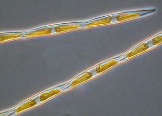
Phaeodactylum tricornutum is a diatom. It is the only species in the genus Phaeodactylum. Unlike other diatoms, P. tricornutum can exist in different morphotypes and changes in cell shape can be stimulated by environmental conditions. This feature can be used to explore the molecular basis of cell shape control and morphogenesis. Unlike most diatoms, P. tricornutum can grow in the absence of silicon and can survive without making silicified frustules. This provides opportunities for experimental exploration of silicon-based nanofabrication in diatoms.

<i>Asterionella</i> Genus of single-celled organisms

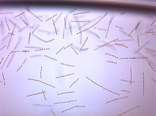
Asterionella is a genus of pennate freshwater diatoms. They are frequently found in star-shaped colonies of individuals.

<span class="mw-page-title-main">Photosynthetic picoplankton</span> Group of photosynthetic plankton

Photosynthetic picoplankton or picophytoplankton is the fraction of the photosynthetic phytoplankton of cell sizes between 0.2 and 2 µm. It is especially important in the central oligotrophic regions of the world oceans that have very low concentration of nutrients.

Auxospores are specialised cells in diatoms that are produced at key stages in their cell cycle or life history. Auxospores typically play a role in growth processes, sexual reproduction or dormancy.

<i>Thalassiosira pseudonana</i> Species of single-celled organism

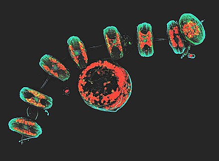
Thalassiosira pseudonana is a species of marine centric diatoms. It was chosen as the first eukaryotic marine phytoplankton for whole genome sequencing. T. pseudonana was selected for this study because it is a model for diatom physiology studies, belongs to a genus widely distributed throughout the world's oceans, and has a relatively small genome at 34 mega base pairs. Scientists are researching on diatom light absorption, using the marine diatom of Thalassiosira. The diatom requires a high enough concentration of CO2 in order to utilize C4 metabolism (Clement et al. 2015).

<i>Chaetoceros</i> Genus of single-celled organisms

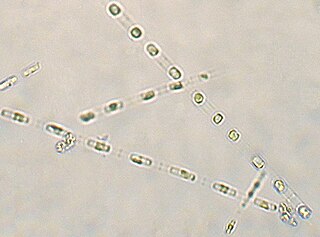
Chaetoceros is a genus of diatoms in the family Chaetocerotaceae, first described by the German naturalist C. G. Ehrenberg in 1844. Species of this genus are mostly found in marine habitats, but a few species exist in freshwater. It is arguably the common and most diverse genus of marine planktonic diatoms, with over 200 accepted species. It is the type genus of its family.

<span class="mw-page-title-main">Ochrophyte</span> Phylum of algae

Ochrophytes, also known as heterokontophytes or stramenochromes, are a group of algae. They are the photosynthetic stramenopiles, a group of eukaryotes, organisms with a cell nucleus, characterized by the presence of two unequal flagella, one of which has tripartite hairs called mastigonemes. In particular, they are characterized by photosynthetic organelles or plastids enclosed by four membranes, with membrane-bound compartments called thylakoids organized in piles of three, chlorophyll a and c as their photosynthetic pigments, and additional pigments such as β-carotene and xanthophylls. Ochrophytes are one of the most diverse lineages of eukaryotes, containing ecologically important algae such as brown algae and diatoms. They are classified either as phylum Ochrophyta or Heterokontophyta, or as subphylum Ochrophytina within phylum Gyrista. Their plastids are of red algal origin.

<i>Pseudo-nitzschia</i> Genus of marine planktonic diatoms

Pseudo-nitzschia is a marine planktonic diatom genus that accounts for 4.4% of pennate diatoms found worldwide. Some species are capable of producing the neurotoxin domoic acid (DA), which is responsible for the neurological disorder in humans known as amnesic shellfish poisoning (ASP). Currently, 58 species are known, 28 of which have been shown to produce DA. It was originally hypothesized that only dinoflagellates could produce harmful algal toxins, but a deadly bloom of Pseudo-nitzschia occurred in 1987 in the bays of Prince Edward Island, Canada, and led to an outbreak of ASP. Over 100 people were affected by this outbreak after consuming contaminated mussels; three people died. Since this event, no additional deaths have been attributed to ASP, though the prevalence of toxic diatoms and DA has increased worldwide. This anomaly is likely due to increased awareness of harmful algal blooms (HABs) and their implications for human and ecosystem health.

<span class="mw-page-title-main">Marine microorganisms</span> Any life form too small for the naked human eye to see that lives in a marine environment

Marine microorganisms are defined by their habitat as microorganisms living in a marine environment, that is, in the saltwater of a sea or ocean or the brackish water of a coastal estuary. A microorganism is any microscopic living organism or virus, which is invisibly small to the unaided human eye without magnification. Microorganisms are very diverse. They can be single-celled or multicellular and include bacteria, archaea, viruses, and most protozoa, as well as some fungi, algae, and animals, such as rotifers and copepods. Many macroscopic animals and plants have microscopic juvenile stages. Some microbiologists also classify viruses as microorganisms, but others consider these as non-living.

Thalassiosira weissflogii is a species of centric diatoms, a unicellular microalga. It is found in marine environments and also in inland waters in many parts of the world. It is actively studied because it may use C4-plant style strategies to increase its photosynthetic efficiency.

<i>Ditylum brightwellii</i> Species of diatom

Ditylum brightwellii is a species of cosmopolitan marine centric diatoms. It is a unicellular photosynthetic autotroph that has the ability to divide rapidly and contribute to spring phytoplankton blooms.

<span class="mw-page-title-main">Thalassiosirales</span> Order of single-celled organisms

Thalassiosirales is an order of centric diatoms. The order currently contains 471 species. Species in the order Thalassiosirales are common in brackish, nearshore, and open-ocean habitats, with approximately the same number of freshwater and marine species.

<span class="mw-page-title-main">Thalassiosiraceae</span> Family of single-celled organisms

Thalassiosiraceae is a family of diatoms in the order Thalassiosirales. The family of Thalassiosiraceae have the unique quality of having a flat valve face. These diatoms are common in brackish, nearshore, and open-ocean habitats, with approximately the same number of freshwater and marine species. Thalassiosiraceae are a centric diatom full of fultoportula. These can often be mistaken for Areola. These belong to many diatom families and can be found in different forms such as the different Areolae that can be found on Navicula or Gomphoneis known as lineolate and punctate. Unlike naviculaceae who are symmetrical in shape some Thalassiosiraceae take on being tangentially undulate.

<i>Skeletonema</i> Genus of single-celled organisms

Skeletonema is a genus of diatoms in the family Skeletonemataceae. It is the type genus of its family. The genus Skeletonema was established by R. K. Greville in 1865 for a single species, S. barbadense, found in the Barbados deposit [Jung 2009]. These diatoms are photosynthetic organisms, meaning they obtain carbon dioxide from their surrounding environment and produce oxygen along with other byproducts. Reproduce sexually and asexually [Guiry 2011]. Skeletonema belong to the morphological category referred to as centric diatoms. These are classified by having valves with radial symmetry and the cells lack significant motility [Horner 2002]. Skeletonema are cylindrical shaped with a silica frustule. Cells are joined by long marginal processes to form a filament [Horner 2002]. Their length ranges from 2-61 micrometers, with a diameter ranging from 2-21 micrometers [Hasle 1997]. They are found typically in the neritic zone of the ocean and are highly populous in coastal systems [Jung 2009]. The genus is considered cosmopolitan, showing a wide range of tolerance for salinity and temperature [Hasle 1973]. For example, they have been found in various aquatic environments such as brackish or freshwater. Skeletonema are found worldwide excluding Antarctic waters [Hevia-Orube 2016]. Some harmful effects these diatoms may have on an ecosystem are attributed to large blooming events which may cause hypoxic events in coastal systems. Additionally, they are known to cause water discoloration [Kraberg 2010].

<i>Cyclotella</i> Genus of diatoms

Cyclotella is a genus of diatoms often found in oligotrophic environments, both marine and fresh water. It is in the family Stephanodiscaceae and the order Thalassiosirales. The genus was first discovered in the mid-1800s and since then has become an umbrella genus for nearly 100 different species, the most well-studied and the best known being Cyclotella meneghiniana. Despite being among the most dominant genera in low-productivity environments, it is relatively understudied.

<span class="mw-page-title-main">Marine protists</span> Protists that live in saltwater or brackish water

Marine protists are defined by their habitat as protists that live in marine environments, that is, in the saltwater of seas or oceans or the brackish water of coastal estuaries. Life originated as marine single-celled prokaryotes and later evolved into more complex eukaryotes. Eukaryotes are the more developed life forms known as plants, animals, fungi and protists. Protists are the eukaryotes that cannot be classified as plants, fungi or animals. They are mostly single-celled and microscopic. The term protist came into use historically as a term of convenience for eukaryotes that cannot be strictly classified as plants, animals or fungi. They are not a part of modern cladistics because they are paraphyletic.

<span class="mw-page-title-main">Protist shell</span> Protective shell of a type of eukaryotic organism

Many protists have protective shells or tests, usually made from silica (glass) or calcium carbonate (chalk). Protists are a diverse group of eukaryote organisms that are not plants, animals, or fungi. They are typically microscopic unicellular organisms that live in water or moist environments.

<span class="mw-page-title-main">Tatiana Rynearson</span> American oceanographer

Tatiana Rynearson is an American oceanographer who is a professor at the University of Rhode Island. Her research considers plankton diversity and abundance. Rynearson has been on several research cruises, including trips to the North Sea, Puget Sound, the Gulf of Mexico and the North Atlantic.

Algal viruses are the viruses infecting algae, which are photosynthetic single-celled eukaryotes. As of 2020, there were 61 viruses known to infect algae. Algae are integral components of aquatic food webs and drive nutrient cycling, so the viruses infecting algal populations also impacts the organisms and nutrient cycling systems that depend on them. Thus, these viruses can have significant, worldwide economic and ecological effects. Their genomes varied between 4.4 to 560 kilobase pairs (kbp) long and used double-stranded Deoxyribonucleic Acid (dsDNA), double-stranded Ribonucleic Acid (dsRNA), single-stranded Deoxyribonucleic Acid (ssDNA), and single-stranded Ribonucleic Acid (ssRNA). The viruses ranged between 20 and 210 nm in diameter. Since the discovery of the first algae-infecting virus in 1979, several different techniques have been used to find new viruses infecting algae and it seems that there are many algae-infecting viruses left to be discovered

References

  1. 1 2 3 4 Dreux Chappell, P., Whitney, L. A. P., Haddock, T. L., Menden-Deuer, S., Roy, E. G., Wells, M. L., & Jenkins, B. D. (2013). Thalassiosira spp. community composition shifts in response to chemical and physical forcing in the northeast Pacific Ocean. Frontiers in Microbiology, 4(SEP), 273. https://doi.org/10.3389/fmicb.2013.00273
  2. 1 2 3 4 5 6 Armbrust, E. V. (2004). The Genome of the Diatom Thalassiosira pseudonana: Ecology, Evolution, and Metabolism. Science, 306(5693), 79–86. https://doi.org/10.1126/science.1101156
  3. Poulsen, N., Chesley, P. M., & Kröger, N. (2006). Molecular genetic manipulation of the diatom Thalassiosira pseudonana (Bacillariophyceae). Journal of Phycology, 42(5), 1059–1065. https://doi.org/10.1111/j.1529-8817.2006.00269.x
  4. 1 2 Sumper, M., & Brunner, E. (2008, May 23). Silica biomineralisation in diatoms: The model organism Thalassiosira pseudonana. ChemBioChem. https://doi.org/10.1002/cbic.200700764
  5. 1 2 Miranda, S. V., Guiry, M. D., & Guiry, G. (2015, June 19). Thalassiosira Cleve, 1873 : Algaebase. Retrieved March 30, 2020, from https://www.algaebase.org/search/genus/detail/?genus_id=43768&-session=abv4:AC1F24C807fd0012FBNw3B92519A
  6. Burkhardt, Lotte (2022). Eine Enzyklopädie zu eponymischen Pflanzennamen [Encyclopedia of eponymic plant names](pdf) (in German). Berlin: Botanic Garden and Botanical Museum, Freie Universität Berlin. doi:10.3372/epolist2022. ISBN   978-3-946292-41-8. S2CID   246307410 . Retrieved January 27, 2022.
  7. (Bacillariophyta) species rarely recorded in Brazilian coastal waters. Braz. J. Biol (Vol. 69).
  8. Dyhrman, S. T., Jenkins, B. D., Rynearson, T. A., Saito, M. A., Mercier, M. L., Alexander, H., … Heithoff, A. (2012). The transcriptome and proteome of the diatom thalassiosira pseudonana reveal a diverse phosphorus stress response. PLoS ONE, 7(3), e33768. https://doi.org/10.1371/journal.pone.0033768
  9. Mock, T., Samanta, M. P., Iverson, V., Berthiaume, C., Robison, M., Holtermann, K., … Armbrust, E. V. (2008). Whole-genome expression profiling of the marine diatom Thalassiosira pseudonana identifies genes involved in silicon bioprocesses. Proceedings of the National Academy of Sciences of the United States of America, 105(5), 1579–1584. https://doi.org/10.1073/pnas.0707946105
  10. Longnecker, Krista; Kido Soule, Melissa C.; Kujawinski, Elizabeth B. (2015-01-20). "Dissolved organic matter produced by Thalassiosira pseudonana". Marine Chemistry. 168: 114–123. doi:10.1016/j.marchem.2014.11.003. hdl: 1912/7175 . ISSN   0304-4203.
  11. 1 2 The Diatom - Thalassiosira. (n.d.). Retrieved February 27, 2020, from https://algaeresearchsupply.com/pages/the-diatom-thalassiosira
  12. 1 2 Harris, A., Medlin, L., Lewis, J., Jones, K., D Harris, A. S., Medlin, L. K., & Jones, K. J. (1995). Thalassiosira species (Bacillariophyceae) from a Scottish sea-loch. European Journal of Phycology, 30(2), 117–148. https://doi.org/10.1080/09670269500650881
  13. Chansawang, Narin; Obara, Boguslaw; Geider, Richard J.; Laissue, Pierre Philippe (2015). "Three-Dimensional Visualisation and Quantification of Lipids in Microalgae Using Confocal Laser Scanning Microscopy". Hydrocarbon and Lipid Microbiology Protocols. Springer Protocols Handbooks. pp. 145–161. doi:10.1007/8623_2015_142. ISBN   978-3-662-49132-4.
  14. Hoppenrath, M., Beszteri, B., Drebes, G., Halliger, H., Van Beusekom, J. E. E., Janisch, S., & Wiltshire, K. H. (2007). Thalassiosira species (Bacillariophyceae, Thalassiosirales) in the North Sea at Helgoland (German Bight) and Sylt (North Frisian Wadden Sea)-a first approach to assessing diversity. European Journal of Phycology, 42(3), 271–288. https://doi.org/10.1080/09670260701352288
  15. Moore, E. R., Bullington, B. S., Weisberg, A. J., Jiang, Y., Chang, J., & Halsey, K. H. (2017). Morphological and transcriptomic evidence for ammonium induction of sexual reproduction in Thalassiosira pseudonana and other centric diatoms. PLOS ONE, 12(7), e0181098. https://doi.org/10.1371/journal.pone.0181098
  16. Armbrust, E. V. (1999). Identification of a new gene family expressed during the onset of sexual reproduction in the centric diatom Thalassiosira weissflogii. Applied and Environmental Microbiology, 65(7), 3121–3128. https://doi.org/10.1128/aem.65.7.3121-3128.1999
  17. Alverson, A. J. (2014). Timing marine–freshwater transitions in the diatom order Thalassiosirales. Paleobiology, 40(1), 91–101. https://doi.org/10.1666/12055
  18. Delalat, B., Sheppard, V. C., Rasi Ghaemi, S., Rao, S., Prestidge, C. A., McPhee, G., … Voelcker, N. H. (2015). Targeted drug delivery using genetically engineered diatom biosilica. Nature Communications, 6(1), 1–11. https://doi.org/10.1038/ncomms9791
  19. D’Ippolito, G., Sardo, A., Paris, D., Vella, F. M., Adelfi, M. G., Botte, P., … Fontana, A. (2015). Potential of lipid metabolism in marine diatoms for biofuel production. Biotechnology for Biofuels, 8(1), 28. https://doi.org/10.1186/s13068-015-0212-4

Further references