Venipuncture

Last updated
Still photo of a venipuncture procedure Venipuncture using a BD Vacutainer.JPG
Still photo of a venipuncture procedure
Video of a venipuncture procedure

In medicine, venipuncture or venepuncture is the process of obtaining intravenous access for the purpose of venous blood sampling (also called phlebotomy ) or intravenous therapy. In healthcare, this procedure is performed by medical laboratory scientists, medical practitioners, some EMTs, paramedics, phlebotomists, dialysis technicians, and other nursing staff. [1] In veterinary medicine, the procedure is performed by veterinarians and veterinary technicians.

Contents

It is essential to follow a standard procedure for the collection of blood specimens to get accurate laboratory results. Any error in collecting the blood or filling the test tubes may lead to erroneous laboratory results. [2]

Venipuncture is one of the most routinely performed invasive procedures and is carried out for any of five reasons:

  1. to obtain blood for diagnostic purposes;
  2. to monitor levels of blood components; [3]
  3. to administer therapeutic treatments including medications, nutrition, or chemotherapy;
  4. to remove blood due to excess levels of iron or erythrocytes (red blood cells); or
  5. to collect blood for later uses, mainly transfusion either in the donor or in another person.

Blood analysis is an important diagnostic tool available to clinicians within healthcare.

Blood is most commonly obtained from the superficial veins of the upper limb. [1] The median cubital vein, which lies within the cubital fossa anterior to the elbow, is close to the surface of the skin without many large nerves positioned nearby. Other veins that can be used in the cubital fossa for venipuncture include the cephalic, basilic, and median antebrachial veins. [4]

Minute quantities of blood may be taken by fingerstick sampling and collected from infants by means of a heelprick or from scalp veins with a winged infusion needle.

Phlebotomy (incision into a vein) is also the treatment of certain diseases such as hemochromatosis and primary and secondary polycythemia.

Complications

A 1996 study of blood donors (a larger needle is used in blood donation than in routine venipuncture) found that 1 in 6,300 donors sustained a nerve injury. [5]

Risk and side affects can include a variety of things. Dizziness, sweating, and a drop in your heart rate and blood pressure. [6]

Equipment

An elastic tourniquet used to temporarily restrict blood flow. The tourniquet distends the veins, making them more palpable and visible. Prameta tourniquet-4981.jpg
An elastic tourniquet used to temporarily restrict blood flow. The tourniquet distends the veins, making them more palpable and visible.

There are many ways in which blood can be drawn from a vein, and the method used depends on the person's age, the equipment available, and the type of tests required.

Most blood collection in the US, UK, Canada and Hong Kong is done with an evacuated tube system. Two common systems are Vacutainer (Becton, Dickinson and company) and Vacuette (Greiner Bio-One). The equipment consists of a plastic adapter, also known as a tube or needle holder/hub, a hypodermic needle and a vacuum tube. Under certain circumstances, a syringe may be used, often with a butterfly needle, which is a plastic catheter attached to a short needle. In the developing world, the evacuated tube system is the preferred method of drawing blood.[ citation needed ]

With evacuated or vacuum tubes

Vacutainer blood vials Vacutainer blood bottles.jpg
Vacutainer blood vials

Greiner Bio-One manufactured the first ever plastic evacuated blood collection tube in 1985 under the VACUETTE brand name. Today, many companies sell vacuum tubes as the patent for this device is now in the public domain. These tubes are manufactured with a specific volume of gas removed from the sealed tube. When a needle from a hub or transfer device is inserted into the stopper, the tube's vacuum automatically pulls in the required volume of blood.[ citation needed ]

The basic Evacuated Tube System (ETS) consists of a needle, a tube holder, and the evacuated tubes. The needle is attached to the tube holder by the phlebotomist prior to collection, or may come from the manufacturer as one unit. The needle protrudes through the end of the tube holder, and has a needle on each end. After first cleaning the venipuncture site and applying a tourniquet, the phlebotomist uncaps the needle attached to the tube holder, inserts the needle into the vein, then slides evacuated tubes into the tube holder, where the tube's stopper is pierced by the back end of the needle. The vacuum in the tube then automatically draws the needed blood directly from the vein. Multiple vacuum tubes can be attached to and removed in turn from a single needle, allowing multiple samples to be obtained from a single procedure. This is possible due to the multiple sample sleeve, which is a flexible rubber fitting over the posterior end of the needle cannula which seals the needle until it is pushed out of the way. This keeps blood from freely draining out of the back of the needle inserted in the vein, as each test tube is removed and the next impaled. OSHA safety regulations require that needles or tube holders come equipped with a safety device to cover the needle after the procedure to prevent accidental needle stick injury. [7]

Fittings and adapters used to fill evacuated tubes from butterfly needle kits and syringes are also available.[ citation needed ]

There are several needle gauges for a phlebotomist to choose from. The most commonly used are as follows: a 21g (green top) needle, a 22g (black top) needle, a 21g (green label) butterfly needle, a 23g (light blue label) butterfly needle, and a 25g (orange or dark blue label) butterfly needle (however this needle is only used in pediatrics or extreme cases as it is so small that it can often result in hemolyzing the blood sample). There are also a variety of tube and bottle sizes and volumes for different test requirements. [8]

Additives and order of draw

The test tubes in which blood is collected may contain one or more of several additives. In general, tests requiring whole blood call for blood samples collected in test tubes containing some form of the anticoagulant EDTA. EDTA chelates calcium to prevent clotting. EDTA is preferred for hematology tests because it does minimum damage to cell morphology. Sodium citrate is the anticoagulant used in specimens collected for coagulation tests. The majority of chemistry and immunology tests are performed on serum, which is produced by clotting and then separating the blood specimen via centrifuge. These specimens are collected in either a non-additive tube or one containing a clotting activator. This clotting activator can interfere with some assays, and so a plain tube is recommended in these cases, but will delay testing. Tubes containing lithium heparin or sodium heparin are also commonly used for a variety of chemistry tests, as they do not require clotting and can be centrifuged immediately after collection. A combination of sodium fluoride and potassium oxalate is used for glucose tests, as these additives both prevent clotting and stop glycolosis, so that blood glucose levels are preserved after collection. [9] Another specialty tube is an opaque amber colored tube used to collect blood for light sensitive analytes, such as bilirubin.[ citation needed ]

Test tubes are labeled with the additive they contain, but the stopper on each tube is color coded according to additive as well. While colors vary between manufacturers, stopper colors generally are associated with each additive as listed below. Because the additives from each tube can be left on the needle used to fill the tubes, they must be drawn in a specific order to ensure that cross contamination will not negatively affect testing of the samples if multiple tubes are to be drawn at once. The "order of draw" varies by collection method. Below in the order of draw generally required for the Evacuated Tube System (ETS) collection method are the most common tubes, listing additive and color: [10] [11]

Vacutainer/sample tube types for venipuncture/phlebotomy   edit
Tube cap color or type in order of drawAdditiveUsage and comments
Blood culture bottleSodium polyanethol sulfonate (anticoagulant) and growth media for microorganisms Usually drawn first for minimal risk of contamination. [12] Two bottles are typically collected in one blood draw; one for aerobic organisms and one for anaerobic organisms. [13]
Blue

("light blue")

Sodium citrate

(weak calcium chelator/anticoagulant)

Coagulation tests such as prothrombin time (PT) and partial thromboplastin time (PTT) and thrombin time (TT). Tube must be filled 100%.
Plain redNo additiveSerum: Total complement activity, cryoglobulins
Gold (sometimes red and grey "tiger top" [14] )Clot activator and serum separating gel [15] Serum-separating tube (SST): Tube inversions promote clotting. Most chemistry, endocrine and serology tests, including hepatitis and HIV.
OrangeClot activator and serum separating gel [16] Rapid serum-separating tube (RST).
Dark greenSodium heparin (anticoagulant)Chromosome testing, HLA typing, ammonia, lactate
Light greenLithium heparin (anticoagulant)

Plasma separator gel

Plasma . Tube inversions prevent clotting
Lavender ("purple") EDTA (chelator / anticoagulant) Whole blood : CBC, ESR, Coombs test, platelet antibodies, flow cytometry, blood levels of tacrolimus and cyclosporin
PinkK2 EDTA (chelator / anticoagulant) Blood typing and cross-matching, direct Coombs test, HIV viral load
Royal blue

("navy")

EDTA (chelator / anticoagulant)Trace elements, heavy metals, most drug levels, toxicology
TanSodium EDTA (chelator / anticoagulant) Lead
Gray

Fluoride Oxalate

Glucose , lactate, [17] toxicology [18]
Yellow Acid-citrate-dextrose A (anticoagulant) Tissue typing, DNA studies, HIV cultures
Pearlescent ("white")Separating gel and (K2)EDTA PCR for adenovirus, toxoplasma and HHV-6
Black Sodium Citrate Paediatric ESR
QuantiFERON

Grey, Green, Yellow, Purple

QuantiFERON

1. Grey (nil) tube 2. Green (TB1 antigen) tube 3. Yellow (TB2 antigen) tube 4. Purple (mitogen) tube

Tuberculosis

In children

Use of lidocaine iontophoresis is effective for reducing pain and alleviating distress during venipuncture in children. [19] A needle-free powder lignocaine delivery system has been shown to decrease the pain of venipuncture in children. [20] [21] Rapid dermal anesthesia can be achieved by local anesthetic infiltration, but it may evoke anxiety in children frightened by needles or distort the skin, making vascular access more difficult and increasing the risk of needle exposure to health care workers. Dermal anesthesia can also be achieved without needles by the topical application of local anesthetics or by lidocaine iontophoresis. By contrast, noninvasive dermal anesthesia can be established in 5–15 min without distorting underlying tissues by lidocaine iontophoresis, where a direct electric current facilitates dermal penetration of positively charged lidocaine molecules when placed under the positive electrode.[ citation needed ]

One study concluded that the iontophoretic administration of lidocaine was safe and effective in providing dermal anesthesia for venipuncture in children 6–17 years old. This technique may not be applicable to all children. Future studies may provide information on the minimum effective iontophoretic dose for dermal anesthesia in children and the comparison of the anesthetic efficacy and satisfaction of lidocaine iontophoresis with topical anesthetic creams and subcutaneous infiltration. [19]

Non-pharmacological treatments for pain associated with venipuncture in children includes hypnosis and distraction. These treatments reduced self reported pain and when combined with cognitive-behavioural therapy (CBT) the reduction of pain was even greater. Other interventions have not been found to be effective and these are suggestion, blowing out air, and distraction with parent coaching did not differ from control for pain and distress. [22]

With needle and syringe

Some health care workers prefer to use a syringe-needle technique for venipuncture. Sarstedt manufactures a blood-drawing system (S-Monovette) that uses this principle. [23] This method can be preferred on the elderly, those with cancer, severe burns, obesity, or where the veins are unreliable or fragile. Because syringes are manually operated, the amount of suction applied may be easily controlled. This is particularly helpful when veins are small which may collapse under the suction of an evacuated tube. In children or other circumstances where the quantity of blood gained may be limited it can be helpful to know how much blood can be obtained before distributing it amongst the various additives that the laboratory will require. Another alternative is drawing blood from indwelling cannulae. [24]

Blood cultures

There are times when a blood culture collection is required. The culture will determine if there are pathogens in the blood. Normally blood is sterile. When drawing blood from cultures use a sterile solution such as Betadine rather than alcohol. This is done using sterile gloves, while not wiping away the surgical solution, touching the puncture site, or in any way compromising the sterile process. It is vital that the procedure is performed in as sterile a manner as possible as the persistent presence of skin commensals in blood cultures could indicate endocarditis but they are most often found as contaminants. It is encouraged to use an abrasive method of skin preparation. This removes the upper layers of dead skin cells along with their contaminating bacteria. [25] Povidone-iodine has traditionally been used but in the UK a 2% chlorhexidine in 70% ethanol or isopropyl alcohol solution is preferred and time must be allowed for it to dry. The tops of any containers used when drawing a blood culture should also be disinfected using a similar solution. Some labs will actively discourage iodine use where iodine is thought to degrade the rubber stopper through which blood enters the bottle, thus allowing contaminates to enter the container.[ citation needed ]

The blood is collected into special transport bottles, which are like vacuum tubes but shaped differently. The blood culture bottle contains transport media to preserve any microorganisms present while they are being transported to the laboratory for cultures. Because it is unknown whether the pathogens are anaerobic (living without oxygen) or aerobic (living with oxygen), blood is collected to test for both. The aerobic bottle is filled first, and then the anaerobic bottle is filled. However, if the collection is performed using a syringe, the anaerobic bottle is filled first. If a butterfly collection kit is used, the aerobic bottle is filled first, so that any air in the tubing is released into the oxygen-containing bottle.[ citation needed ]

Specially designed blood culture collection bottles eliminate the need for either the syringe or butterfly collection method. These specially designed bottles have long necks that fit into the evacuated tubes holders that are use for regular venipuncture collection. These bottles also allow for collection of other blood specimens via evacuated tubes, to be collected without additional venipuncture.[ citation needed ]

The amount of blood that is collected is critical for the optimal recovery of microorganisms. Up to 10mL of blood is typical, but can vary according to the recommends of the manufacturer of the collection bottle. Collection from infants and children are 1 to 5 mL. If too little blood is collected, the ratio of blood-to-nutrient broth will inhibit the growth of microorganisms. If too much blood is collected, there is the risk of a hospital-induced anemia and the ratio of blood-to-nutrient broth will tilt in the opposite direction, which also is not conductive to optimal growth.[ citation needed ]

The bottles are then incubated in specialized units for 24 hours before a lab technician studies and/or tests it. This step allows the very small numbers of bacteria (potentially 1 or 2 organisms) to multiply to a level which is sufficient for identification +/-antibiotic resistance testing. Modern blood culture bottles have an indicator in the base which changes color in the presence of bacterial growth and can be read automatically by machine. (For this reason the barcoded stickers found on these bottles should not be removed as they are used by the laboratory's automated systems.)[ citation needed ]

Taking blood samples from animals

Blood samples from living laboratory animals may be collected using following methods: [26] [27]

The volume of the blood sample collection is very important in experimental animals. All nonterminal blood collection without replacement of fluids is limited up to 10% of total circulating blood volume in healthy, normal, adult animals on a single occasion and collection may be repeated after three to four weeks. In case repeated blood samples are required at short intervals, a maximum of 0.6 ml/kg/day or 1.0% of an animal's total blood volume can be removed every 24 hours. The estimated blood volume in adult animals is 55 to 70 ml/kg body weight. Care should be taken for older and obese animals. If blood collection volume exceeds more than 10% of total blood volume, fluid replacement may be required. Lactated Ringer's solution (LRS) is recommended as the best fluid replacement by National Institutes of Health (NIH). If the volume of blood collection exceeds more than 30% of the total circulatory blood volume, adequate care should be taken so that the animal does not develop hypovolemia. [27]

Blood alcohol tests

It is generally not advisable to use isopropyl alcohol to cleanse the venipuncture site when obtaining a specimen for a blood alcohol test. This has been related largely to the potential legal implications associated with the use of alcohol-based cleaners that could theoretically impact analysis. Numerous police alcohol collection kits have been marketed that incorporate a sodium fluoride/potassium oxalate preservative and non-alcohol-based cleansing agents to ensure proper collection. Using soap and hot water or a povidone-iodine swab are advisable alternatives to isopropyl alcohol in this case.[ citation needed ]

See also

Related Research Articles

<span class="mw-page-title-main">Phlebotomy</span> Medical procedure involving an incision in a vein

Phlebotomy is the process of making a puncture in a vein, usually in the arm, with a cannula for the purpose of drawing blood. The procedure itself is known as a venipuncture, which is also used for intravenous therapy. A person who performs a phlebotomy is called a phlebotomist, although most doctors, nurses, and other technicians can also carry out a phlebotomy. In contrast, phlebectomy is the removal of a vein.

<span class="mw-page-title-main">Local anesthetic</span> Medications to reversibly block pain

A local anesthetic (LA) is a medication that causes absence of all sensation in a specific body part without loss of consciousness, as opposed to a general anesthetic, which eliminates all sensation in the entire body and causes unconsciousness. Local anesthetics are most commonly used to eliminate pain during or after surgery. When it is used on specific nerve pathways, paralysis also can be induced.

<span class="mw-page-title-main">Vacutainer</span> Type of blood collection equipment for medical testing

A vacutainer blood collection tube is a sterile glass or plastic test tube with a colored rubber stopper creating a vacuum seal inside of the tube, facilitating the drawing of a predetermined volume of liquid. Vacutainer tubes may contain additives designed to stabilize and preserve the specimen prior to analytical testing. Tubes are available with a safety-engineered stopper, with a variety of labeling options and draw volumes. The color of the top indicates the additives in the vial.

<span class="mw-page-title-main">Hypodermic needle</span> Device to inject substances into the circulatory system

A hypodermic needle, one of a category of medical tools which enter the skin, called sharps, is a very thin, hollow tube with one sharp tip. It is commonly used with a syringe, a hand-operated device with a plunger, to inject substances into the body or extract fluids from the body. Large-bore hypodermic intervention is especially useful in catastrophic blood loss or treating shock.

<span class="mw-page-title-main">Pleurisy</span> Disease of the lungs

Pleurisy, also known as pleuritis, is inflammation of the membranes that surround the lungs and line the chest cavity (pleurae). This can result in a sharp chest pain while breathing. Occasionally the pain may be a constant dull ache. Other symptoms may include shortness of breath, cough, fever, or weight loss, depending on the underlying cause. Pleurisy can be caused by a variety of conditions, including viral or bacterial infections, autoimmune disorders, and pulmonary embolism.

<span class="mw-page-title-main">Cannula</span> Tube surgically implanted in the body

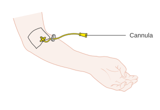
A cannula is a tube that can be inserted into the body, often for the delivery or removal of fluid or for the gathering of samples. In simple terms, a cannula can surround the inner or outer surfaces of a trocar needle thus extending the effective needle length by at least half the length of the original needle. Its size mainly ranges from 14 to 26 gauge. Different-sized cannula have different colours as coded.

<span class="mw-page-title-main">Blood test</span> Laboratory analysis performed on a blood sample

A blood test is a laboratory analysis performed on a blood sample that is usually extracted from a vein in the arm using a hypodermic needle, or via fingerprick. Multiple tests for specific blood components, such as a glucose test or a cholesterol test, are often grouped together into one test panel called a blood panel or blood work. Blood tests are often used in health care to determine physiological and biochemical states, such as disease, mineral content, pharmaceutical drug effectiveness, and organ function. Typical clinical blood panels include a basic metabolic panel or a complete blood count. Blood tests are also used in drug tests to detect drug abuse.

<span class="mw-page-title-main">Serum-separating tube</span>

Serum-separating tubes, also known as serum separator tubes or SSTs, are test tubes used in clinical chemistry tests requiring blood serum.

<span class="mw-page-title-main">Blood culture</span> Test to detect bloodstream infections

A blood culture is a medical laboratory test used to detect bacteria or fungi in a person's blood. Under normal conditions, the blood does not contain microorganisms: their presence can indicate a bloodstream infection such as bacteremia or fungemia, which in severe cases may result in sepsis. By culturing the blood, microbes can be identified and tested for resistance to antimicrobial drugs, which allows clinicians to provide an effective treatment.

<span class="mw-page-title-main">Bone marrow examination</span> Form of pathologic analysis

Bone marrow examination refers to the pathologic analysis of samples of bone marrow obtained by bone marrow biopsy and bone marrow aspiration. Bone marrow examination is used in the diagnosis of a number of conditions, including leukemia, multiple myeloma, lymphoma, anemia, and pancytopenia. The bone marrow produces the cellular elements of the blood, including platelets, red blood cells and white blood cells. While much information can be gleaned by testing the blood itself, it is sometimes necessary to examine the source of the blood cells in the bone marrow to obtain more information on hematopoiesis; this is the role of bone marrow aspiration and biopsy.

<span class="mw-page-title-main">Fear of needles</span> Phobia of injections or needles

Fear of needles, known in medical literature as needle phobia, is the extreme fear of medical procedures involving injections or hypodermic needles. This can lead to avoidance of medical care and vaccine hesitancy.

Infiltration is the diffusion or accumulation of foreign substances in amounts excess of the normal. The material collected in those tissues or cells is called infiltrate.

<span class="mw-page-title-main">Radial artery puncture</span>

Radial artery puncture is a medical procedure performed to obtain a sample of arterial blood for gas analysis. A needle is inserted into the radial artery and spontaneously fills with blood. The syringe is either prepacked with a small amount of heparin to prevent coagulation, or must be heparinised, by drawing up a small amount of heparin and squirting it out again.. The available evidence suggests that the use of local anaesthesia, prior to arterial puncture, does not reduce the perceived pain of the procedure.

The Keidel vacuum tube was a type of blood collecting device, first manufactured by Hynson, Wescott and Dunning in around 1922. This vacuum was one of the first evacuated systems, predating the more well known Vacutainer. Its primary use was to test for syphilis and typhoid fever.

<span class="mw-page-title-main">Winged infusion set</span> Specialized venipuncture needle

A winged infusion set—also known as "butterfly" or "scalp vein" set—is a device specialized for venipuncture: i.e. for accessing a superficial vein or artery for either intravenous injection or phlebotomy. It consists, from front to rear, of a hypodermic needle, two bilateral flexible "wings", flexible small-bore transparent tubing, and lastly a connector. This connector attaches to another device: e.g. syringe, vacuum tube holder/hub, or extension tubing from an infusion pump or gravity-fed infusion/transfusion bag/bottle.

Veterinary anesthesia is a specialization in the veterinary medicine field dedicated to the proper administration of anesthetic agents to non-human animals to control their consciousness during procedures. A veterinarian or a Registered Veterinary Technician administers these drugs to minimize stress, destructive behavior, and the threat of injury to both the patient and the doctor. The duration of the anesthesia process goes from the time before an animal leaves for the visit to the time after the animal reaches home after the visit, meaning it includes care from both the owner and the veterinary staff. Generally, anesthesia is used for a wider range of circumstances in animals than in people not only due to their inability to cooperate with certain diagnostic or therapeutic procedures, but also due to their species, breed, size, and corresponding anatomy. Veterinary anesthesia includes anesthesia of the major species: dogs, cats, horses, cattle, sheep, goats, and pigs, as well as all other animals requiring veterinary care such as birds, pocket pets, and wildlife.

Dental anesthesia is the application of anesthesia to dentistry. It includes local anesthetics, sedation, and general anesthesia.

While needle phobia is not age-specific, it is more common in children than in adults. The latest research from all fields indicates that needle-fear is predominant among children fears with some research claiming that up to 93% of children experience [needle-related] stress." Many studies have been performed investigating psychosocial methods of helping children cope with their fear. Current research in this area has investigated several types of non-invasive treatments to aid children in their needle phobia. These can be categorized into distraction techniques and other methods. These techniques offer safer, cheaper alternatives to drug or anesthetic treatments.

<span class="mw-page-title-main">Sampling (medicine)</span> Collection of bodily substances for medical assessment

In medicine, sampling is gathering of matter from the body to aid in the process of a medical diagnosis and/or evaluation of an indication for treatment, further medical tests or other procedures. In this sense, the sample is the gathered matter, and the sampling tool or sampler is the person or material to collect the sample.

Suprapubic aspiration is a procedure to take a urine sample. It involves putting a needle through the skin just above the pubic bone into the bladder. It is typically used as a method to collect urine in child less than 2 years of age who is not yet toilet trained in an effort to diagnose a urinary tract infection.

References

  1. 1 2 Higgins, Dan (28 September 2004). "Venepuncture". Nursing Times . 100 (39): 30–1. PMID   15500234. Archived from the original on Jan 2, 2020.
  2. Dg, Dayyal (22 May 2018). "Procedures for the collection of blood for hemotological investigations". BioScience Pakistan. ISSN   2521-5760. Archived from the original on Sep 18, 2020.
  3. Lavery, I; Ingram, P (August 2005). "Venepuncture: Best Practice". Nurs Stand. 19 (49): 55–65. doi:10.7748/ns2005.08.19.49.55.c3936. PMID   16134421.
  4. Ialongo, Cristiano; Bernardini, Sergio (2016-02-15). "Phlebotomy, a bridge between laboratory and patient". Biochemia Medica. 26 (1): 17–33. doi:10.11613/BM.2016.002. ISSN   1330-0962. PMC   4783087 . PMID   26981016.
  5. Japenga, Ann (30 May 2006). "'A Little Sting' Can Become a Debilitating Injury". The New York Times. Retrieved 25 March 2020.
  6. Nelson, Angela. "What Is Phlebotomy?". WebMD. Retrieved 2024-02-01.
  7. McCall, Ruth (2020). Phlebotomy Essentials (7th ed.). Wolters Kluwer. p. 193. ISBN   9781496387073.
  8. Perry, Sally (2007). "Ask the Lab: Hemolyze blood specimens". Nursing.advanceweb.com. Retrieved 2013-12-29.
  9. 1 2 Castellini MA, Castellini JM, Kirby VL (1992). "Effects of standard anticoagulants and storage procedures on plasma glucose values in seals". J Am Vet Med Assoc. 201 (1): 145–8. PMID   1644639.
  10. "Collection". www.osfhealthcare.org. Retrieved 2019-12-08.
  11. Christopher Parker (2019-01-31). "Phlebotomy Services". University of North Carolina School of Medicine .
  12. Pagana, KD; Pagana, TJ; Pagana, TN (19 September 2014). Mosby's Diagnostic and Laboratory Test Reference - E-Book. Elsevier Health Sciences. p. xiii. ISBN   978-0-323-22592-2.
  13. "Chapter 3.4.1: Blood cultures; general detection and interpretation". Clinical Microbiology Procedures Handbook. Wiley. 6 August 2020. ISBN   978-1-55581-881-4.
  14. "Test Tube Guide and Order of Draw" (PDF). Guthrie Laboratory Services. June 2019.
  15. "Specimen requirements/containers". Pathology & Laboratory Medicine, UCI School of Medicine. Retrieved 2020-09-10.
  16. "Proper Order of Draw" (PDF). Memorial Lab Services. Retrieved 2023-06-15.
  17. Amitava Dasgupta; Jorge L. Sepulveda (20 July 2019). Accurate Results in the Clinical Laboratory: A Guide to Error Detection and Correction. Elsevier Science. p. 131. ISBN   978-0-12-813777-2.
  18. "What types of samples will the Toxicology Lab analyze?". Oklahoma State Bureau of Investigation. Retrieved 2024-01-24.
  19. 1 2 Rose, John B.; Galinkin, Jeffrey L.; Jantzen, Ellen C.; Chiavacci, Rosetta M. (2002). "A Study of Lidocaine Iontophoresis for Pediatric Venipuncture". Anesthesia & Analgesia. 94 (4): 867–71, table of contents. doi: 10.1097/00000539-200204000-00017 . PMID   11916787. S2CID   25591907.
  20. Lacroix, Helene (October 2008). "A needle-free powder lignocaine delivery system reduced the pain of venipuncture in children". Evidence-Based Nursing. 11 (4): 106. doi:10.1136/ebn.11.4.106. PMID   18815318. S2CID   44888650.
  21. Robertson, J (January 2006). "Liposomal lidocaine improved intravenous cannulation success rates in children". Evid-Based Nurs. 9 (1): 10. doi:10.1136/ebn.9.1.10. PMID   16437788. S2CID   45422187.
  22. Robertson, J (July 2007). "Review: Distraction, Hypnosis, and Combined Cognitive-Behavioural Interventions Reduce Needle Related Pain and Distress in Children and Adolescents". Evid-Based Nurs. 10 (3): 75. doi:10.1136/ebn.10.3.75. PMID   17596380. S2CID   34364928.
  23. "Blood collection with the S-Monovette" (PDF). Retrieved 27 May 2020.
  24. Lesser, Finnian D; Lanham, David A; Davis, Daniel (6 May 2020). "Blood sampled from existing peripheral IV cannulae yields results equivalent to venepuncture: a systematic review". JRSM Open. 11 (5). doi: 10.1177/2054270419894817 . PMC   7236571 . PMID   32523703.
  25. Garcia, RA; Spitzer, ED; Beaudry, J; Beck, C; Diblasi, R; Gilleeny-Blabac, M; Haugaard, C; Heuschneider, S; Kranz, BP; McLean, K; Morales, KL; Owens, S; Paciella, ME; Torregrosa, E (November 2015). "Multidisciplinary Team Review of Best Practices for Collection and Handling of Blood Cultures to Determine Effective Interventions for Increasing the Yield of True-positive Bacteremias, Reducing Contamination, and Eliminating False-positive Central Line-associated Bloodstream Infections". Am J Infect Control. 43 (11): 1222–37. doi:10.1016/j.ajic.2015.06.030. PMID   26298636.
  26. Hoff, J. (2000). "Methods of Blood Collection in the Mouse" (PDF). Lab Animal. 29 (10): 47–53. Archived from the original (PDF) on 2012-09-15. Retrieved 2013-12-29.
  27. 1 2 Parasuraman, S; Raveendran, R; Kesavan, R (2010). "Blood sample collection in small laboratory animals". Journal of Pharmacology and Pharmacotherapeutics. 1 (2): 87–93. doi: 10.4103/0976-500X.72350 . PMC   3043327 . PMID   21350616.