3D Slicer

Last updated
Original author(s) The Slicer Community
Stable release
5.6.2 / 5 April 2024;6 days ago (2024-04-05)
Repository github.com/Slicer/Slicer
Written in C++, Python, Qt
Operating system Linux, macOS, Windows
Size 200MB
Available inEnglish
Type Scientific visualization and image computing
License BSD-style
Website www.slicer.org

3D Slicer (Slicer) is a free and open source software package for image analysis [1] [2] and scientific visualization. Slicer is used in a variety of medical applications, including autism, multiple sclerosis, systemic lupus erythematosus, prostate cancer, lung cancer, breast cancer, schizophrenia, orthopedic biomechanics, COPD, cardiovascular disease and neurosurgery. [3]

Contents

About

3D Slicer is a free open source software (BSD-style license) that is a flexible, modular platform for image analysis and visualization. 3D Slicer is extended to enable development of both interactive and batch processing tools for a variety of applications. [4]

3D Slicer provides image registration, processing of DTI (diffusion tractography), an interface to external devices for image guidance support, and GPU-enabled volume rendering, among other capabilities. 3D Slicer has a modular organization that allows the addition of new functionality and provides a number of generic features not available in competing tools.[ citation needed ]

The interactive visualization capabilities of 3D Slicer include the ability to display arbitrarily oriented image slices, build surface models from image labels, and hardware accelerated volume rendering.[ citation needed ] 3D Slicer also supports a rich set of annotation features (fiducials and measurement widgets, customized color maps).[ citation needed ]

Slicer's capabilities include: [5]

Slicer is compiled for use on multiple computing platforms, including Windows, Linux, and macOS.

Slicer is distributed under a BSD style, free, open source license. The license has no restrictions on use of the software in academic or commercial projects. However, no claims are made on the software being useful for any particular task. It is entirely the responsibility of the user to ensure compliance with local rules and regulations. The slicer has not been formally approved for clinical use by the FDA in the US or by any other regulatory body elsewhere.

History

Slicer started as a master's thesis project between the Surgical Planning Laboratory at the Brigham and Women's Hospital and the MIT Artificial Intelligence Laboratory in 1998. [6] 3D Slicer version 2 has been downloaded several thousand times. In 2007 a completely revamped version 3 of Slicer was released. The next major refactoring of Slicer was initiated in 2009, which transitioned the GUI of Slicer from using KWWidgets to Qt. Qt-enabled Slicer version 4 was released in 2011. [7] As of 2022, Slicer 4 had been downloaded over one million times by users around the world. [8]

Slicer software has enabled a variety of research publications, all aimed at improving image analysis. [9]

This significant software project has been enabled by the participation of several large-scale NIH funded efforts, including the NA-MIC, NAC, BIRN, CIMIT, Harvard Catalyst and NCIGT communities. The funding support comes from several federal funding sources, including NCRR, NIBIB, NIH Roadmap, NCI, NSF and the DOD.

Users

Slicer's platform provides functionalities for segmentation, registration and three-dimensional visualization of multimodal image data, as well as advanced image analysis algorithms for diffusion tensor imaging, functional magnetic resonance imaging and image-guided radiation therapy. Standard image file formats are supported, and the application integrates interface capabilities to biomedical research software.

Slicer has been used in a variety of clinical research. In image-guided therapy research, Slicer is frequently used to construct and visualize collections of MRI data that are available pre- and intra-operatively to allow for the acquiring of spatial coordinates for instrument tracking. [10] In fact, Slicer has already played such a pivotal role in image-guided therapy, it can be considered as growing up alongside that field, with over 200 publications referencing Slicer since 1998.

In addition to producing 3D models from conventional MRI images, Slicer has also been used to present information derived from fMRI (using MRI to assess blood flow in the brain related to neural or spinal cord activity), [11] DTI (using MRI to measure the restricted diffusion of water in imaged tissue), [12] and electrocardiography. [13] For example, Slicer's DTI package allows the conversion and analysis of DTI images. The results of such analysis can be integrated with the results from analysis of morphologic MRI, MR angiograms and fMRI. Other uses of Slicer include paleontology [14] and neurosurgery planning. [15]

There is an active community at Slicer's Discourse server. [16]

Developers

The Slicer Developer Orientation offers resources for developers new to the platform. Slicer development is coordinated on the Slicer Discourse forum, and a summary of development statistics is available on Ohloh. [17]

3D Slicer is built on VTK, a pipeline-based graphical library that is widely used in scientific visualization and ITK, a framework widely used for the development of image segmentation and image registration. In version 4, the core application is implemented in C++, and the API is available through a Python wrapper to facilitate rapid, iterative development and visualization in the included Python console. The user interface is implemented in Qt, and may be extended using either C++ or Python. [18]

Slicer supports several types of modular development. Fully interactive, custom interfaces may be written in C++ or Python. Command-line programs in any language may be wrapped using a light-weight XML specification, from which a graphical interface is automatically generated.

For modules that are not distributed in the Slicer core application, a system is available to automatically build and distribute for selective download from within Slicer. This mechanism facilitates the incorporation of code with different license requirements from the permissive BSD-style license used for the Slicer core.

The Slicer build process utilizes CMake to automatically build prerequisite and optional libraries (excluding Qt). The core development cycle incorporates automatic testing, as well as incremental and nightly builds on all platforms, monitored using an online dashboard.

Slicer's development is managed primarily through its GitHub repository. [19]

External dependencies

See also

Notes

1. ^ For a list of publications citing Slicer usage since 1998, visit: slicer.org Archived 2016-03-29 at the Wayback Machine

Related Research Articles

<span class="mw-page-title-main">Scientific visualization</span> Interdisciplinary branch of science concerned with presenting scientific data visually

Scientific visualization is an interdisciplinary branch of science concerned with the visualization of scientific phenomena. It is also considered a subset of computer graphics, a branch of computer science. The purpose of scientific visualization is to graphically illustrate scientific data to enable scientists to understand, illustrate, and glean insight from their data. Research into how people read and misread various types of visualizations is helping to determine what types and features of visualizations are most understandable and effective in conveying information.

<span class="mw-page-title-main">Volume rendering</span> Representing a 3D-modeled object or dataset as a 2D projection

In scientific visualization and computer graphics, volume rendering is a set of techniques used to display a 2D projection of a 3D discretely sampled data set, typically a 3D scalar field.

<span class="mw-page-title-main">PyQt</span> Python GUI library

PyQt is a Python binding of the cross-platform GUI toolkit Qt, implemented as a Python plug-in. PyQt is free software developed by the British firm Riverbank Computing. It is available under similar terms to Qt versions older than 4.5; this means a variety of licenses including GNU General Public License (GPL) and commercial license, but not the GNU Lesser General Public License (LGPL). PyQt supports Microsoft Windows as well as various kinds of UNIX, including Linux and MacOS.

<span class="mw-page-title-main">Analysis of Functional NeuroImages</span>

Analysis of Functional NeuroImages (AFNI) is an open-source environment for processing and displaying functional MRI data—a technique for mapping human brain activity.

<span class="mw-page-title-main">VTK</span> Free software software system for 3D computer graphics, image processing and visualization

The Visualization Toolkit (VTK) is a free software system for 3D computer graphics, image processing and scientific visualization.

ITK is a cross-platform, open-source application development framework widely used for the development of image segmentation and image registration programs. Segmentation is the process of identifying and classifying data found in a digitally sampled representation. Typically the sampled representation is an image acquired from such medical instrumentation as CT or MRI scanners. Registration is the task of aligning or developing correspondences between data. For example, in the medical environment, a CT scan may be aligned with an MRI scan in order to combine the information contained in both.

<span class="mw-page-title-main">FreeSurfer</span> Brain imaging software package

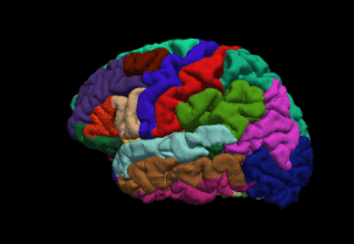
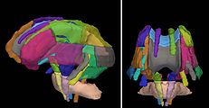
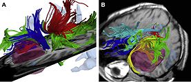
FreeSurfer is a brain imaging software package originally developed by Bruce Fischl, Anders Dale, Martin Sereno, and Doug Greve. Development and maintenance of FreeSurfer is now the primary responsibility of the Laboratory for Computational Neuroimaging at the Athinoula A. Martinos Center for Biomedical Imaging. FreeSurfer contains a set of programs with a common focus of analyzing magnetic resonance imaging (MRI) scans of brain tissue. It is an important tool in functional brain mapping and contains tools to conduct both volume based and surface based analysis. FreeSurfer includes tools for the reconstruction of topologically correct and geometrically accurate models of both the gray/white and pial surfaces, for measuring cortical thickness, surface area and folding, and for computing inter-subject registration based on the pattern of cortical folds.

<span class="mw-page-title-main">ParaView</span> Scientific visualization software

ParaView is an open-source multiple-platform application for interactive, scientific visualization. It has a client–server architecture to facilitate remote visualization of datasets, and generates level of detail (LOD) models to maintain interactive frame rates for large datasets. It is an application built on top of the Visualization Toolkit (VTK) libraries. ParaView is an application designed for data parallelism on shared-memory or distributed-memory multicomputers and clusters. It can also be run as a single-computer application.

<span class="mw-page-title-main">ITK-SNAP</span> Medical imaging software

ITK-SNAP is an interactive software application that allows users to navigate three-dimensional medical images, manually delineate anatomical regions of interest, and perform automatic image segmentation. The software was designed with the audience of clinical and basic science researchers in mind, and emphasis has been placed on having a user-friendly interface and maintaining a limited feature set to prevent feature creep. ITK-SNAP is most frequently used to work with magnetic resonance imaging (MRI), cone-beam computed tomography (CBCT) and computed tomography (CT) data sets.

<span class="mw-page-title-main">VisIt</span>

VisIt is an open-source interactive parallel visualization and graphical analysis tool for viewing scientific data. It can be used to visualize scalar and vector fields defined on 2D and 3D structured and unstructured meshes. VisIt was designed to handle big data set sizes in the terascale range and small data sets in the kilobyte range.

<span class="mw-page-title-main">GIMIAS</span>

GIMIAS is a workflow-oriented environment focused on biomedical image computing and simulation. The open-source framework is extensible through plug-ins and is focused on building research and clinical software prototypes. Gimias has been used to develop clinical prototypes in the fields of cardiac imaging and simulation, angiography imaging and simulation, and neurology

<span class="mw-page-title-main">MeVisLab</span>

MeVisLab is a cross-platform application framework for medical image processing and scientific visualization. It includes advanced algorithms for image registration, segmentation, and quantitative morphological and functional image analysis. An IDE for graphical programming and rapid user interface prototyping is available.

<span class="mw-page-title-main">Point Cloud Library</span> Open-source algorithm library

The Point Cloud Library (PCL) is an open-source library of algorithms for point cloud processing tasks and 3D geometry processing, such as occur in three-dimensional computer vision. The library contains algorithms for filtering, feature estimation, surface reconstruction, 3D registration, model fitting, object recognition, and segmentation. Each module is implemented as a smaller library that can be compiled separately. PCL has its own data format for storing point clouds - PCD, but also allows datasets to be loaded and saved in many other formats. It is written in C++ and released under the BSD license.

<span class="mw-page-title-main">IMOD (software)</span>

IMOD is an open-source, cross-platform suite of modeling, display and image processing programs used for 3D reconstruction and modeling of microscopy images with a special emphasis on electron microscopy data. IMOD has been used across a range of scales from macromolecule structures to organelles to whole cells and can also be used for optical sections. IMOD includes tools for image reconstruction, image segmentation, 3D mesh modeling and analysis of 2D and 3D data.

<span class="mw-page-title-main">Amira (software)</span> Software platform for 3D and 4D data visualization

Amira is a software platform for visualization, processing, and analysis of 3D and 4D data. It is being actively developed by Thermo Fisher Scientific in collaboration with the Zuse Institute Berlin (ZIB), and commercially distributed by Thermo Fisher Scientific — together with its sister software Avizo.

Vaa3D is an Open Source visualization and analysis software suite created mainly by Hanchuan Peng and his team at Janelia Research Campus, HHMI and Allen Institute for Brain Science. The software performs 3D, 4D and 5D rendering and analysis of very large image data sets, especially those generated using various modern microscopy methods, and associated 3D surface objects. This software has been used in several large neuroscience initiatives and a number of applications in other domains. In a recent Nature Methods review article, it has been viewed as one of the leading open-source software suites in the related research fields. In addition, research using this software was awarded the 2012 Cozzarelli Prize from the National Academy of Sciences.

<span class="mw-page-title-main">Ron Kikinis</span> American physician and scientist (born 1956)

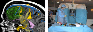
Ron Kikinis is an American physician and scientist best known for his research in the fields of imaging informatics, image guided surgery, and medical image computing. He is a professor of radiology at Harvard Medical School. Kikinis is the founding director of the Surgical Planning Laboratory in the Department of Radiology at Brigham and Women's Hospital, in Boston, Massachusetts. He is the vice-chair for Biomedical Informatics Research in the Department of Radiology.

<span class="mw-page-title-main">Studierfenster</span>

Studierfenster or StudierFenster (SF) is a free, non-commercial open science client/server-based medical imaging processing online framework. It offers capabilities, like viewing medical data (computed tomography (CT), magnetic resonance imaging (MRI), etc.) in two- and three-dimensional space directly in the standard web browsers, like Google Chrome, Mozilla Firefox, Safari, and Microsoft Edge. Other functionalities are the calculation of medical metrics (dice score and Hausdorff distance), manual slice-by-slice outlining of structures in medical images (segmentation), manual placing of (anatomical) landmarks in medical image data, viewing medical data in virtual reality, a facial reconstruction and registration of medical data for augmented reality, one click showcases for COVID-19 and veterinary scans, and a Radiomics module.

References

  1. Golby, Alexandra J. (2015-05-05). Image-Guided Neurosurgery. Academic Press. ISBN   9780128011898. Archived from the original on 2022-08-14. Retrieved 2020-06-03.
  2. Pieper, S.; Halle, M.; Kikinis, R. (2004). 3D SLICER. Proceedings of the 1st IEEE International Symposium on Biomedical Imaging: From Nano to Macro 2004 (Report). pp. 1:632–635.
  3. Adriaan, Germain (2011-08-16). 3dslicer. Brev Publishing. ISBN   9786136666464. Archived from the original on 2020-09-15. Retrieved 2022-08-14.
  4. "3D Slicer image computing platform". www.slicer.org. Archived from the original on 2000-10-18. Retrieved 2017-09-20.
  5. Pieper, S.; Lorensen, B.; Schroeder, W.; Kikinis, R. (2006). The NA-MIC Kit: ITK, VTK, Pipelines, Grids and 3D Slicer as an Open Platform for the Medical Image Computing Community. Proceedings of the 3rd IEEE International Symposium on Biomedical Imaging: From Nano to Macro 2006 (Report). pp. 1:698–701.
  6. Hirayasu, Y; Shenton, ME; Salisbury, DF; Dickey, CC; Fischer, IA; Mazzoni, P; Kisler, T; Arakaki, H; Kwon, JS; Anderson, JE; Yurgelun-Todd, D; Tohen, M; McCarley, RW (1998). "Lower left temporal lobe MRI volumes in patients with first-episode schizophrenia compared with psychotic patients with first-episode affective disorder and normal subjects". The American Journal of Psychiatry. 155 (10): 1384–91. doi:10.1176/ajp.155.10.1384. PMID   9766770. S2CID   14136755.
  7. Fedorov; Beichel; Kalpathy-Cramer; Finet; Fillion-Robin; Pujol; Bauer; Jennings; Fennessy; Sonka; Buatti; Aylward; Miller; Pieper; Kikinis (2012). "3D Slicer as an image computing platform for the Quantitative Imaging Network". Magnetic Resonance Imaging. 30 (9): 1323–41. doi:10.1016/j.mri.2012.05.001. PMC   3466397 . PMID   22770690.
  8. Butler, James (10 March 2022). "Slicer 4 reaches 1 million downloads". 3D Slicer. Archived from the original on 2022-09-12. Retrieved 2022-08-14.
  9. "Literature Search showing over 14,500 publications that reference 3D Slicer on Google Scholar". Archived from the original on 2009-07-01. Retrieved 2019-12-09.
  10. Hata, N; Piper, S; Jolesz, FA; Tempany, CM; Black, PM; Morikawa, S; Iseki, H; Hashizume, M; Kikinis, R (2007). "Application of open source image guided therapy software in MR-guided therapies". Medical Image Computing and Computer-Assisted Intervention. 10 (Pt 1): 491–8. doi: 10.1007/978-3-540-75757-3_60 . PMID   18051095.
  11. Archip, N; Clatz, O; Whalen, S; Kacher, D; Fedorov, A; Kot, A; Chrisochoides, N; Jolesz, F; Golby, A; Black, PM; Warfield, SK (2007). "Non-rigid alignment of pre-operative MRI, fMRI, and DT-MRI with intra-operative MRI for enhanced visualization and navigation in image-guided neurosurgery". NeuroImage. 35 (2): 609–24. doi:10.1016/j.neuroimage.2006.11.060. PMC   3358788 . PMID   17289403.
  12. Ziyan, U; Tuch, D; Westin, CF (2006). "Segmentation of thalamic nuclei from DTI using spectral clustering". Medical Image Computing and Computer-Assisted Intervention. 9 (Pt 2): 807–14. doi: 10.1007/11866763_99 . PMID   17354847.
  13. Verhey, JF; Nathan, NS; Rienhoff, O; Kikinis, R; Rakebrandt, F; D'ambra, MN (2006). "Finite-element-method (FEM) model generation of time-resolved 3D echocardiographic geometry data for mitral-valve volumetry". BioMedical Engineering OnLine. 5: 17. doi: 10.1186/1475-925X-5-17 . PMC   1421418 . PMID   16512925.
  14. "The Open Source Paleontologist: 3D Slicer: The Tutorial Part VI". 5 March 2009. Archived from the original on 23 November 2020. Retrieved 6 May 2010.
  15. "Picasa-Webalben - András Jakab". Archived from the original on 2015-10-01. Retrieved 2010-05-06.
  16. "3D Slicer Forum". 3D Slicer. Archived from the original on 2022-05-31. Retrieved 2022-05-31.
  17. "Archive for the 'marching cubes' Category". biomedical optics. Archived from the original on 2008-09-22. Retrieved 2017-09-20.
  18. Detection and Quantification of Small Changes in MRI Volumes. 2014. p. 18.
  19. "Slicer/Slicer". GitHub . 4 June 2022. Archived from the original on 31 May 2022. Retrieved 31 May 2022.