An integrated development environment (IDE) is a software application that provides comprehensive facilities for software development. An IDE normally consists of at least a source-code editor, build automation tools, and a debugger. Some IDEs, such as IntelliJ IDEA, Eclipse and Lazarus contain the necessary compiler, interpreter or both; others, such as SharpDevelop, NetBeans do not.

A picture archiving and communication system (PACS) is a medical imaging technology which provides economical storage and convenient access to images from multiple modalities. Electronic images and reports are transmitted digitally via PACS; this eliminates the need to manually file, retrieve, or transport film jackets, the folders used to store and protect X-ray film. The universal format for PACS image storage and transfer is DICOM. Non-image data, such as scanned documents, may be incorporated using consumer industry standard formats like PDF, once encapsulated in DICOM. A PACS consists of four major components: The imaging modalities such as X-ray plain film (PF), computed tomography (CT) and magnetic resonance imaging (MRI), a secured network for the transmission of patient information, workstations for interpreting and reviewing images, and archives for the storage and retrieval of images and reports. Combined with available and emerging web technology, PACS has the ability to deliver timely and efficient access to images, interpretations, and related data. PACS reduces the physical and time barriers associated with traditional film-based image retrieval, distribution, and display.
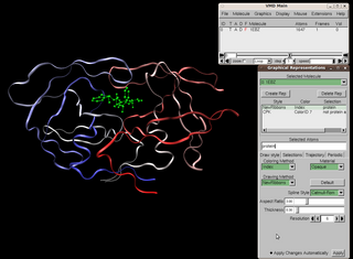
Visual Molecular Dynamics (VMD) is a molecular modelling and visualization computer program. VMD is developed mainly as a tool to view and analyze the results of molecular dynamics simulations. It also includes tools for working with volumetric data, sequence data, and arbitrary graphics objects. Molecular scenes can be exported to external rendering tools such as POV-Ray, RenderMan, Tachyon, Virtual Reality Modeling Language (VRML), and many others. Users can run their own Tcl and Python scripts within VMD as it includes embedded Tcl and Python interpreters. VMD runs on Unix, Apple Mac macOS, and Microsoft Windows. VMD is available to non-commercial users under a distribution-specific license which permits both use of the program and modification of its source code, at no charge.

jEdit is a free software text editor available under GPL-2.0-or-later. It is written in Java and runs on any operating system with Java support, including BSD, Linux, macOS and Windows.

OsiriX is an image processing application for the Apple MacOS operating system dedicated to DICOM images produced by equipment. OsiriX is complementary to existing viewers, in particular to nuclear medicine viewers. It can also read many other file formats: TIFF, JPEG, PDF, AVI, MPEG and QuickTime. It is fully compliant with the DICOM standard for image communication and image file formats. OsiriX is able to receive images transferred by DICOM communication protocol from any PACS or medical imaging modality.
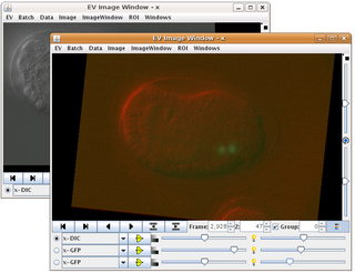
CellProfiler is free, open-source software designed to enable biologists without training in computer vision or programming to quantitatively measure phenotypes from thousands of images automatically. Advanced algorithms for image analysis are available as individual modules that can be placed in sequential order together to form a pipeline; the pipeline is then used to identify and measure biological objects and features in images, particularly those obtained through fluorescence microscopy.

Aptana, Inc. is a company that makes web application development tools for use with a variety of programming languages. Aptana's main products include Aptana Studio, Aptana Cloud and Aptana Jaxer.
Endrov is an open-source plugin architecture aimed for image analysis and data processing. Being based on Java, it is portable and can both be run locally and as an applet. It grew out of the need for an advanced open source software that can cope with complex spatio-temporal image data, mainly obtained from microscopes in biological research. It lends much of the philosophy from ImageJ but aims to supersede it by having a more modern design.
3D Slicer (Slicer) is a free and open source software package for image analysis and scientific visualization. Slicer is used in a variety of medical applications, including autism, multiple sclerosis, systemic lupus erythematosus, prostate cancer, lung cancer, breast cancer, schizophrenia, orthopedic biomechanics, COPD, cardiovascular disease and neurosurgery.

Mango is a non-commercial software for viewing, editing and analyzing volumetric medical images. Mango is written in Java, and distributed freely in precompiled versions for Linux, Mac OS and Microsoft Windows. It supports NIfTI, ANALYZE, NEMA and DICOM formats and is able to load and save 2D, 3D and 4D images.

Fiji is an open source image processing package based on ImageJ2.
KNIME, the Konstanz Information Miner, is a free and open-source data analytics, reporting and integration platform. KNIME integrates various components for machine learning and data mining through its modular data pipelining "Building Blocks of Analytics" concept. A graphical user interface and use of JDBC allows assembly of nodes blending different data sources, including preprocessing, for modeling, data analysis and visualization without, or with only minimal, programming.
GIMIAS is a workflow-oriented environment focused on biomedical image computing and simulation. The open-source framework is extensible through plug-ins and is focused on building research and clinical software prototypes. Gimias has been used to develop clinical prototypes in the fields of cardiac imaging and simulation, angiography imaging and simulation, and neurology
Orthanc is a standalone DICOM server. It is designed to improve the DICOM flows in hospitals and to support research about the automated analysis of medical images. Orthanc lets its users focus on the content of the DICOM files, hiding the complexity of the DICOM format and of the DICOM protocol. It is licensed under the GPLv3.

Ginkgo CADx is a multi platform DICOM viewer (*.dcm) and dicomizer. Ginkgo CADx is licensed under LGPL license, being an open source project with an Open core approach. The goal of Ginkgo CADx project is to develop an open source professional DICOM workstation.