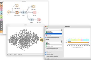
Orange is an open-source data visualization, machine learning and data mining toolkit. It features a visual programming front-end for explorative qualitative data analysis and interactive data visualization.
ITK is a cross-platform, open-source application development framework widely used for the development of image segmentation and image registration programs. Segmentation is the process of identifying and classifying data found in a digitally sampled representation. Typically the sampled representation is an image acquired from such medical instrumentation as CT or MRI scanners. Registration is the task of aligning or developing correspondences between data. For example, in the medical environment, a CT scan may be aligned with an MRI scan in order to combine the information contained in both.

ImageJ is a Java-based image processing program developed at the National Institutes of Health and the Laboratory for Optical and Computational Instrumentation. Its first version, ImageJ 1.x, is developed in the public domain, while ImageJ2 and the related projects SciJava, ImgLib2, and SCIFIO are licensed with a permissive BSD-2 license. ImageJ was designed with an open architecture that provides extensibility via Java plugins and recordable macros. Custom acquisition, analysis and processing plugins can be developed using ImageJ's built-in editor and a Java compiler. User-written plugins make it possible to solve many image processing and analysis problems, from three-dimensional live-cell imaging to radiological image processing, multiple imaging system data comparisons to automated hematology systems. ImageJ's plugin architecture and built-in development environment has made it a popular platform for teaching image processing.
CellProfiler is free, open-source software designed to enable biologists without training in computer vision or programming to quantitatively measure phenotypes from thousands of images automatically. Advanced algorithms for image analysis are available as individual modules that can be placed in sequential order together to form a pipeline; the pipeline is then used to identify and measure biological objects and features in images, particularly those obtained through fluorescence microscopy.

ParaView is an open-source multiple-platform application for interactive, scientific visualization. It has a client–server architecture to facilitate remote visualization of datasets, and generates level of detail (LOD) models to maintain interactive frame rates for large datasets. It is an application built on top of the Visualization Toolkit (VTK) libraries. ParaView is an application designed for data parallelism on shared-memory or distributed-memory multicomputers and clusters. It can also be run as a single-computer application.
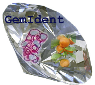
GemIdent is an interactive image recognition program that identifies regions of interest in images and photographs. It is specifically designed for images with few colors, where the objects of interest look alike with small variation. For example, color image segmentation of:

VisTrails is a scientific workflow management system developed at the Scientific Computing and Imaging Institute at the University of Utah that provides support for data exploration and visualization. It is written in Python and employs Qt via PyQt bindings. The system is open source, released under the GPL v2 license. The pre-compiled versions for Windows, Mac OS X, and Linux come with an installer and several packages, including VTK, matplotlib, and ImageMagick. VisTrails also supports user-defined packages.

Fiji is an open source image processing package based on ImageJ2.

In computer science, Orfeo Toolbox (OTB) is a software library for processing images from Earth observation satellites.
KNIME, the Konstanz Information Miner, is a free and open-source data analytics, reporting and integration platform. KNIME integrates various components for machine learning and data mining through its modular data pipelining "Building Blocks of Analytics" concept. A graphical user interface and use of JDBC allows assembly of nodes blending different data sources, including preprocessing, for modeling, data analysis and visualization without, or with minimal, programming.

Spyder is an open-source cross-platform integrated development environment (IDE) for scientific programming in the Python language. Spyder integrates with a number of prominent packages in the scientific Python stack, including NumPy, SciPy, Matplotlib, pandas, IPython, SymPy and Cython, as well as other open-source software. It is released under the MIT license.

IMOD is an open-source, cross-platform suite of modeling, display and image processing programs used for 3D reconstruction and modeling of microscopy images with a special emphasis on electron microscopy data. IMOD has been used across a range of scales from macromolecule structures to organelles to whole cells and can also be used for optical sections. IMOD includes tools for image reconstruction, image segmentation, 3D mesh modeling and analysis of 2D and 3D data.
VIGRA is the abbreviation for "Vision with Generic Algorithms". It is a free open-source computer vision library which focuses on customizable algorithms and data structures. VIGRA component can be easily adapted to specific needs of target application without compromising execution speed, by using template techniques similar to those in the C++ Standard Template Library.
Vaa3D is an Open Source visualization and analysis software suite created mainly by Hanchuan Peng and his team at Janelia Research Campus, HHMI and Allen Institute for Brain Science. The software performs 3D, 4D and 5D rendering and analysis of very large image data sets, especially those generated using various modern microscopy methods, and associated 3D surface objects. This software has been used in several large neuroscience initiatives and a number of applications in other domains. In a recent Nature Methods review article, it has been viewed as one of the leading open-source software suites in the related research fields. In addition, research using this software was awarded the 2012 Cozzarelli Prize from the National Academy of Sciences.

The Aphelion Imaging Software Suite is a software suite that includes three base products - Aphelion Lab, Aphelion Dev, and Aphelion SDK for addressing image processing and image analysis applications. The suite also includes a set of extension programs to implement specific vertical applications that benefit from imaging techniques.

Keras is an open-source library that provides a Python interface for artificial neural networks. Keras was first independent software, then integrated into TensorFlow library, and later supporting more. "Keras 3 is a full rewrite of Keras [can be used] as a low-level cross-framework language to develop custom components such as layers, models, or metrics that can be used in native workflows in JAX, TensorFlow, or PyTorch — with one codebase." Keras 3 will be the default Keras version for TensorFlow 2.16 onwards, but Keras 2 can still be used.

SimpleITK is a simplified, open-source interface to the Insight Segmentation and Registration Toolkit (ITK). The SimpleITK image analysis library is available in multiple programming languages including C++, Python, R, Java, C#, Lua, Ruby and Tcl. Binary distributions are available for all three major operating systems.
Medical open network for AI (MONAI) is an open-source, community-supported framework for Deep learning (DL) in healthcare imaging. MONAI provides a collection of domain-optimized implementations of various DL algorithms and utilities specifically designed for medical imaging tasks. MONAI is used in research and industry, aiding the development of various medical imaging applications, including image segmentation, image classification, image registration, and image generation.
Anna Kreshuk is a group leader at the European Molecular Biology Laboratory in Heidelberg, Germany. She joined the Cell Biology and Biophysics Unit in July 2018, where her group employs machine learning to develop automated methods to help biologists speed up image analysis.