Alcohol-related brain damage

Last updated

Alcohol-related brain damage [1] alters both the structure and function of the brain as a result of the direct neurotoxic effects of alcohol intoxication or acute alcohol withdrawal. Increased alcohol intake is associated with damage to brain regions including the frontal lobe, [2] limbic system, and cerebellum, [3] with widespread cerebral atrophy, or brain shrinkage caused by neuron degeneration. This damage can be seen on neuroimaging scans. [4]

Contents

Frontal lobe damage becomes the most prominent as alcoholics age and can lead to impaired neuropsychological performance in areas such as problem solving, good judgment, and goal-directed behaviors. [2] Impaired emotional processing results from damage to the limbic system. This may lead to troubles recognizing emotional facial expressions and "interpreting nonverbal emotional cues". [2]

Binge drinking, or heavy episodic drinking, can lead to damage in the limbic system that occurs after a relatively short period of time. This brain damage increases the risk of alcohol-related dementia, and abnormalities in mood and cognitive abilities. Binge drinkers also have an increased risk of developing chronic alcoholism. Alcoholism is a chronic relapsing disorder that can include extended periods of abstinence followed by relapse to heavy drinking. It is also associated with many other health problems including memory disorders, high blood pressure, muscle weakness, heart problems, anaemia, low immune function, liver disease, disorders of the digestive system, and pancreatic problems. It has also been correlated with depression, unemployment, and family problems with an increased risk of domestic abuse.

Gender and parental history of alcoholism and binge drinking has an influence on susceptibility to alcohol dependence as higher levels are typically seen in males and in those with a family history. [5]

Prevalence

Nearly half of American alcoholics exhibit "neuropsychological disabilities [that] can range from mild to severe" [2] with approximately two million requiring lifelong care after developing permanent and debilitating conditions. Prolonged alcohol abstinence can lead to an improvement in these disabilities. For those with mild impairments, some improvement has been seen within a year, but this can take much longer in those with higher severity damage. [2]

Populations at risk

Adolescents and genetic factors

The impulsivity and sensation seeking seen in adolescence may lead to increased alcohol intake and more frequent binge drinking episodes leaving adolescents particularly at risk for alcoholism. The still developing brain of adolescents is more vulnerable to the damaging neurotoxic and neurodegenerative effects of alcohol. [6] "High impulsivity has [also] been found in families with alcoholism, suggestive of a genetic link. Thus, the genetics of impulsivity overlaps with genetic risks for alcohol use disorder and possibly alcohol neurodegeneration". [6]

There is also a genetic risk for proinflammatory cytokine mediated alcohol-related brain damage. There is evidence that variants of these genes are involved not only in contributing to brain damage but also to impulsivity and alcohol abuse. All three of these genetic traits contribute heavily to an alcohol use disorder. [6]

Neurological deficits

Brain anatomy Brain Anatomy Striatum.png
Brain anatomy

Alcoholics can typically be divided into two categories, uncomplicated and complicated. [3] Uncomplicated alcoholics do not have nutritional deficiency states or liver disease, but have a reduction in overall brain volume due to white matter cerebral atrophy. The severity of atrophy sustained from alcohol consumption is proportional to the rate and amount of alcohol consumed during a person's life. [7] Complicated alcoholics may have liver damage that impacts brain structure and function and nutritional deficiencies "that can cause severe brain damage and dysfunction". [3] [7]

Pathophysiology

Adolescents are much more vulnerable to alcohol-related brain damage in the form of persistent changes in neuroimmune signalling from binge drinking. [8] The endocrine system includes the hypothalamic–pituitary–adrenal axis, the hypothalamic–pituitary–gonadal axis, the hypothalamic–pituitary–thyroid axis, the hypothalamic–pituitary–growth hormone/insulin-like growth factor-1 axis, and the hypothalamic–posterior pituitary axis, as well as other sources of hormones, such as the endocrine pancreas and endocrine adipose tissue. Alcohol abuse disrupts all of these systems and causes hormonal disturbances that may result in various disorders, such as stress intolerance, reproductive dysfunction, thyroid problems, immune abnormalities, and psychological and behavioral disorders. [9]

The cerebral atrophy that alcoholics often present with is due to alcohol induced neurotoxicity. [6] [10] Evidence of neurodegeneration can be supported by an increased microglia density and expression of proinflammatory cytokines in the brain. Animal studies find that heavy and regular binge drinking causes neurodegeneration in corticolimbic brain regions areas which are involved in learning and spatial memory. The corticolimbic brain regions affected include the olfactory bulb, piriform cortex, perirhinal cortex, entorhinal cortex, and the hippocampal dentate gyrus. It was found that a heavy two-day drinking binge caused extensive neurodegeneration in the entorhinal cortex with resultant learning deficits in rats. [5]

It is unclear how the frequency and length of these binge drinking sessions impacts brain damage in humans. Humans who drank at least 100 drinks (male) or 80 drinks (female) per month (concentrated to 21 occasions or less per month) throughout a three-year period had impaired decision-making skills compared to non-binge drinkers. [5] An MRI brain scan found that levels of N-acetylaspartate (NAA), a metabolite biomarker for neural integrity, was lower in binge drinkers. Additionally, abnormal brain metabolism, a loss of white brain matter in the frontal lobe, and higher parietal gray matter NAA levels were found. This shows a correlation between binge drinking, poor executive functioning, and working memory. A decrease in frontal lobe NAA levels is associated with impaired executive functioning and processing speed in neuro-performance tests. [5]

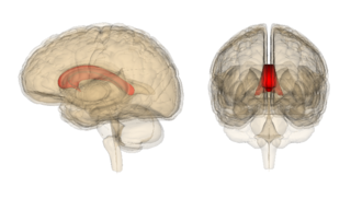
The volume of the corpus callosum, a large white matter tract that connects the two cerebral hemispheres, is shown to decrease with alcohol abuse due to a loss of myelination. This integration between the two cerebral hemispheres and cognitive function is affected. A limited amount of myelin can be restored with alcohol abstinence, leading to transient neurological deficits. [7]

Alcohol abuse affects neurons in the frontal cortex that typically have a large soma, or cell body. This type of neuron is more susceptible to Alzheimer's disease and normal aging. Research is still being conducted to determine whether there is a direct link between excessive alcohol consumption and Alzheimer's disease. [7]

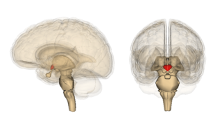
Higher order functioning of the cerebral cortex is organized by the cerebellum. In those with cerebral atrophy, Purkinje cells, or the cerebellar output neurons, in the vermis are reduced in number by 43%. [7] This large reduction in Purkinje cells causes a decrease in high order cerebral cortex organization. The cerebellum is also responsible for refining crude motor output from the primary motor cortex. When this refinement is missing, symptoms such as unsteadiness and ataxia [7] will present. A potential cause of chronic alcoholic cerebellar dysfunction is an alteration of GABA-A receptor. This dysfunction causes an increase in the neurotransmitter GABA in cerebellar Purkinje cells, granule cells, and interneurons leading to a disruption in normal cell signaling. [7]

GABA-A receptor embedded in cell membrane Cell GABA Receptor.png
GABA-A receptor embedded in cell membrane

Kindling and excitotoxicity

Binge drinkers and alcoholics who go through multiple detoxifications show prefrontal cortex dysfunction, as it is known that alcohol has long-term effects on prefrontal cortex function, leading to impairments in executive control tasks. Animal studies show that repeated alcohol withdrawals are associated with a significantly impaired ability to learn new information. [11] Alcohol's acute effects on GABAergic enhancement and NMDA suppression cause alcohol induced neurotoxicity and kindling, or worsening of alcohol withdrawal symptoms with each subsequent withdrawal period. This may cause CNS depression leading to acute tolerance to these withdrawal effects. This tolerance is followed by a damaging rebound effect during withdrawal. This rebound causes hyperexcitability of neurotransmission systems. If this hyperexcitability state occurs multiple times, kindling and neurotoxicity can occur leading to increased alcohol-related brain damage. Damaging excitotoxicity may also occur as a result of repeated withdrawals. Similar to people who have gone through multiple detoxifications, binge drinkers show a higher rate of emotional disturbance due to these damaging effects. [11]

Thiamine deficiency

Thiamine is a vitamin your body needs for growth, development, and cellular function, as well as converting food into energy. Thiamine is naturally present in some foods, added to some food products, and available as a dietary supplement. [12] A nutritional deficiency in thiamine can worsen alcohol-related brain damage. There is a genetic component to thiamine deficiency that causes intestinal malabsorption. [13] A nutritional vitamin deficiency state that is caused by thiamine deficiency which is seen most commonly in alcoholics leads to Wernicke's encephalopathy and Alcoholic Korsakoff syndrome (AKS) which frequently occur simultaneously, known as Wernicke–Korsakoff syndrome (WKS). This disorder is preventable through supplementation of the diet by thiamine and an awareness by health professionals to treat 'at risk' patients with thiamine. [13] Thiamine deficiency may occur in upwards of 80% of patients with alcoholism however, only ≈13% of such individuals develop WKS, raising the possibility that a genetic predisposition to WKS may exist in some individuals. [14] [15] Lesions, or brain abnormalities, are typically located in the diencephalon which result in anterograde and retrograde amnesia, or memory loss. [15]

Neuroimaging

Neuroimaging is used to study the effect that alcohol has on the brain. The two main imaging methods are hemodynamic and electromagnetic. These techniques have allowed for the study of the functional, biochemical, and anatomical changes of the brain due to prolonged alcohol abuse. [2] Neuroimaging provides valuable information in determining the risk an individual has for developing alcohol dependence and the efficacy of potential treatment. [2] [16]

Hemodynamic methods

Parasagittal render of human brain with MRI MRI brain.jpg
Parasagittal render of human brain with MRI

Hemodynamic methods record changes in blood volume, blood flow, blood oxygenation, and energy metabolism to produce images. [2] Positron emission tomography (PET) and single-photon emission computed tomography (SPECT) are common techniques that require the injection of a radioactively labeled molecule, such as glucose, to allow for proper visualization. After injection, the patient is then observed while performing mental tasks, such as a memory task. PET and SPECT studies have confirmed and expanded previous findings stating that the prefrontal cortex is particularly susceptible to decreased metabolism in alcohol abusing patients. [2]

Magnetic resonance imaging (MRI) and functional magnetic resonance imaging (fMRI) are other commonly used tenichiques. These methods are noninvasive, and have no radioactive risk involved. The fMRI method records the metabolic changes in a particular brain structure or region during a mental task. To detect damage to white matter, the standard MRI is not sufficient. An MRI derivative technique known as diffusion tensor imaging (DTI) is used to determine the orientation and integrity of specific nerve pathways, allowing the detection of damage. [2] When imaging those with alcoholism, the DTI results show that heavy drinking disrupts the microstructure of nerve fibers. [2] Another MRI derivative technique, magnetic resonance spectroscopy imaging (MRSI), can provide further information about the brain's neurochemistry and can detect the distribution of certain metabolites, neurotransmitters, and alcohol.[ citation needed ]

Electromagnetic methods

Man ready for EEG recording EEG recording.jpg
Man ready for EEG recording

While the hemo-dynamic methods are effective for observing spatial and chemical changes, they cannot show the time course of these changes. Electromagnetic imaging methods are capable of capturing real-time changes in the brain's electrical currents. [17] Electroencephalography (EEG) imaging utilizes small electrodes that are attached to the scalp. The recordings are averaged by a technique known as event-related potentials (ERP). This is done to determine the time sequence of activity after being exposed to a stimulus, such as a word or image. [2] Magnetoencephalography (MEG) is another imaging method that utilizes sensors. This measures the magnetic field created as a result of the brain's electrical activity. These techniques are noninvasive, harmless, and provide a large amount of detail regarding the order and timing of electrical activity. The poor spatial imaging of these methods are a large downside.[ citation needed ]

These neuroimaging methods have found that alcohol alters the nervous system on multiple levels. [2] This includes impairment of lower order brainstem functions and higher order functioning, such as problem solving. These methods have also shown differences in electrical brain activity and responsiveness when comparing alcohol-dependent and healthy individuals. [2]

Clinical applications

In Korsakoff patients, MRI shows atrophy of the thalamus and mamillary bodies. PET showed decreased metabolism, and therefore decreased activity in the thalamus and other diencephalon structures. [13] Uncomplicated alcoholics, those with chronic Wernicke's encephalopathy (WE), and Korsakoff psychosis showed significant neuronal loss in the frontal cortex, white matter, hippocampus, and basal forebrain. [13] Uncomplicated alcoholics were seen to have a shrinkage in raphe neurons, the mamillary bodies, and the thalamus. [13]

Treatment and prevention

Alcohol-related brain damage can have drastic effects on the individuals affected and their loved ones. The options for treatment are very limited compared to other disorders. Although limited, most patients with alcohol-related cognitive deficits experienced slight improvement of their symptoms over the first two to three months of treatment. [7] Others have said to see increase in cerebral metabolism as soon as one month after treatment. [2]

Education on the prevention of alcoholism is the best supported method of avoiding alcohol-related brain damage. [7] By providing information that studies have found on risk factors and the mechanisms of damage, the efforts to find an effective treatment may increase. This may also reduce mortality by influencing doctors to pay closer attention to the warning signs. [7]

Related Research Articles

<span class="mw-page-title-main">Alcoholism</span> Problematic excessive alcohol consumption

Alcoholism is the continued drinking of alcohol despite it causing problems. Some definitions require evidence of dependence and withdrawal. Problematic use of alcohol has been mentioned in the earliest historical records, the World Health Organization (WHO) estimated there were 283 million people with alcohol use disorders worldwide as of 2016. The term alcoholism was first coined in 1852, but alcoholism and alcoholic are stigmatizing and discourage seeking treatment, so clinical diagnostic terms such as alcohol use disorder or alcohol dependence are used instead.

<span class="mw-page-title-main">Alcohol abuse</span> Substance abuse of alcoholic beverages

Alcohol abuse encompasses a spectrum of alcohol-related substance abuse, ranging from the consumption of more than 2 drinks per day on average for men, or more than 1 drink per day on average for women, to binge drinking or alcohol use disorder.

<span class="mw-page-title-main">Brain damage</span> Destruction or degeneration of brain cells

Neurotrauma, brain damage or brain injury (BI) is the destruction or degeneration of brain cells. Brain injuries occur due to a wide range of internal and external factors. In general, brain damage refers to significant, undiscriminating trauma-induced damage.

<span class="mw-page-title-main">Wernicke–Korsakoff syndrome</span> Combined presence of Wernickes encephalopathy (WE) and Korsakoffs syndrome

Wernicke-Korsakoff syndrome (WKS) is the combined presence of Wernicke encephalopathy (WE) and alcoholic Korsakoff syndrome. Due to the close relationship between these two disorders, people with either are usually diagnosed with WKS as a single syndrome. It mainly causes vision changes, ataxia and impaired memory.

<span class="mw-page-title-main">Korsakoff syndrome</span> Mental illness caused by a lack of thiamine in the brain

Korsakoff syndrome (KS) is a disorder of the central nervous system characterized by amnesia, deficits in explicit memory, and confabulation. This neurological disorder is caused by a deficiency of thiamine (vitamin B1) in the brain, and it is typically associated with and exacerbated by the prolonged, excessive ingestion of alcohol. Korsakoff syndrome is often accompanied by Wernicke encephalopathy; this combination is called Wernicke–Korsakoff syndrome.

The health effects of long-term alcohol consumption on health vary depending on the amount of ethanol consumed. Even light drinking poses health risks, but small amounts of alcohol may also have health benefits. Chronic heavy drinking causes severe health consequences which outweigh any potential benefits.

<span class="mw-page-title-main">Frontal lobe</span> Part of the brain

The frontal lobe is the largest of the four major lobes of the brain in mammals, and is located at the front of each cerebral hemisphere. It is parted from the parietal lobe by a groove between tissues called the central sulcus and from the temporal lobe by a deeper groove called the lateral sulcus. The most anterior rounded part of the frontal lobe is known as the frontal pole, one of the three poles of the cerebrum.

<span class="mw-page-title-main">Wernicke encephalopathy</span> Medical condition

Wernicke encephalopathy (WE), also Wernicke's encephalopathy, or wet brain is the presence of neurological symptoms caused by biochemical lesions of the central nervous system after exhaustion of B-vitamin reserves, in particular thiamine (vitamin B1). The condition is part of a larger group of thiamine deficiency disorders that includes beriberi, in all its forms, and alcoholic Korsakoff syndrome. When it occurs simultaneously with alcoholic Korsakoff syndrome it is known as Wernicke–Korsakoff syndrome.

<span class="mw-page-title-main">Alcoholic polyneuropathy</span> Medical condition

Alcoholic polyneuropathy is a neurological disorder in which peripheral nerves throughout the body malfunction simultaneously. It is defined by axonal degeneration in neurons of both the sensory and motor systems and initially occurs at the distal ends of the longest axons in the body. This nerve damage causes an individual to experience pain and motor weakness, first in the feet and hands and then progressing centrally. Alcoholic polyneuropathy is caused primarily by chronic alcoholism; however, vitamin deficiencies are also known to contribute to its development. This disease typically occurs in chronic alcoholics who have some sort of nutritional deficiency. Treatment may involve nutritional supplementation, pain management, and abstaining from alcohol.

<span class="mw-page-title-main">Fetal alcohol spectrum disorder</span> Group of conditions resulting from maternal alcohol consumption during pregnancy

Fetal alcohol spectrum disorders (FASDs) are a group of conditions that can occur in a person who is exposed to alcohol during gestation, as a result of their mother drinking alcohol during pregnancy. The several forms of the condition are: fetal alcohol syndrome (FAS), partial fetal alcohol syndrome (pFAS), alcohol-related neurodevelopmental disorder (ARND), and neurobehavioral disorder associated with prenatal alcohol exposure (ND-PAE). Other terms used are fetal alcohol effects (FAE), partial fetal alcohol effects (PFAE), alcohol-related birth defects (ARBD), and static encephalopathy, but these terms have fallen out of favor and are no longer considered part of the spectrum.

Cerebral atrophy is a common feature of many of the diseases that affect the brain. Atrophy of any tissue means a decrement in the size of the cell, which can be due to progressive loss of cytoplasmic proteins. In brain tissue, atrophy describes a loss of neurons and the connections between them. Brain atrophy can be classified into two main categories: generalized and focal atrophy. Generalized atrophy occurs across the entire brain whereas focal atrophy affects cells in a specific location. If the cerebral hemispheres are affected, conscious thought and voluntary processes may be impaired.

<span class="mw-page-title-main">Thiamine deficiency</span> Human disease

Thiamine deficiency is a medical condition of low levels of thiamine (vitamin B1). A severe and chronic form is known as beriberi. The name beriberi is possibly in the 18th century borrowed from the Sinhalese phrase බැරි බැරි (bæri bæri, “I cannot, I cannot”) due to weakness. The two main types in adults are wet beriberi and dry beriberi. Wet beriberi affects the cardiovascular system, resulting in a fast heart rate, shortness of breath, and leg swelling. Dry beriberi affects the nervous system, resulting in numbness of the hands and feet, confusion, trouble moving the legs, and pain. A form with loss of appetite and constipation may also occur. Another type, acute beriberi, found mostly in babies, presents with loss of appetite, vomiting, lactic acidosis, changes in heart rate, and enlargement of the heart.

<span class="mw-page-title-main">Marchiafava–Bignami disease</span> Medical condition

Marchiafava–Bignami disease is a progressive neurological disease of alcohol use disorder, characterized by corpus callosum demyelination and necrosis and subsequent atrophy. The disease was first described in 1903 by the Italian pathologists Amico Bignami and Ettore Marchiafava in an Italian Chianti drinker. In this autopsy, Marchiafava and Bignami noticed that the middle two-thirds of the corpus callosum were necrotic. It is very difficult to diagnose and there is no specific treatment. Until 2008 only around 300 cases had been reported. If caught early enough, most patients survive.

Alcohol-related dementia (ARD) is a form of dementia caused by long-term, excessive consumption of alcohol, resulting in neurological damage and impaired cognitive function.

<span class="mw-page-title-main">Alcohol withdrawal syndrome</span> Medical condition

Alcohol withdrawal syndrome (AWS) is a set of symptoms that can occur following a reduction in alcohol use after a period of excessive use. Symptoms typically include anxiety, shakiness, sweating, vomiting, fast heart rate, and a mild fever. More severe symptoms may include seizures, and delirium tremens (DTs); which can be fatal in untreated patients. Symptoms start at around 6 hours after last drink. Peak incidence of seizures occurs at 24-36 hours and peak incidence of delirium tremens is at 48-72 hours.

<span class="mw-page-title-main">Binge drinking</span> Form of excessive alcohol intake

Binge drinking, or heavy episodic drinking, is drinking alcoholic beverages with an intention of becoming intoxicated by heavy consumption of alcohol over a short period of time, but definitions vary considerably.

<span class="mw-page-title-main">Nutritional neuroscience</span> Scientific discipline

Nutritional neuroscience is the scientific discipline that studies the effects various components of the diet such as minerals, vitamins, protein, carbohydrates, fats, dietary supplements, synthetic hormones, and food additives have on neurochemistry, neurobiology, behavior, and cognition.

Kindling due to substance withdrawal is the neurological condition which results from repeated withdrawal episodes from sedative–hypnotic drugs such as alcohol and benzodiazepines.

While researchers have found that moderate alcohol consumption in older adults is associated with better cognition and well-being than abstinence, excessive alcohol consumption is associated with widespread and significant brain lesions. Other data – including investigated brain-scans of 36,678 UK Biobank participants – suggest that even "light" or "moderate" consumption of alcohol by itself harms the brain, such as by reducing brain grey matter volume. This may imply that alternatives and generally aiming for lowest possible consumption could usually be the advisable approach.

<span class="mw-page-title-main">Cerebellar degeneration</span> Medical condition

Cerebellar degeneration is a condition in which cerebellar cells, otherwise known as neurons, become damaged and progressively weaken in the cerebellum. There are two types of cerebellar degeneration; paraneoplastic cerebellar degeneration, and alcoholic or nutritional cerebellar degeneration. As the cerebellum contributes to the coordination and regulation of motor activities, as well as controlling equilibrium of the human body, any degeneration to this part of the organ can be life-threatening. Cerebellar degeneration can result in disorders in fine movement, posture, and motor learning in humans, due to a disturbance of the vestibular system. This condition may not only cause cerebellar damage on a temporary or permanent basis, but can also affect other tissues of the central nervous system, those including the cerebral cortex, spinal cord and the brainstem.

References

  1. Zahr, Natalie M.; Kaufman, Kimberley L.; Harper, Clive G. (May 2011). "Clinical and pathological features of alcohol-related brain damage". Nature Reviews Neurology. 7 (5): 284–294. doi:10.1038/nrneurol.2011.42. ISSN   1759-4758. PMC   8121189 . PMID   21487421.
  2. 1 2 3 4 5 6 7 8 9 10 11 12 13 14 15 Oscar-Berman, Marlene (June 2003). "Alcoholism and the Brain". Alcohol Research & Health. 27 (2): 125–133.
  3. 1 2 3 Sutherland, Greg (1 January 2014). "Neuropathology of alcoholism". Alcohol and the Nervous System. Handbook of Clinical Neurology. Vol. 125. pp. 603–615. doi:10.1016/B978-0-444-62619-6.00035-5. hdl: 2123/19684 . ISBN   9780444626196. ISSN   0072-9752. PMID   25307599.
  4. Nutt, David; Hayes, Alexandra; Fonville, Leon; Zafar, Rayyan; Palmer, Emily O.C.; Paterson, Louise; Lingford-Hughes, Anne (4 November 2021). "Alcohol and the Brain". Nutrients. 13 (11): 3938. doi: 10.3390/nu13113938 . ISSN   2072-6643. PMC   8625009 . PMID   34836193.
  5. 1 2 3 4 Courtney, Kelly E.; Polich, John (January 2009). "Binge Drinking in Young Adults: Data, Definitions, and Determinants". Psychological Bulletin. 135 (1): 142–156. doi:10.1037/a0014414. ISSN   0033-2909. PMC   2748736 . PMID   19210057.
  6. 1 2 3 4 Crews, Fulton Timm; Boettiger, Charlotte Ann (September 2009). "Impulsivity, Frontal Lobes and Risk for Addiction". Pharmacology Biochemistry and Behavior. 93 (3): 237–247. doi:10.1016/j.pbb.2009.04.018. ISSN   0091-3057. PMC   2730661 . PMID   19410598.
  7. 1 2 3 4 5 6 7 8 9 10 Harper, Clive (15 January 2009). "The Neuropathology of Alcohol-Related Brain Damage". Alcohol and Alcoholism. 44 (2): 136–140. doi: 10.1093/alcalc/agn102 . PMID   19147798.
  8. Crews FT, Vetreno RP (2014). "Neuroimmune basis of alcoholic brain damage". Int. Rev. Neurobiol. International Review of Neurobiology. 118: 315–57. doi:10.1016/B978-0-12-801284-0.00010-5. ISBN   9780128012840. PMC   5765863 . PMID   25175868.
  9. Rachdaoui, Nadia; Sarkar, Dipak K. (2017). "Effects of Alcohol on the Endocrine System". Endocrinology and Metabolism Clinics of North America. 42 (3): 593–615. doi:10.1016/j.ecl.2013.05.008. ISSN   0889-8529. PMC   5513689 . PMID   28988577.
  10. Monnig, Mollie A.; Tonigan, J. Scott; Yeo, Ronald A.; Thoma, Robert J.; McCrady, Barbara S. (May 2013). "White Matter Volume in Alcohol Use Disorders: A Meta-Analysis". Addiction Biology. 18 (3): 581–592. doi:10.1111/j.1369-1600.2012.00441.x. ISSN   1355-6215. PMC   3390447 . PMID   22458455.
  11. 1 2 Stephens, David N; Duka, Theodora (12 October 2008). "Cognitive and emotional consequences of binge drinking: role of amygdala and prefrontal cortex". Philosophical Transactions of the Royal Society B: Biological Sciences. 363 (1507): 3169–3179. doi:10.1098/rstb.2008.0097. ISSN   0962-8436. PMC   2607328 . PMID   18640918.
  12. "Office of Dietary Supplements - Thiamine". ods.od.nih.gov. Retrieved 11 January 2023.
  13. 1 2 3 4 5 Harper, Clive (1 February 2005). "Ethanol and brain damage". Current Opinion in Pharmacology. 5 (1): 73–78. doi:10.1016/j.coph.2004.06.011. ISSN   1471-4892. PMID   15661629.
  14. Zahr, Natalie M.; Kaufman, Kimberley L.; Harper, Clive G. (May 2011). "Clinical and pathological features of alcohol-related brain damage". Nature Reviews Neurology. 7 (5): 284–294. doi:10.1038/nrneurol.2011.42. ISSN   1759-4758. PMC   8121189 . PMID   21487421.
  15. 1 2 Arts, Nicolaas JM; Walvoort, Serge JW; Kessels, Roy PC (27 November 2017). "Korsakoff's syndrome: a critical review". Neuropsychiatric Disease and Treatment. 13: 2875–2890. doi: 10.2147/NDT.S130078 . ISSN   1176-6328. PMC   5708199 . PMID   29225466.
  16. Fritz, Michael; Klawonn, Anna M.; Zahr, Natalie M. (May 2022). "Neuroimaging in alcohol use disorder: From mouse to man". Journal of Neuroscience Research. 100 (5): 1140–1158. doi:10.1002/jnr.24423. ISSN   0360-4012. PMC   6810809 . PMID   31006907.
  17. Biasiucci, Andrea; Franceschiello, Benedetta; M. Murray, Micah (2019). "Electroencephalography". Current Biology. 29 (3) (3rd ed.). Elsevier (published 4 February 2019): R80–R85. Bibcode:2019CBio...29..R80B. doi: 10.1016/j.cub.2018.11.052 . ISSN   0960-9822. PMID   30721678. S2CID   208786878.