Channel blocker

Last updated
Tetrodotoxin, an example of a channel block molecule. Tetrodotoxin.png
Tetrodotoxin, an example of a channel block molecule.

A channel blocker is the biological mechanism in which a particular molecule is used to prevent the opening of ion channels in order to produce a physiological response in a cell. Channel blocking is conducted by different types of molecules, such as cations, anions, amino acids, and other chemicals. These blockers act as ion channel antagonists, preventing the response that is normally provided by the opening of the channel.

Contents

Ion channels permit the selective passage of ions through cell membranes by utilizing proteins that function as pores, which allow for the passage of electrical charge in and out of the cell. [1] These ion channels are most often gated, meaning they require a specific stimulus to cause the channel to open and close. These ion channel types regulate the flow of charged ions across the membrane and therefore mediate membrane potential of the cell.

Molecules that act as channel blockers are important in the field of pharmacology, as a large portion of drug design is the use of ion channel antagonists in regulating physiological response. The specificity of channel block molecules on certain channels makes it a valuable tool in the treatment of numerous disorders. [2] [3]

Background

Ion channels

Example of voltage-dependent potassium ion channel in relation to changing ion concentrations

To comprehend the mechanism of channel blockers, it is critical to understand the composition of ion channels. Their main function is to contribute to the resting membrane potential of a cell via the flow of ions through a cell membrane. To accomplish this task, ions must be able to cross the hydrophobic region of a lipid bilayer membrane, an unfavorable process. To assist in ion transport, ion channels form a hydrophilic pore through the membrane which allows for the usually unfavorable transfer of hydrophilic molecules. [4] Various ion channels have varying mechanisms of function. They include:

Molecules that act as ion channel blockers can be used in relation to any of these various channels. For example, sodium channels, which are essential to the production of action potentials, are affected by many different toxins. Tetrodotoxin (TTX), a toxin found in pufferfish, completely blocks sodium ion transportation by blocking the selectivity filter region of the channel. [5] Much of the structure of the pores of ion channels has been elucidated from studies that used toxins to inhibit channel function. [6] [7] [8]

Identity

Tools such as X-ray crystallography and electrophysiology have been essential in locating the binding sites of open channel block molecules. By studying the biological and chemical makeup of ion channels, researchers can determine the makeup of the molecules that bind to certain regions. X-ray crystallography provides a structural image of the channel and molecule in question. [9] Determining the hydrophobicity of channel domains through hydrophobicity plots also provides clues to the chemical makeup of the molecule and why it binds to a certain region. For example, if a protein binds to a hydrophobic region of the channel (and therefore, has a transmembrane region), the molecule in question might be composed of the amino acids alanine, leucine, or phenylalanine, as they are all hydrophobic themselves. [10] Electrophysiology is also an important tool in identifying channel structure, as analyzing the ionic factors that lead to channel activation can be critical to understanding the inhibiting actions of open channel block molecules. [3] [9]

Physiology

This diagram of a NMDA receptor shows the binding points for a diverse array of molecules which can affect the receptor function. Legend: 1. Cell membrane 2. Channel blocked by Mg at the block site (3) 3. Block site by Mg 4. Hallucinogen compounds binding site 5. Binding site for Zn 6. Binding site for agonists(glutamate) and/or antagonist ligands(APV) 7. Glycosylation sites 8. Proton binding sites 9. Glycine binding sites 10. Polyamines binding site 11. Extracellular space 12. Intracellular space NMDA receptor.jpg
This diagram of a NMDA receptor shows the binding points for a diverse array of molecules which can affect the receptor function. Legend: 1. Cell membrane 2. Channel blocked by Mg at the block site (3) 3. Block site by Mg 4. Hallucinogen compounds binding site 5. Binding site for Zn 6. Binding site for agonists(glutamate) and/or antagonist ligands(APV) 7. Glycosylation sites 8. Proton binding sites 9. Glycine binding sites 10. Polyamines binding site 11. Extracellular space 12. Intracellular space

Receptor antagonist

Channel blockers are antagonists for the respective ion channels. Many channels have binding spots for regulatory elements which can promote or repress normal function depending on the requirements within the cell and organism. The normal function of agonist binding is the generation of cellular changes leading to various downstream effects; these effects range from altering membrane potential to initiation of signaling cascades. [11] Conversely, when open channel blockers bind to the cell they prevent the normal function of agonist binding. For example, voltage-gated channels open and close based on membrane potential and are critical in the generation of action potentials by their allowance of ions to flow down established gradients. However, open channels blockers can bind to these channels to prevent ions from flowing, thus inhibiting the initiation of an action potential. [12]

Specificity of molecules

Many different organic compounds can act as channel blockers despite channel specificity. Channels have evolved structures that, due to their membrane spanning regions, can discriminate between various ions or compounds. For example, some objects are too large for to fit into channels that are structurally specified to transport smaller objects, such as a potassium ion attempting to fit into a sodium channel. Conversely, some objects are too small to be properly stabilized by certain channel pores, such as a sodium ion attempting to pass through a potassium channel. [11] [13] In both cases, channel flux is not permitted. However, as long as a particular compound possesses adequate chemical affinity to a channel, that compound may be able to bind and block the channel pore. For example, TTX can bind and inactivate voltage-gated sodium channels, despite the fact that TTX is much larger and chemically different than sodium ions. Given the disparities in size and chemical properties between TTX and a sodium ion, this is an example of structure being used to block usually specific channels. [14]

Kinetics

A channel block can be induced by many different types of organic compounds as long as they can bind to some portion of the target channel's pore. The kinetics of channel blockers are primarily understood though their use as anesthetics. Local anesthetics work by inducing a phasic block state in the targeted neurons. [13] Initially, open channel blockers do not effectively prevent action potentials, as few channels are blocked and the blocker itself can be released from the channel either quickly or slowly depending on its characteristics. However, phasic blocks occur as repeated depolarization increases blockers’ affinity for channels in the neuron. The combination of an increase in available channels and the change in channel conformation to increase blocker binding affinity are responsible for this action. [13] [15] [16]

Clinical significance

Therapeutic uses

Various neurodegenerative diseases have been associated with excessive NMDA receptor activation meant to mediate calcium dependent neurotoxicity. Researchers have examined many different NMDA antagonists and their therapeutic efficacy, none of which have concluded to be both safe and effective. [17] For years, researchers have been investigating the effects of an open channel block, memantine, as a treatment option for neurotoxicity. They have hypothesized that the faster blocking and unblocking rates, and overall kinetics, of memantine could be the underlying reason for the clinical tolerance. [17] [3] As an uncompetitive antagonist, memantine should bring NMDA levels close to normal despite high glutamate concentration. Based on this information, researchers have speculated that someday memantine could be used as an open channel block to prevent increasing glutamate levels associated with neurotoxicity with few to no side effects compared to other treatment options. [17]

Alzheimer's disease

Alzheimer's disease, a specific neurodegenerative disorder, is linked to glutaminergic neurotransmission interruptions that are believed to result in the staple cognitive symptoms of Alzheimer's. [18] [2] [3] Researchers suggest that noncompetitive NMDA receptor agonists can be used to aid in the management of these symptoms without producing severe side effects. [18] As one of the only drugs approved for Alzheimer's treatment, memantine has been shown to allow excitatory post-synaptic currents to remain unaffected while decreasing the incidence and amplitude of inhibitory post-synaptic currents. [19] Evidence supports the hypothesis that both the strong voltage dependency and fast kinetics of memantine may be responsible for the decreased side effects and cognitive progress. [20]

Cystic fibrosis

Cystic fibrosis is a progressive, genetic disease that is linked to CF transmembrane regulator (CFTR) dysfunction. [21] Blockage of this channel by certain cytoplasmic, negatively-charged substances results in reduced chloride ion and bicarbonate anion transport, as well as reduced fluid and salt secretion. This results in a buildup of thick mucus, which is characteristic of cystic fibrosis. [21]

Pharmacology

Anesthetics

Channel blockers are essential in the field of anesthetics. Sodium channel inhibitors are used as both antiepileptics and antiarrhythmics, as they can inhibit the hyper-excitable tissues in a patient. [22] Introducing specific sodium channel blockers into a tissue allows for the preferential binding of the blocker to sodium channels, which results in an ultimate inhibition of the flow of sodium into the tissue. Over time, this mechanism leads to an overall decrease in tissue excitation. Prolonged hyperpolarization interrupts normal channel recovery and allows for constant inhibition, providing dynamic control of the anesthetics in a given setting. [22]

Alzheimer's disease

Excessive exposure to glutamate leads to neurotoxicity in patients with Alzheimer's disease. Specifically, over-activation of NMDA-type glutamate receptors have been linked to neural cell excitotoxicity and cell death. [18] [2] A potential solution to this is a decrease in NMDA receptor activity, without interfering so drastically as to cause clinical side effects. [23]

In an attempt to prevent further neurodegeneration, researchers have used memantine, an open channel block, as a form of treatment. Thus far, the use of memantine in patients with Alzheimer's disease quickly results in clinical progress across many different symptoms. Memantine is thought to work effectively due to its ability to quickly modify its kinetics, which prevents buildup in the channel and allows normal synaptic transmission. Other channel blockers have been found to block all NMDA receptor activity, leading to adverse clinical side effects. [3]

CFTR channel dysfunction

Cystic Fibrosis transmembrane regulators (CFTRs) function in chloride ion, bicarbonate anion, and fluid transport. [24] They are expressed primarily in apical membranes of epithelial cells in respiratory, pancreatic, gastrointestinal, and reproductive tissues. [21] [24] Abnormally-elevated CFTR function results in excessive fluid secretion. High-affinity CFTR inhibitors, such as CFTRinh-172 and GlyH-101, have been shown to be efficient in treatment of secretory diarrheas. [25] [26] Theoretically, CFTR channel blockers may also be useful as male contraceptives. CFTR channels mediate bicarbonate anion entry which is essential for sperm capacitation. [27]

Various types of substances have been known to block CFTR chloride ion channels. Some of the best-known and studied substances include sulfonylureas, arylaminobenzenoates, and disulfonic stilbenes. [28] [29] [30] These blockers are side-dependent as they enter the pore exclusively from the cytoplasmic side, voltage-dependent as hyperpolarized membrane potentials favor negatively-charged substance entry into the pore from the cytoplasmic side, and chloride ion concentration-dependent as high extracellular chloride ions electrostatically repel negatively-charged blockers back into the cytoplasm. [31]

Types

There are several different major classes of channel blockers, including:

The following types which act on ligand-gated ion channels (LGICs) via binding to their pore also exist:

Channel blockers are also known to act at AMPA receptors, Glycine receptors, Kainate receptors, P2X receptors and Zinc (Zn2+)-activated channels. The type of inhibition mediated by channel blockers may be referred to as noncompetitive or uncompetitive.

See also

Related Research Articles

<span class="mw-page-title-main">Ion channel</span> Pore-forming membrane protein

Ion channels are pore-forming membrane proteins that allow ions to pass through the channel pore. Their functions include establishing a resting membrane potential, shaping action potentials and other electrical signals by gating the flow of ions across the cell membrane, controlling the flow of ions across secretory and epithelial cells, and regulating cell volume. Ion channels are present in the membranes of all cells. Ion channels are one of the two classes of ionophoric proteins, the other being ion transporters.

<span class="mw-page-title-main">NMDA receptor</span> Glutamate receptor and ion channel protein found in nerve cells

The N-methyl-D-aspartatereceptor (also known as the NMDA receptor or NMDAR), is a glutamate receptor and ion channel found in neurons. The NMDA receptor is one of three types of ionotropic glutamate receptors, the other two being AMPA and kainate receptors. Depending on its subunit composition, its ligands are glutamate and glycine (or D-serine). However, the binding of the ligands is typically not sufficient to open the channel as it may be blocked by Mg2+ ions which are only removed when the neuron is sufficiently depolarized. Thus, the channel acts as a “coincidence detector” and only once both of these conditions are met, the channel opens and it allows positively charged ions (cations) to flow through the cell membrane. The NMDA receptor is thought to be very important for controlling synaptic plasticity and mediating learning and memory functions.

A membrane transport protein is a membrane protein involved in the movement of ions, small molecules, and macromolecules, such as another protein, across a biological membrane. Transport proteins are integral transmembrane proteins; that is they exist permanently within and span the membrane across which they transport substances. The proteins may assist in the movement of substances by facilitated diffusion, active transport, osmosis, or reverse diffusion. The two main types of proteins involved in such transport are broadly categorized as either channels or carriers. Examples of channel/carrier proteins include the GLUT 1 uniporter, sodium channels, and potassium channels. The solute carriers and atypical SLCs are secondary active or facilitative transporters in humans. Collectively membrane transporters and channels are known as the transportome. Transportomes govern cellular influx and efflux of not only ions and nutrients but drugs as well.

<span class="mw-page-title-main">Membrane potential</span> Type of physical quantity

Membrane potential is the difference in electric potential between the interior and the exterior of a biological cell. That is, there is a difference in the energy required for electric charges to move from the internal to exterior cellular environments and vice versa, as long as there is no acquisition of kinetic energy or the production of radiation. The concentration gradients of the charges directly determine this energy requirement. For the exterior of the cell, typical values of membrane potential, normally given in units of milli volts and denoted as mV, range from –80 mV to –40 mV.

<span class="mw-page-title-main">Cardiac action potential</span> Biological process in the heart

The cardiac action potential is a brief change in voltage across the cell membrane of heart cells. This is caused by the movement of charged atoms between the inside and outside of the cell, through proteins called ion channels. The cardiac action potential differs from action potentials found in other types of electrically excitable cells, such as nerves. Action potentials also vary within the heart; this is due to the presence of different ion channels in different cells.

Inorganic ions in animals and plants are ions necessary for vital cellular activity. In body tissues, ions are also known as electrolytes, essential for the electrical activity needed to support muscle contractions and neuron activation. They contribute to osmotic pressure of body fluids as well as performing a number of other important functions. Below is a list of some of the most important ions for living things as well as examples of their functions:

<span class="mw-page-title-main">End-plate potential</span>

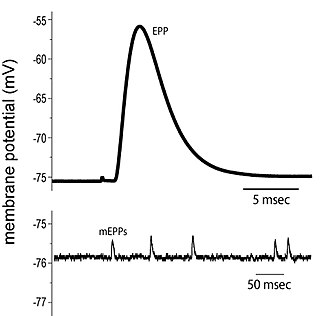
End plate potentials (EPPs) are the voltages which cause depolarization of skeletal muscle fibers caused by neurotransmitters binding to the postsynaptic membrane in the neuromuscular junction. They are called "end plates" because the postsynaptic terminals of muscle fibers have a large, saucer-like appearance. When an action potential reaches the axon terminal of a motor neuron, vesicles carrying neurotransmitters are exocytosed and the contents are released into the neuromuscular junction. These neurotransmitters bind to receptors on the postsynaptic membrane and lead to its depolarization. In the absence of an action potential, acetylcholine vesicles spontaneously leak into the neuromuscular junction and cause very small depolarizations in the postsynaptic membrane. This small response (~0.4mV) is called a miniature end plate potential (MEPP) and is generated by one acetylcholine-containing vesicle. It represents the smallest possible depolarization which can be induced in a muscle.

<span class="mw-page-title-main">Voltage-gated ion channel</span> Type of ion channel transmembrane protein

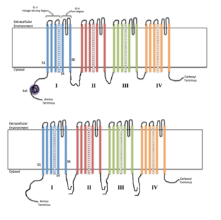
Voltage-gated ion channels are a class of transmembrane proteins that form ion channels that are activated by changes in the electrical membrane potential near the channel. The membrane potential alters the conformation of the channel proteins, regulating their opening and closing. Cell membranes are generally impermeable to ions, thus they must diffuse through the membrane through transmembrane protein channels. They have a crucial role in excitable cells such as neuronal and muscle tissues, allowing a rapid and co-ordinated depolarization in response to triggering voltage change. Found along the axon and at the synapse, voltage-gated ion channels directionally propagate electrical signals. Voltage-gated ion-channels are usually ion-specific, and channels specific to sodium (Na+), potassium (K+), calcium (Ca2+), and chloride (Cl) ions have been identified. The opening and closing of the channels are triggered by changing ion concentration, and hence charge gradient, between the sides of the cell membrane.

<span class="mw-page-title-main">Ligand-gated ion channel</span> Type of ion channel transmembrane protein

Ligand-gated ion channels (LICs, LGIC), also commonly referred to as ionotropic receptors, are a group of transmembrane ion-channel proteins which open to allow ions such as Na+, K+, Ca2+, and/or Cl to pass through the membrane in response to the binding of a chemical messenger (i.e. a ligand), such as a neurotransmitter.

A calcium channel is an ion channel which shows selective permeability to calcium ions. It is sometimes synonymous with voltage-gated calcium channel, which are a type of calcium channel regulated by changes in membrane potential. Some calcium channels are regulated by the binding of a ligand. Other calcium channels can also be regulated by both voltage and ligands to provide precise control over ion flow. Some cation channels allow calcium as well as other cations to pass through the membrane.

<span class="mw-page-title-main">Chloride channel</span> Class of transport proteins

Chloride channels are a superfamily of poorly understood ion channels specific for chloride. These channels may conduct many different ions, but are named for chloride because its concentration in vivo is much higher than other anions. Several families of voltage-gated channels and ligand-gated channels have been characterized in humans.

Neuropharmacology is the study of how drugs affect function in the nervous system, and the neural mechanisms through which they influence behavior. There are two main branches of neuropharmacology: behavioral and molecular. Behavioral neuropharmacology focuses on the study of how drugs affect human behavior (neuropsychopharmacology), including the study of how drug dependence and addiction affect the human brain. Molecular neuropharmacology involves the study of neurons and their neurochemical interactions, with the overall goal of developing drugs that have beneficial effects on neurological function. Both of these fields are closely connected, since both are concerned with the interactions of neurotransmitters, neuropeptides, neurohormones, neuromodulators, enzymes, second messengers, co-transporters, ion channels, and receptor proteins in the central and peripheral nervous systems. Studying these interactions, researchers are developing drugs to treat many different neurological disorders, including pain, neurodegenerative diseases such as Parkinson's disease and Alzheimer's disease, psychological disorders, addiction, and many others.

Molecular neuroscience is a branch of neuroscience that observes concepts in molecular biology applied to the nervous systems of animals. The scope of this subject covers topics such as molecular neuroanatomy, mechanisms of molecular signaling in the nervous system, the effects of genetics and epigenetics on neuronal development, and the molecular basis for neuroplasticity and neurodegenerative diseases. As with molecular biology, molecular neuroscience is a relatively new field that is considerably dynamic.

The induction of NMDA receptor-dependent long-term potentiation (LTP) in chemical synapses in the brain occurs via a fairly straightforward mechanism. A substantial and rapid rise in calcium ion concentration inside the postsynaptic cell is most possibly all that is required to induce LTP. But the mechanism of calcium delivery to the postsynaptic cell in inducing LTP is more complicated.

<span class="mw-page-title-main">Glutamate receptor</span> Cell-surface proteins that bind glutamate and trigger changes which influence the behavior of cells

Glutamate receptors are synaptic and non synaptic receptors located primarily on the membranes of neuronal and glial cells. Glutamate is abundant in the human body, but particularly in the nervous system and especially prominent in the human brain where it is the body's most prominent neurotransmitter, the brain's main excitatory neurotransmitter, and also the precursor for GABA, the brain's main inhibitory neurotransmitter. Glutamate receptors are responsible for the glutamate-mediated postsynaptic excitation of neural cells, and are important for neural communication, memory formation, learning, and regulation.

Sodium channels are integral membrane proteins that form ion channels, conducting sodium ions (Na+) through a cell's membrane. They belong to the superfamily of cation channels.

<span class="mw-page-title-main">Epithelial sodium channel</span> Group of membrane proteins

The epithelial sodium channel(ENaC), (also known as amiloride-sensitive sodium channel) is a membrane-bound ion channel that is selectively permeable to sodium ions (Na+). It is assembled as a heterotrimer composed of three homologous subunits α or δ, β, and γ, These subunits are encoded by four genes: SCNN1A, SCNN1B, SCNN1G, and SCNN1D. The ENaC is involved primarily in the reabsorption of sodium ions at the collecting ducts of the kidney's nephrons. In addition to being implicated in diseases where fluid balance across epithelial membranes is perturbed, including pulmonary edema, cystic fibrosis, COPD and COVID-19, proteolyzed forms of ENaC function as the human salt taste receptor.

Two-pore channels (TPCs) are eukaryotic intracellular voltage-gated and ligand gated cation selective ion channels. There are two known paralogs in the human genome, TPC1s and TPC2s. In humans, TPC1s are sodium selective and TPC2s conduct sodium ions, calcium ions and possibly hydrogen ions. Plant TPC1s are non-selective channels. Expression of TPCs are found in both plant vacuoles and animal acidic organelles. These organelles consist of endosomes and lysosomes. TPCs are formed from two transmembrane non-equivalent tandem Shaker-like, pore-forming subunits, dimerized to form quasi-tetramers. Quasi-tetramers appear very similar to tetramers, but are not quite the same. Some key roles of TPCs include calcium dependent responses in muscle contraction(s), hormone secretion, fertilization, and differentiation. Disorders linked to TPCs include membrane trafficking, Parkinson's disease, Ebola, and fatty liver.

Light-gated ion channels are a family of ion channels regulated by electromagnetic radiation. Other gating mechanisms for ion channels include voltage-gated ion channels, ligand-gated ion channels, mechanosensitive ion channels, and temperature-gated ion channels. Most light-gated ion channels have been synthesized in the laboratory for study, although two naturally occurring examples, channelrhodopsin and anion-conducting channelrhodopsin, are currently known. Photoreceptor proteins, which act in a similar manner to light-gated ion channels, are generally classified instead as G protein-coupled receptors.

<span class="mw-page-title-main">Gating (electrophysiology)</span>

In electrophysiology, the term gating refers to the opening (activation) or closing of ion channels. This change in conformation is a response to changes in transmembrane voltage.

References

  1. "Medical Definition of Ion channel". MedicineNet. Retrieved 2017-03-20.
  2. 1 2 3 Kocahan S, Doğan Z (February 2017). "Mechanisms of Alzheimer's Disease Pathogenesis and Prevention: The Brain, Neural Pathology, N-methyl-D-aspartate Receptors, Tau Protein and Other Risk Factors". Clinical Psychopharmacology and Neuroscience. 15 (1): 1–8. doi:10.9758/cpn.2017.15.1.1. PMC   5290713 . PMID   28138104.
  3. 1 2 3 4 5 Lipton SA (January 2004). "Failures and successes of NMDA receptor antagonists: molecular basis for the use of open-channel blockers like memantine in the treatment of acute and chronic neurologic insults". NeuroRx. Neuroprotection. 1 (1): 101–10. doi:10.1602/neurorx.1.1.101. PMC   534915 . PMID   15717010.
  4. Ahern CA, Payandeh J, Bosmans F, Chanda B (January 2016). "The hitchhiker's guide to the voltage-gated sodium channel galaxy". The Journal of General Physiology. 147 (1): 1–24. doi:10.1085/jgp.201511492. PMC   4692491 . PMID   26712848.
  5. Moore JW, Blaustein MP, Anderson NC, Narahashi T (May 1967). "Basis of tetrodotoxin's selectivity in blockage of squid axons". The Journal of General Physiology. 50 (5): 1401–11. doi:10.1085/jgp.50.5.1401. PMC   2225715 . PMID   6033592.
  6. Stevens M, Peigneur S, Tytgat J (2011-11-09). "Neurotoxins and their binding areas on voltage-gated sodium channels". Frontiers in Pharmacology. 2: 71. doi: 10.3389/fphar.2011.00071 . PMC   3210964 . PMID   22084632.
  7. Miller C (December 1988). "Competition for block of a Ca2(+)-activated K+ channel by charybdotoxin and tetraethylammonium". Neuron. 1 (10): 1003–6. doi:10.1016/0896-6273(88)90157-2. PMID   2483092. S2CID   32262373.
  8. Aiyar J, Withka JM, Rizzi JP, Singleton DH, Andrews GC, Lin W, Boyd J, Hanson DC, Simon M, Dethlefs B (November 1995). "Topology of the pore-region of a K+ channel revealed by the NMR-derived structures of scorpion toxins". Neuron. 15 (5): 1169–81. doi: 10.1016/0896-6273(95)90104-3 . PMID   7576659.
  9. 1 2 Findeisen F, Campiglio M, Jo H, Abderemane-Ali F, Rumpf CH, Pope L, Rossen ND, Flucher BE, DeGrado WF, Minor DL (March 2017). "Stapled Voltage-Gated Calcium Channel (CaV) α-Interaction Domain (AID) Peptides Act As Selective Protein-Protein Interaction Inhibitors of CaV Function". ACS Chemical Neuroscience. 8 (6): 1313–1326. doi:10.1021/acschemneuro.6b00454. PMC   5481814 . PMID   28278376.
  10. Phoenix DA, Harris F (2002-01-01). "The hydrophobic moment and its use in the classification of amphiphilic structures (review)". Molecular Membrane Biology. 19 (1): 1–10. doi: 10.1080/09687680110103631 . PMID   11989818. S2CID   19943697.
  11. 1 2 Jackson MB (February 2010). "Open channel block and beyond". The Journal of Physiology. 588 (Pt 4): 553–4. doi:10.1113/jphysiol.2009.183210. PMC   2828128 . PMID   20173077.
  12. Ahern CA, Payandeh J, Bosmans F, Chanda B (January 2016). "The hitchhiker's guide to the voltage-gated sodium channel galaxy". The Journal of General Physiology. 147 (1): 1–24. doi:10.1085/jgp.201511492. PMC   4692491 . PMID   26712848.
  13. 1 2 3 Butterworth JF, Strichartz GR (April 1990). "Molecular mechanisms of local anesthesia: a review". Anesthesiology. 72 (4): 711–34. doi: 10.1097/00000542-199004000-00022 . PMID   2157353. S2CID   8277924.
  14. Evans MH (September 1969). "Mechanism of saxitoxin and tetrodotoxin poisoning". British Medical Bulletin. 25 (3): 263–7. doi:10.1093/oxfordjournals.bmb.a070715. PMID   5812102.
  15. Mert T, Gunes Y, Guven M, Gunay I, Ozcengiz D (March 2002). "Comparison of nerve conduction blocks by an opioid and a local anesthetic". European Journal of Pharmacology. 439 (1–3): 77–81. doi:10.1016/S0014-2999(02)01368-7. PMID   11937095.
  16. Mitolo-Chieppa D, Carratu MR (May 1983). "Anaesthetic drugs: electrophysiological bases of their conduction blocking effect". Pharmacological Research Communications. 15 (5): 439–50. doi:10.1016/s0031-6989(83)80064-2. PMID   6351107.
  17. 1 2 3 Chen HS, Pellegrini JW, Aggarwal SK, Lei SZ, Warach S, Jensen FE, Lipton SA (November 1992). "Open-channel block of N-methyl-D-aspartate (NMDA) responses by memantine: therapeutic advantage against NMDA receptor-mediated neurotoxicity". The Journal of Neuroscience. 12 (11): 4427–36. doi:10.1523/JNEUROSCI.12-11-04427.1992. PMC   6576016 . PMID   1432103.
  18. 1 2 3 Müller WE, Mutschler E, Riederer P (July 1995). "Noncompetitive NMDA receptor antagonists with fast open-channel blocking kinetics and strong voltage-dependency as potential therapeutic agents for Alzheimer's dementia". Pharmacopsychiatry. 28 (4): 113–24. doi:10.1055/s-2007-979603. PMID   7491365. S2CID   260240191.
  19. Povysheva NV, Johnson JW (December 2016). "Effects of memantine on the excitation-inhibition balance in prefrontal cortex". Neurobiology of Disease. 96: 75–83. doi:10.1016/j.nbd.2016.08.006. PMC   5102806 . PMID   27546057.
  20. Dominguez, Evangelyn; Chin, Ting-Yu; Chen, Chih-Ping; Wu, Tzong-Yuan (2011-12-01). "Management of moderate to severe Alzheimer's disease: Focus on memantine". Taiwanese Journal of Obstetrics and Gynecology. 50 (4): 415–423. doi: 10.1016/j.tjog.2011.10.004 . ISSN   1028-4559. PMID   22212311.
  21. 1 2 3 Lubamba B, Dhooghe B, Noel S, Leal T (October 2012). "Cystic fibrosis: insight into CFTR pathophysiology and pharmacotherapy". Clinical Biochemistry. 45 (15): 1132–44. doi:10.1016/j.clinbiochem.2012.05.034. PMID   22698459.
  22. 1 2 Ramos E, O'leary ME (October 2004). "State-dependent trapping of flecainide in the cardiac sodium channel". The Journal of Physiology. 560 (Pt 1): 37–49. doi:10.1113/jphysiol.2004.065003. PMC   1665201 . PMID   15272045.
  23. Lipton SA (May 2007). "Pathologically-activated therapeutics for neuroprotection: mechanism of NMDA receptor block by memantine and S-nitrosylation". Current Drug Targets. 8 (5): 621–32. doi:10.2174/138945007780618472. PMID   17504105.
  24. 1 2 Frizzell RA, Hanrahan JW (June 2012). "Physiology of epithelial chloride and fluid secretion". Cold Spring Harbor Perspectives in Medicine. 2 (6): a009563. doi:10.1101/cshperspect.a009563. PMC   3367533 . PMID   22675668.
  25. Muanprasat C, Sonawane ND, Salinas D, Taddei A, Galietta LJ, Verkman AS (August 2004). "Discovery of glycine hydrazide pore-occluding CFTR inhibitors: mechanism, structure-activity analysis, and in vivo efficacy". The Journal of General Physiology. 124 (2): 125–37. doi:10.1085/jgp.200409059. PMC   2229623 . PMID   15277574.
  26. Ma T, Thiagarajah JR, Yang H, Sonawane ND, Folli C, Galietta LJ, Verkman AS (December 2002). "Thiazolidinone CFTR inhibitor identified by high-throughput screening blocks cholera toxin-induced intestinal fluid secretion". The Journal of Clinical Investigation. 110 (11): 1651–8. doi:10.1172/JCI16112. PMC   151633 . PMID   12464670.
  27. Chen H, Ruan YC, Xu WM, Chen J, Chan HC (2012-11-01). "Regulation of male fertility by CFTR and implications in male infertility". Human Reproduction Update. 18 (6): 703–13. doi: 10.1093/humupd/dms027 . PMID   22709980.
  28. Schultz BD, DeRoos AD, Venglarik CJ, Singh AK, Frizzell RA, Bridges RJ (August 1996). "Glibenclamide blockade of CFTR chloride channels". The American Journal of Physiology. 271 (2 Pt 1): L192-200. doi:10.1152/ajplung.1996.271.2.L192. PMID   8770056.
  29. Zhang ZR, Zeltwanger S, McCarty NA (May 2000). "Direct comparison of NPPB and DPC as probes of CFTR expressed in Xenopus oocytes". The Journal of Membrane Biology. 175 (1): 35–52. doi:10.1007/s002320001053. PMID   10811966. S2CID   19341540.
  30. Linsdell P, Hanrahan JW (November 1996). "Disulphonic stilbene block of cystic fibrosis transmembrane conductance regulator Cl- channels expressed in a mammalian cell line and its regulation by a critical pore residue". The Journal of Physiology. 496 (Pt 3): 687–93. doi:10.1113/jphysiol.1996.sp021719. PMC   1160856 . PMID   8930836.
  31. Linsdell P (February 2014). "Cystic fibrosis transmembrane conductance regulator chloride channel blockers: Pharmacological, biophysical and physiological relevance". World Journal of Biological Chemistry. 5 (1): 26–39. doi: 10.4331/wjbc.v5.i1.26 . PMC   3942540 . PMID   24600512.