Desmosine

Last updated
Desmosine
Desmosine Structural Formulae V.1.svg
Names
IUPAC name
6-{4-[(4S)-4-Amino-4-carboxybutyl]-3,5-bis[(3S)-3-amino-3-carboxypropyl]-1-pyridiniumyl}-L-norleucine
Identifiers
3D model (JSmol)
ChemSpider
PubChem CID
UNII
  • InChI=1S/C24H39N5O8/c25-17(21(30)31)5-1-2-11-29-12-14(7-9-19(27)23(34)35)16(4-3-6-18(26)22(32)33)15(13-29)8-10-20(28)24(36)37/h12-13,17-20H,1-11,25-28H2,(H3-,30,31,32,33,34,35,36,37)/p+1 Yes check.svgY
    Key: VEVRNHHLCPGNDU-MUGJNUQGSA-O X mark.svgN
  • O=C(O)[C@@H](N)CCCC[n+]1cc(c(c(c1)CC[C@@H](C(=O)O)N)CCC[C@@H](C(=O)O)N)CC[C@@H](C(=O)O)N
Properties
C24H40N5O8
Molar mass 526.611 g·mol−1
Except where otherwise noted, data are given for materials in their standard state (at 25 °C [77 °F], 100 kPa).
X mark.svgN  verify  (what is  Yes check.svgYX mark.svgN ?)

Desmosine is an amino acid found uniquely in elastin, a protein found in connective tissue such as skin, lungs, and elastic arteries.

Contents

Desmosine is a component of elastin and cross links with its isomer, isodesmosine, giving elasticity to the tissue. Detection of desmosine in urine, plasma or sputum samples can be a marker for elastin breakdown due to high elastase activity related to certain diseases. [1] [2]

Structure

Desmosine and its isomer isodesmosine are both composed of four lysine residues, allowing for bonding to multiple peptide chains. The four lysine groups combine to form a pyridinium nucleus, which can be reduced to neutralize positive charge associated, and increase the hydrophobicity. The four lysines form side chains around the pyridinium nucleus with exposed carboxyl groups. The difference between desmosines and isodesmosines are an exchange of a lysine side chain on carbon 1 with a proton on carbon 5. [3] Desmosine is associated with alanine, bonding with it on the N terminal side. It is this alanine association that allows it to bond well with pairs of tropoelastin, to form elastin and elastin networks. [4]

Desmosine and isodesmosine are unable to be differentiated thus far because of the lack of technology. The differentiation would be helpful in order to understand desmosine and its properties better. Currently, mass spectrometry is used and aids in the release of characteristic fragments which would help with differentiation, especially in larger peptides.[ citation needed ]

Synthesis

Desmosine has pathways for form multiple conformations of itself, both through biosynthesis and through man-made systems.[ citation needed ]

Biosynthesis

The formation of desmosines occurs within the formation of precursor tropoelastin. The tropoelastin initially lacks any of these complex binding molecules, and has a similar make up to that of the final stage elastin, however it contains a greater amount of lysine side chains, which directly corresponds with desmosines later found. These precursor molecules are processed through Dehydrogenation, along with dihydroD, and ultimately form elastin bound with desmosine. [5] Through the Lysyl oxidase enzyme, lysyl c- amino groups is oxidized, forming allysine. This spontaneously condenses with other allysine molecules to form a bifunctional cross-link, allysine aldol, or with a c-amino group of lysine, forming dehydrolysinonorleucine. These compounds are then further condensed to form a tetrafunctional pyridinium cross-links of desmosines and isodesmosines. [3] These reactions occur with lysines in areas of high alanine, due to alanine having a small side chain that won't block the enzyme binding to the lysine groups.

Lab Synthesis

Desmosines can be synthesized in a lab through a few methods, like palladium catalyzed cross-coupling reactions. The various treatments can create slightly different confirmations. [6]

Process of Desmosine Synthesis through biological pathways, allowing creation of pyridinium ring from lysyl side chains Desmosine Synthesis from proto-elastin Chains.png
Process of Desmosine Synthesis through biological pathways, allowing creation of pyridinium ring from lysyl side chains

Bonding

Desmosine bonding in alanine rich areas, which allow for enzymes to bond and create the unique double crosslink that sets desmosine apart. SJ DESMOSINE BONDING.png
Desmosine bonding in alanine rich areas, which allow for enzymes to bond and create the unique double crosslink that sets desmosine apart.

Some models of bonding for desmosines, created through the study of bovine ligament elastin, suggest a combination of desmosine and secondary cross-linking to bind together peptide chains. This model has desmosine bonding near an alanine on the peptide chain, then to 3 other amino acids on the 2 peptide chains, despite being able to bond to up to 4 chains. It has been suggested that the secondary cross-linking occurs with either desmosine or lysinonorleucine, which maintains an alpha helix conformation in alanine rich sections on peptides. [3]

Both isodesmosine and desmosine can have similar bonding sites in elastin, though it rarely shown this way in nature. They more often will appear in close proximity to each other on the peptide chain. [3]

Bonding in elastin/collagen

Desmosine is found to have a hydrogen bond donor count of eight and a hydrogen bond acceptor count of twelve. [7]

Function

Elastin, a protein in the extracellular matrix, provides elasticity and its soluble precursor is tropoelastin. When elastin cross links it produces desmosine and isodesmosine. [8] When desmosine is mentioned, it is usually grouped with isodesmosine, the other tetrafunctional amino acid that is specific to elastin.

Demosine can not only be found in elastin, but also in urine, plasma, sputum, and there are different ways to identify and measure these quantities. [9] This means that it is used as a biomarker for elastin degradation which can be a detection for chronic obstructive pulmonary disease (COPD). Desmosine is a potential biomarker for matrix degradation.

Mass spectrometry of desmosine.
.mw-parser-output cite.citation{font-style:inherit;word-wrap:break-word}.mw-parser-output .citation q{quotes:"\"""\"""'""'"}.mw-parser-output .citation:target{background-color:rgba(0,127,255,0.133)}.mw-parser-output .id-lock-free a,.mw-parser-output .citation .cs1-lock-free a{background:url("//upload.wikimedia.org/wikipedia/commons/6/65/Lock-green.svg")right 0.1em center/9px no-repeat}.mw-parser-output .id-lock-limited a,.mw-parser-output .id-lock-registration a,.mw-parser-output .citation .cs1-lock-limited a,.mw-parser-output .citation .cs1-lock-registration a{background:url("//upload.wikimedia.org/wikipedia/commons/d/d6/Lock-gray-alt-2.svg")right 0.1em center/9px no-repeat}.mw-parser-output .id-lock-subscription a,.mw-parser-output .citation .cs1-lock-subscription a{background:url("//upload.wikimedia.org/wikipedia/commons/a/aa/Lock-red-alt-2.svg")right 0.1em center/9px no-repeat}.mw-parser-output .cs1-ws-icon a{background:url("//upload.wikimedia.org/wikipedia/commons/4/4c/Wikisource-logo.svg")right 0.1em center/12px no-repeat}.mw-parser-output .cs1-code{color:inherit;background:inherit;border:none;padding:inherit}.mw-parser-output .cs1-hidden-error{display:none;color:#d33}.mw-parser-output .cs1-visible-error{color:#d33}.mw-parser-output .cs1-maint{display:none;color:#3a3;margin-left:0.3em}.mw-parser-output .cs1-format{font-size:95%}.mw-parser-output .cs1-kern-left{padding-left:0.2em}.mw-parser-output .cs1-kern-right{padding-right:0.2em}.mw-parser-output .citation .mw-selflink{font-weight:inherit}
doi:10.1007/s13361-014-1075-9 Fingerprinting Desmosine-Containing Elastin Peptides.png
Mass spectrometry of desmosine. doi : 10.1007/s13361-014-1075-9

Desmosine and Isodesmosine are unable to be differentiated thus far because of the lack of technology. The differentiation would be helpful in order to understand desmosine and its properties better. Currently, mass spectrometry is used and aids in the release of characteristic fragments which would help with differentiation, especially in larger peptides.

Material properties

The molecular weight of this rare amino acid that is found in elastin is 526.611 g/mol. [7] The desmosine pyridinium ring has three allysyl side chains and one unaltered lysyl side chain. It has been tested to show that the pyridinium core of Desmosine remains intact even at very high collision energies.

Current usage in medicine

Desmosine is currently used as a biomarker in the medical field. It is measured in order to monitor elastin breakdown. Since it is connected to the degradation of elastin, it can be used to identify COPD. Desmosine is one of the oldest biomarkers and was developed in the 1960s, but the first time it was correlated to lung elastin content was in the 80s through urinary excretion. Biomarkers are judged in 6 ways: [9]

  1. Biomarkers should be central to the pathophysiological process
  2. They should be a ‘‘true’’ surrogate end-point
  3. Biomarkers should be stable and vary with disease progression only
  4. The severity of the condition should relate to the concentration of the Biomarker
  5. Progression should be predicted
  6. Effective treatment should show change

Even though desmosine can check-off the first three it cannot check off the rest. And this is why research is being done to further the validation of using desmosine as a biomarker for certain diseases like COPD.

Application of desmosine

Organization of elastin (red) and cell nuclei (blue) in both young and old SDFT and CDET, two equine tendons. With increasing age, there is a distinct decrease in elastin content in the SDFT. Elastin in Horse Tendons.png
Organization of elastin (red) and cell nuclei (blue) in both young and old SDFT and CDET, two equine tendons. With increasing age, there is a distinct decrease in elastin content in the SDFT.

Because desmosine is most prevalent in mature elastin, it can be consistently located and measured in urine samples after elastin breakdown in the human body. [11] [10] Desmosine does not exist elsewhere within the body, nor can it be sourced from elsewhere outside the body, which isolates it as a key marker for elastin breakdown. [11] Indeed, desmosine "has been studied as a marker of elastin breakdown in several chronic pulmonary conditions, including chronic obstructive pulmonary disease (COPD), cystic fibrosis, and chronic tobacco use." [11] In one study, hyperoxic mice that formed alveoli as a result of lung maturation also showed drastic changes in collagen and elastin within the lungs, as well as a change in cross-linking. [12] In another study, deceased patients with acute respiratory distress syndrome (ARDS) were reported to have higher concentrations of desmosine in their urine than those patients who survived ARDS, and higher concentrations of desmosine revealed that "more severe damage to the extracellular matrix occurred in the most critically ill [ acute lung injury] patients." [11]

However, it has been argued in the same study that desmosine does "not correlate well with markers of disease severity," correlating only weakly with age. [11] Instead, it is suggested "that desmosine may be more useful in understanding the pathogenesis of ALI and less useful as a marker of disease severity.” [11] The current standard for measuring lung disease progression, for example, is measured through the forced expiratory volume in one second (FEV1) compared to the maximum lung capacity; [9] in other words, the volume of air a person can exhale from full lungs in one second compared to their maximum lung capacity. This method, while simple and physiologically thorough, has biological limitations, [9] and so a superior biological marker is being sought after. Desmosine has been studied as one such biological marker, with studies in the 1980s to link urinary desmosine concentration with elastin breakdown in the lungs. [9] Though large amounts of data have been collected with regards to desmosine's potential as a replacement biological marker in determining disease progression, some believe there is still insufficient evidence for desmosine to meet and fill this need. [9]

In orthopedics, one study examined equine tendons and how their increasing stiffness and fatigue with age was due to fragmentation of the elastin in the tendons. [10] The superficial digital flexor tendon (SDFT) and the common digital extensor tendon (CDET) were analyzed for elastin composition, comparing older tendons to younger ones. [10] While both the CDET and the SDFT are positional tendons, enabling muscles to move the skeleton, the SDFT also stores energy and is far more elastic than the CDET due to "specialization of the [ interfascicular matrix] to enable repeated interfascicular sliding and recoil." [10] Desmosine concentrations were reported to be far greater in new tendons than in tendons that had partially degraded, suggesting that not only is there fragmentation of tendon elastin with age, but also a smaller total composition of elastin within the SDFT, though this was not true in the case of the CDET examined. [10]

Research has also been performed to determine the cross-linking structure of elastin, in an effort to better understand the relationship between elastin and pertinent diseases, such as cystic fibrosis, chronic obstructive pulmonary disease (COPD), and aortic aneurysms. [4] A study was conducted to find this structure through synthesis of a cyclic peptide containing desmosine, to partially mimic elastin in the hopes of running mass spectrometry on the peptide to reveal the cross-linking structure. [4] The elastin mimic was eventually synthesized successfully, and though work has not yet been done to clarify the cross-linking structure of elastin, preliminary mass spectrometry demonstrated the presence of the expected ion formed from the chemical reactions used. [4]

Related Research Articles

Collagen is the main structural protein in the extracellular matrix found in the body's various connective tissues. As the main component of connective tissue, it is the most abundant protein in mammals, making up from 25% to 35% of the whole-body protein content. Collagen consists of amino acids bound together to form a triple helix of elongated fibril known as a collagen helix. It is mostly found in connective tissue such as cartilage, bones, tendons, ligaments, and skin.

<span class="mw-page-title-main">Protein primary structure</span> Linear sequence of amino acids in a peptide or protein

Protein primary structure is the linear sequence of amino acids in a peptide or protein. By convention, the primary structure of a protein is reported starting from the amino-terminal (N) end to the carboxyl-terminal (C) end. Protein biosynthesis is most commonly performed by ribosomes in cells. Peptides can also be synthesized in the laboratory. Protein primary structures can be directly sequenced, or inferred from DNA sequencess.

<span class="mw-page-title-main">Proteolysis</span> Breakdown of proteins into smaller polypeptides or amino acids

Proteolysis is the breakdown of proteins into smaller polypeptides or amino acids. Uncatalysed, the hydrolysis of peptide bonds is extremely slow, taking hundreds of years. Proteolysis is typically catalysed by cellular enzymes called proteases, but may also occur by intra-molecular digestion.

<span class="mw-page-title-main">Trypsin</span> Family of digestive enzymes

Trypsin is an enzyme in the first section of the small intestine that starts the digestion of protein molecules by cutting long chains of amino acids into smaller pieces. It is a serine protease from the PA clan superfamily, found in the digestive system of many vertebrates, where it hydrolyzes proteins. Trypsin is formed in the small intestine when its proenzyme form, the trypsinogen produced by the pancreas, is activated. Trypsin cuts peptide chains mainly at the carboxyl side of the amino acids lysine or arginine. It is used for numerous biotechnological processes. The process is commonly referred to as trypsin proteolysis or trypsinization, and proteins that have been digested/treated with trypsin are said to have been trypsinized. Trypsin was discovered in 1876 by Wilhelm Kühne and was named from the Ancient Greek word for rubbing since it was first isolated by rubbing the pancreas with glycerin.

<span class="mw-page-title-main">Post-translational modification</span> Biological processes

Post-translational modification (PTM) is the covalent and generally enzymatic modification of proteins following protein biosynthesis. This process often occurs in the endoplasmic reticulum and the golgi apparatus. Proteins are synthesized by ribosomes translating mRNA into polypeptide chains, which may then undergo PTM to form the mature protein product. PTMs are important components in cell signaling, as for example when prohormones are converted to hormones.

<span class="mw-page-title-main">Proteinogenic amino acid</span> Amino acid that is incorporated biosynthetically into proteins during translation

Proteinogenic amino acids are amino acids that are incorporated biosynthetically into proteins during translation. The word "proteinogenic" means "protein creating". Throughout known life, there are 22 genetically encoded (proteinogenic) amino acids, 20 in the standard genetic code and an additional 2 that can be incorporated by special translation mechanisms.

<span class="mw-page-title-main">Elastin</span> Protein allowing tissue in the body to resume shape after stretching

Elastin is a protein that in humans is encoded by the ELN gene. Elastin is a key component of the extracellular matrix in gnathostomes. It is highly elastic and present in connective tissue allowing many tissues in the body to resume their shape after stretching or contracting. Elastin helps skin to return to its original position when it is poked or pinched. Elastin is also an important load-bearing tissue in the bodies of vertebrates and used in places where mechanical energy is required to be stored.

<span class="mw-page-title-main">DD-transpeptidase</span>

DD-transpeptidase is a bacterial enzyme that catalyzes the transfer of the R-L-αα-D-alanyl moiety of R-L-αα-D-alanyl-D-alanine carbonyl donors to the γ-OH of their active-site serine and from this to a final acceptor. It is involved in bacterial cell wall biosynthesis, namely, the transpeptidation that crosslinks the peptide side chains of peptidoglycan strands.

<span class="mw-page-title-main">Elastic fiber</span> Type of connective tissue in animals

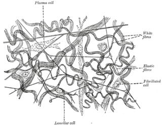
Elastic fibers are an essential component of the extracellular matrix composed of bundles of proteins (elastin) which are produced by a number of different cell types including fibroblasts, endothelial, smooth muscle, and airway epithelial cells. These fibers are able to stretch many times their length, and snap back to their original length when relaxed without loss of energy. Elastic fibers include elastin, elaunin and oxytalan.

A peptide library is a tool for studying proteins. The Peptide libraries contain a great number of peptides that have a systematic combination of amino acids. Usually, the peptide library is synthesized on a solid phase, mostly on resin, which can be made as a flat surface or beads. The peptide library provides a powerful tool for drug design, protein–protein interactions, and other biochemical and pharmaceutical applications.

<span class="mw-page-title-main">Isopeptide bond</span>

An isopeptide bond is a type of amide bond formed between a carboxyl group of one amino acid and an amino group of another. An isopeptide bond is the linkage between the side chain amino or carboxyl group of one amino acid to the α-carboxyl, α-amino group, or the side chain of another amino acid. In a typical peptide bond, also known as eupeptide bond, the amide bond always forms between the α-carboxyl group of one amino acid and the α-amino group of the second amino acid. Isopeptide bonds are rarer than regular peptide bonds. Isopeptide bonds lead to branching in the primary sequence of a protein. Proteins formed from normal peptide bonds typically have a linear primary sequence.

<span class="mw-page-title-main">Lysyl oxidase</span> Mammalian protein found in Homo sapiens

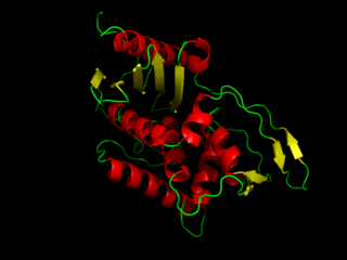
Lysyl oxidase (LOX), also known as protein-lysine 6-oxidase, is an enzyme that, in humans, is encoded by the LOX gene. It catalyzes the conversion of lysine residues into its aldehyde derivative allysine. Allysine form cross-links in extracellular matrix proteins. Inhibition of lysyl oxidase can cause osteolathyrism, but, at the same time, its upregulation by tumor cells may promote metastasis of the existing tumor, causing it to become malignant and cancerous.

<span class="mw-page-title-main">Allysine</span> Chemical compound

Allysine is a derivative of lysine that features a formyl group in place of the terminal amine. The free amino acid does not exist, but the allysine residue does. It is produced by aerobic oxidation of lysine residues by the enzyme lysyl oxidase. The transformation is an example of a post-translational modification. The semi-aldehyde form exists in equilibrium with a cyclic derivative.

<span class="mw-page-title-main">Isodesmosine</span> Chemical compound

Isodesmosine is a lysine derivative found in elastin. Isodesmosine is an isomeric pyridinium-based amino acid resulting from the condensation of four lysine residues between elastin proteins by lysyl-oxidase. These represent ideal biomarkers for monitoring elastin turnover because these special cross-links are only found in mature elastin in mammals.

<span class="mw-page-title-main">GLB1</span> Protein

Galactosidase, beta 1, also known as GLB1, is a protein which in humans is encoded by the GLB1 gene.

<span class="mw-page-title-main">Pyridinoline</span> Chemical compound

Pyridinoline, also known as Hydroxylysylpyridinoline, is a fluorescent cross-linking compound of collagen fibers. Crosslinks in collagen and elastin are derived from lysyl and hydroxylysyl residues, a process catalyzed by lysyl oxidase. Fujimoto and colleagues first described the isolation and characterization of a fluorescent material in bovine Achilles tendon collagen and termed it pyridinoline. It is reported to be present in collagen of bone and cartilage, but is absent in collagen of skin. It is not present in newly synthesized collagen and is formed from aldimine cross-links during maturation of collagen fibers.

<span class="mw-page-title-main">Copper peptide GHK-Cu</span> Chemical compound

Copper peptide GHK-Cu is a naturally occurring copper complex of the tripeptide glycyl-L-histidyl-L-lysine. The tripeptide has strong affinity for copper(II) and was first isolated from human plasma. It can be found also in saliva and urine.

<span class="mw-page-title-main">Non-proteinogenic amino acids</span> Are not naturally encoded in the genome

In biochemistry, non-coded or non-proteinogenic amino acids are distinct from the 22 proteinogenic amino acids which are naturally encoded in the genome of organisms for the assembly of proteins. However, over 140 non-proteinogenic amino acids occur naturally in proteins and thousands more may occur in nature or be synthesized in the laboratory. Chemically synthesized amino acids can be called unnatural amino acids. Unnatural amino acids can be synthetically prepared from their native analogs via modifications such as amine alkylation, side chain substitution, structural bond extension cyclization, and isosteric replacements within the amino acid backbone. Many non-proteinogenic amino acids are important:

Lysine carboxypeptidase is an enzyme. This enzyme catalyses the following chemical reaction:

Elastin-like polypeptides (ELPs) are synthetic biopolymers with potential applications in the fields of cancer therapy, tissue scaffolding, metal recovery, and protein purification. For cancer therapy, the addition of functional groups to ELPs can enable them to conjugate with cytotoxic drugs. Also, ELPs may be able to function as polymeric scaffolds, which promote tissue regeneration. This capacity of ELPs has been studied particularly in the context of bone growth. ELPs can also be engineered to recognize specific proteins in solution. The ability of ELPs to undergo morphological changes at certain temperatures enables specific proteins that are bound to the ELPs to be separated out from the rest of the solution via experimental techniques such as centrifugation.

References

  1. Millipore product catalogue, purified desmosine "Desmosine - Millipore". Archived from the original on 2014-02-24. Retrieved 2014-02-19.
  2. Ma, S; Turino, G. M.; Lin, Y. Y. (2011). "Quantitation of desmosine and isodesmosine in urine, plasma, and sputum by LC-MS/MS as biomarkers for elastin degradation". Journal of Chromatography B. 879 (21): 1893–8. doi:10.1016/j.jchromb.2011.05.011. PMID   21621489.
  3. 1 2 3 4 Mecham, R. P. (1978). "A structural model for desmosine cross-linked peptides". Biochemical Journal. 173 (2): 617–625. doi:10.1042/bj1730617. PMC   1185816 . PMID   697739.
  4. 1 2 3 4 Ogawa, Keita; Hayashi, Takahiro; Lin, Yong Y.; Usuki, Toyonobu (6 July 2017). "Synthesis of desmosine-containing cyclic peptide for the possible elucidation of elastin crosslinking structure". Tetrahedron. 73 (27–28): 3838–3847. doi:10.1016/j.tet.2017.05.045.
  5. Gallop, PM; Blumenfeld, OO; Seifter, S (1972). "Structure and metabolism of connective 801 tissue proteins". Annual Review of Biochemistry. 41: 617–72. doi:10.1146/annurev.bi.41.070172.003153. PMID   4343456.
  6. Watanabe, D. (2017). "Synthesis of desmosine-d4: Improvement of isotopic purity by D-H exchange of amino groups". Tetrahedron Letters. 58 (12): 1194–1197. doi:10.1016/j.tetlet.2017.02.018.
  7. 1 2 National Center for Biotechnology Information. PubChem Compound Database; CID=25435, https://pubchem.ncbi.nlm.nih.gov/compound/25435 (accessed Apr. 13, 2018)
  8. Schräder, C. U., Heinz, A., Majovsky, P., & Schmelzer, C. (2015). Fingerprinting Desmosine-Containing Elastin Peptides. American Society for Mass Spectrometry,26(5), 762-773. doi : 10.1007/s13361-014-1075-9 Link to article: https://link.springer.com/content/pdf/10.1007%2Fs13361-014-1075-9.pdf
  9. 1 2 3 4 5 6 Luisetti, M.; Stolk, J.; Iadarola, P. (2012). "Desmosine, a biomarker for COPD: old and in the way" (PDF). European Respiratory Journal. 39 (4): 797–798. doi: 10.1183/09031936.00172911 . PMID   22467719.
  10. 1 2 3 4 5 6 Goinho, Marta S. C.; Thorpe, Chavaunne T.; Greenwald, Steve E.; Screen, Hazel R. C. (30 August 2017). "Elastin is Localised to the Interfascicular Matrix of Energy Storing Tendons and Becomes Increasingly Disorganised With Ageing". Scientific Reports. 7 (9713): 9713. Bibcode:2017NatSR...7.9713G. doi: 10.1038/s41598-017-09995-4 . PMC   5577209 . PMID   28855560.
  11. 1 2 3 4 5 6 McClintock, Dana E.; Starcher, Barry; Eisner, Mark D.; Thompson, B. Taylor; Hayden, Doug L.; Church, Gwynne D.; Matthay, Michael A. (4 May 2006). "Higher urine desmosine levels are associated with mortality in patients with acute lung injury". American Journal of Physiology. Lung Cellular and Molecular Physiology. 291 (4): L566–L571. doi:10.1152/ajplung.00457.2005. PMC   2765125 . PMID   16698854.
  12. Mižíková, Ivana; Ruiz-Camp, Jordi; Steenbock, Heiko; Madurga, Alicia; Vadász, István; Herold, Susanne; Mayer, Konstantin; Seeger, Werner; Brinckmann, Jürgen; Morty, Rory E. (3 April 2015). "Collagen and elastin cross-linking is altered during aberrant late lung development associated with hyperoxia". American Journal of Physiology. Lung Cellular and Molecular Physiology. 308 (11): L1145–L1148. doi:10.1152/ajplung.00039.2015. PMID   25840994.