Drosophila embryogenesis

Last updated
Ventral view of repeating denticle bands on the cuticle of a 22-hour-old embryo. The head is on the left. DrosophilaKutikula.jpg
Ventral view of repeating denticle bands on the cuticle of a 22-hour-old embryo. The head is on the left.

Drosophila embryogenesis, the process by which Drosophila (fruit fly) embryos form, is a favorite model system for genetics and developmental biology. The study of its embryogenesis unlocked the century-long puzzle of how development was controlled, creating the field of evolutionary developmental biology. [1] The small size, short generation time, and large brood size make it ideal for genetic studies. Transparent embryos facilitate developmental studies. Drosophila melanogaster was introduced into the field of genetic experiments by Thomas Hunt Morgan in 1909.

Contents

Life cycle

Drosophila display a holometabolous method of development, meaning that they have three distinct stages of their post-embryonic life cycle, each with a radically different body plan: larva, pupa and finally, adult. The machinery necessary for the function and smooth transition between these three phases develops during embryogenesis. During embryogenesis, the larval stage fly will develop and hatch at a stage of its life known as the first larval instar. Cells that will produce adult structures are put aside in imaginal discs. During the pupal stage, the larval body breaks down as the imaginal disks grow and produce the adult body. This process is called complete metamorphosis . About 24 hours after fertilization, an egg hatches into a larva, which undergoes three molts taking about 5.5 to 6 days, after which it is called a pupa. The pupa metamorphoses into an adult fly, which takes about 3.5 to 4.5 days. The entire growth process from egg to adult fly takes an estimated 10 to 12 days to complete at 25 °C. [2]

The mother fly produces oocytes that already have anterior-posterior and dorsal-ventral axes defined by maternal activities.

Early embryogenesis, showing the cycles of nuclei divisions in the syncytial blastoderm and the morphogenetic movements of gastrulation.

Embryogenesis in Drosophila is unique among model organisms in that cleavage occurs in a multinucleate syncytium (strictly a coenocyte). Early on, 256 nuclei migrate to the perimeter of the egg, creating the syncytial blastoderm. The germ line segregates from the somatic cells through the formation of pole cells at the posterior end of the embryo. After thirteen mitotic divisions and about 4 hours after fertilization, an estimated 6,000 nuclei accumulate in the unseparated cytoplasm of the oocyte before they migrate to the surface and are encompassed by plasma membranes to form cells surrounding the yolk sac producing a cellular blastoderm.

Like other triploblastic metazoa, gastrulation leads to the formation of three germ layers: the endoderm, mesoderm, and ectoderm. The mesoderm invaginates from the ventral furrow (VF), as does the ectoderm that will give rise to the midgut. The pole cells are internalized by a different route.

Germ band elongation involves many rearrangements of cells, and the appearance of distinct differences in the cells of the three germ bands and various regions of the embryo. The posterior region (including the hindgut) expands and extends towards the anterior pole along the dorsal side of the embryo. At this time, segments of the embryo become visible, creating a striped arrangement along the anterior-posterior axis. The earliest signs of segmentation appear during this phase with the formation of parasegmental furrows. This is also when the tracheal pits form, the first signs of structures for breathing.

Germ band retraction returns the hindgut to the dorsal side of the posterior pole and coincides with overt segmentation. The remaining stages involve the internalization of the nervous system (ectoderm) and the formation of internal organs (mainly mesoderm).

Anterior-posterior axis patterning in Drosophila

The abdominal cuticular segments of the Drosophila embryo consist of repeating denticle bands separated by naked cuticle. DenticleXsection.png
The abdominal cuticular segments of the Drosophila embryo consist of repeating denticle bands separated by naked cuticle.

One of the best understood examples of pattern formation is the patterning along the future head to tail (antero-posterior) axis of the fruit fly Drosophila melanogaster . There are three fundamental types of genes that give way to the developmental structure of the fly: maternal effect genes, segmentation genes, and homeotic genes. The development of Drosophila is particularly well studied, and it is representative of a major class of animals, the insects or insecta. Other multicellular organisms sometimes use similar mechanisms for axis formation, although the relative importance of signal transfer between the earliest cells of many developing organisms is greater than in the example described here.

Maternal effect genes

mRNA distributions Maternal effect mRNAs.svg
mRNA distributions
Protein distributions Drosophila early embryo protein gradients.svg
Protein distributions

The building-blocks of anterior-posterior axis patterning in Drosophila are laid out during egg formation (oogenesis), well before the egg is fertilized and deposited. The maternal effect genes are responsible for the polarity of the egg and of the embryo. The developing egg (oocyte) is polarized by differentially localized mRNA molecules.

The genes that code for these mRNAs, called maternal effect genes, encode for proteins that get translated upon fertilization to establish concentration gradients that span the egg. Bicoid and Hunchback are the maternal effect genes that are most important for patterning of anterior parts (head and thorax) of the Drosophila embryo. Nanos and Caudal are maternal effect genes that are important in the formation of more posterior abdominal segments of the Drosophila embryo. [4] [5]

In embryos from bicoid mutant mothers, the head and thoracic structures are converted to the abdomen making the embryo with posterior structures on both ends, a lethal phenotype. [4]

Cytoskeletal elements such as microtubules are polarized within the oocyte and can be used to allow the localization of mRNA molecules to specific parts of the cell. Maternally synthesized bicoid mRNAs attach to microtubules and are concentrated at the anterior ends of forming Drosophila eggs. In unfertilized eggs, transcripts are still strictly localized at the tip, but immediately after fertilization, a small mRNA gradient is formed in the anterior 20% of the eggs. Another report documents a mRNA gradient up to 40%. nanos mRNA also attaches to a Drosophila egg's cytoskeleton but is concentrated at the posterior end of the egg. hunchback and caudal mRNAs lack special location control systems and are fairly evenly spread throughout the entire interior of the egg cells.

It has been shown that the dsRNA-binding protein STAUFEN (STAU1) is responsible for guiding bicoid, nanos and other proteins, which play a role in forming the anterior-posterior axis, to the correct regions of the embryo to build gradients. When the mRNAs from the maternal effect genes are translated into proteins, a Bicoid protein gradient forms at the anterior end of the egg. Nanos protein forms a gradient at the posterior end. The Bicoid protein blocks translation of caudal mRNA so Caudal protein is of lower concentration at the anterior part of the embryo and at higher concentration at the posterior part of the embryo. This is of opposite direction of the Bicoid protein. The caudal protein then activates later to turn genes on to form the posterior structures during the segmentation phase. Nanos protein creates a posterior-to-anterior slope and is a morphogen that helps in abdomen formation. Nanos protein, in complex with Pumilio protein, binds to the hunchback mRNA and blocks its translation in the posterior end of Drosophila embryos.

The Bicoid, Hunchback, and Caudal proteins are transcription factors. The Bicoid protein is a morphogen as well. The Nanos protein is a translational repressor protein. Bicoid has a DNA-binding homeodomain that binds both DNA and the nanos mRNA. Bicoid binds a specific RNA sequence in the 3′ untranslated region, called the Bicoid 3′-UTR regulatory element, of caudal mRNA and blocks translation.

Hunchback protein levels in the early embryo are significantly augmented by new hunchback gene transcription and translation of the resulting zygotically produced mRNA. During early Drosophila embryogenesis, there are nuclear divisions without cell division. The many nuclei that are produced distribute themselves around the periphery of the cell cytoplasm. Gene expression in these nuclei is regulated by the Bicoid, Hunchback, and Caudal proteins. For example, Bicoid acts as a transcriptional activator of hunchback gene transcription. In order for development to continue, Hunchback is needed in an area that is declining in amount from anterior to posterior. This is created by the Nanos protein whose existence is at a declining slope from posterior to anterior ends.

Gap genes

Gap genes Gap gene expression.svg
Gap genes

The other important function of the gradients of Bicoid, Hunchback, and Caudal proteins is in the transcriptional regulation of other zygotically expressed proteins. Many of these are the protein products derived from members of the "gap" family of developmental control genes. giant, huckebein, hunchback, knirps, Krüppel and tailless are all gap genes . Their expression patterns in the early embryo are determined by the maternal effect gene products and shown in the diagrams on the right side of this page. The gap genes are part of a larger family called the segmentation genes. These genes establish the segmented body plan of the embryo along the anterior-posterior axis. The segmentation genes specify 14 parasegments that are closely related to the final anatomical segments. The gap genes are the first layer of a hierarchical cascade of the segmentation control genes.

Additional segmentation genes

Pair rule Pair rule.svg
Pair rule

Two additional classes of segmentation genes are expressed after the gap gene products. The pair-rule genes are expressed in striped patterns of seven bands perpendicular to the anterior-posterior axis. These patterns of expression are established within the syncytial blastoderm. After these initial patterning events, cell membranes form around the nuclei of the syncytial blastoderm converting it to a cellular blastoderm.

The expression patterns of the final class of segmentation genes, the segment polarity genes, are then fine-tuned by interactions between the cells of adjacent parasegments with genes such as engrailed . The Engrailed protein is a transcription factor that is expressed in one row of cells at the edge of each parasegment. This expression pattern is initiated by the pair-rule genes (like even-skipped) that code for transcription factors that regulate the engrailed gene's transcription in the syncytial blastoderm.

Cells that make Engrailed can make the cell-to-cell signaling protein Hedgehog . The motion of Hedgehog is limited by its lipid modification, and so Hedgehog activates a thin stripe of cells anterior to the Engrailed-expressing cells. Only cells to one side of the Engrailed-expressing cells are competent to respond to Hedgehog because they express the receptor protein Patched. Cells with activated Patched receptor make the Wingless protein. Wingless is a secreted protein that acts on the adjacent rows of cells by activating its cell surface receptor, Frizzled.

Wingless acts on Engrailed-expressing cells to stabilize Engrailed expression after the cellular blastoderm forms. The Naked cuticle protein is induced by Wingless to limit the number of rows of cells that express Engrailed. The short-range, reciprocal signaling by Hedgehog and Wingless, held in check by the Patched and Naked proteins, stabilizes the boundary between each segment. The Wingless protein is called "wingless" because of the phenotype of some wingless mutants. Wingless and Hedgehog also function in multiple tissues later in embryogenesis and also during metamorphosis.

The transcription factors that are coded for by segmentation genes regulate yet another family of developmental control genes, the homeotic selector genes . These genes exist in two ordered groups on Drosophila chromosome 3. The order of the genes on the chromosome reflects the order that they are expressed along the anterior-posterior axis of the developing embryo. The Antennapedia group of homeotic selector genes includes labial, antennapedia, sex combs reduced, deformed, and proboscipedia . Labial and Deformed proteins are expressed in head segments where they activate the genes that define head features. Sex-combs-reduced and Antennapedia specify the properties of thoracic segments. The bithorax group of homeotic selector genes control the specializations of the third thoracic segment and the abdominal segments. Mutations in some homeotic genes can often be lethal and the cycle of life will end at embryogenesis.

In 1995, the Nobel Prize for Physiology or Medicine was awarded for studies concerning the genetic control of early embryonic development to Christiane Nüsslein-Volhard, Edward B. Lewis and Eric Wieschaus. Their research on genetic screening for embryo patterning mutants revealed the role played in early embryologic development by homeobox genes like bicoid. An example of a homeotic mutation is the so-called Antennapedia mutation. In Drosophila, antennae and legs are created by the same basic "program", they only differ in a single transcription factor. If this transcription factor is damaged, the fly grows legs on its head instead of antennae. See images of this "antennapedia" mutant and others, at FlyBase. Another example is in the bithorax complex. If nonlethal mutations occur in this complex, it can cause the fly to have two sets of wings, instead of one pair of wings and one pair of halteres, which aid in balance in flight.

Dorsal-ventral axis

Formation of the dorsal-ventral axis is dependent on the ventral nuclear concentration of a maternally synthesized transcription factor called Dorsal. The determination of the dorsal side of the embryo occurs during oogenesis when the oocyte nucleus moves along microtubules from the posterior to the anterior-dorsal margin of the oocyte. The nucleus expresses a protein called Gurken which is secreted locally and thus only activates follicle cells in the dorsal region by interacting with the Torpedo receptor. This inhibits the production of Pipe protein and thus follicular cells expressing Pipe are on the ventral side. Pipe activates an extracellular protease cascade in the perivitelline space between the follicle cells and the egg which results in the cleavage of the Toll-ligand Spätzle and activation of the Toll signaling cascade on the ventral side. Dorsal protein is present throughout embryonic cytoplasm but bound to Cactus which prevents it from translocating to the nucleus. Toll signaling results in the degradation of Cactus which allows Dorsal to enter the nuclei on the ventral side of the blastoderm. [6] Overall, a difference in the localization of the oocyte nucleus becomes a difference in the signaling state of the surrounding follicle cells which then signal to the resulting blastoderm nuclei.

Once in the nucleus, Dorsal activates different genes depending upon its nuclear concentration. This process sets up a gradient between the ventral and dorsal side of the blastoderm embryo with the repression or induction of Dorsal target genes being differentially regulated. At the ventral end of the embryo, blastoderm nuclei exposed to high concentrations of dorsal protein induce the transcription of the transcription factors twist and snail while repressing zerknüllt and decapentaplegic . This results in the formation of the mesoderm. In the lateral regions of the embryo, low nuclear concentrations of Dorsal lead to the expression of rhomboid which identifies future neuroectoderm. More dorsally, active Dpp signaling represses rhomboid thus confining it to the lateral blastoderm nuclei. At the dorsal side of the embryo, blastoderm nuclei where this is little or no nuclear dorsal protein express zerknüllt, tolloid, and decapentaplegic (Dpp). This leads to the specification of non-neural ectoderm and later in the blastula stage to anmioserosa. The ventral activity of the TGF-β family signaling protein Dpp is maintained by the expression of the secreted Dpp-antagonist Sog (short gastrulation) in the neuroectoderm. Sog binds to and prevents Dpp from diffusing to the ventral side of the embryo and through the cleavage of Sog by Tolloid also enables a sharpening of the Dpp gradient on the dorsal side. The DV axis of Drosophila is due to the interaction of two gradients – a ventral concentration of nuclear Dorsal and a dorsal concentration of Dpp activity. [6]

See also

Related Research Articles

<span class="mw-page-title-main">Blastulation</span> Sphere of cells formed during early embryonic development in animals

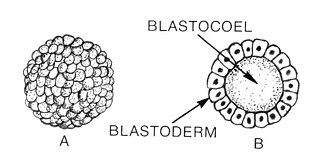
Blastulation is the stage in early animal embryonic development that produces the blastula. In mammalian development the blastula develops into the blastocyst with a differentiated inner cell mass and an outer trophectoderm. The blastula is a hollow sphere of cells known as blastomeres surrounding an inner fluid-filled cavity called the blastocoel. Embryonic development begins with a sperm fertilizing an egg cell to become a zygote, which undergoes many cleavages to develop into a ball of cells called a morula. Only when the blastocoel is formed does the early embryo become a blastula. The blastula precedes the formation of the gastrula in which the germ layers of the embryo form.

A maternal effect is a situation where the phenotype of an organism is determined not only by the environment it experiences and its genotype, but also by the environment and genotype of its mother. In genetics, maternal effects occur when an organism shows the phenotype expected from the genotype of the mother, irrespective of its own genotype, often due to the mother supplying messenger RNA or proteins to the egg. Maternal effects can also be caused by the maternal environment independent of genotype, sometimes controlling the size, sex, or behaviour of the offspring. These adaptive maternal effects lead to phenotypes of offspring that increase their fitness. Further, it introduces the concept of phenotypic plasticity, an important evolutionary concept. It has been proposed that maternal effects are important for the evolution of adaptive responses to environmental heterogeneity.

<span class="mw-page-title-main">Coenocyte</span> Type of cell structure in several groups of organisms

A coenocyte is a multinucleate cell which can result from multiple nuclear divisions without their accompanying cytokinesis, in contrast to a syncytium, which results from cellular aggregation followed by dissolution of the cell membranes inside the mass. The word syncytium in animal embryology is used to refer to the coenocytic blastoderm of invertebrates. A coenocytic colony is referred to as a coenobium, and most coenobia are composed of a distinct number of cells, often as a multiple of two.

Segmentation in biology is the division of some animal and plant body plans into a linear series of repetitive segments that may or may not be interconnected to each other. This article focuses on the segmentation of animal body plans, specifically using the examples of the taxa Arthropoda, Chordata, and Annelida. These three groups form segments by using a "growth zone" to direct and define the segments. While all three have a generally segmented body plan and use a growth zone, they use different mechanisms for generating this patterning. Even within these groups, different organisms have different mechanisms for segmenting the body. Segmentation of the body plan is important for allowing free movement and development of certain body parts. It also allows for regeneration in specific individuals.

<span class="mw-page-title-main">Morphogen</span> Biological substance that guides development by non-uniform distribution

A morphogen is a substance whose non-uniform distribution governs the pattern of tissue development in the process of morphogenesis or pattern formation, one of the core processes of developmental biology, establishing positions of the various specialized cell types within a tissue. More specifically, a morphogen is a signaling molecule that acts directly on cells to produce specific cellular responses depending on its local concentration.

Hox genes, a subset of homeobox genes, are a group of related genes that specify regions of the body plan of an embryo along the head-tail axis of animals. Hox proteins encode and specify the characteristics of 'position', ensuring that the correct structures form in the correct places of the body. For example, Hox genes in insects specify which appendages form on a segment, and Hox genes in vertebrates specify the types and shape of vertebrae that will form. In segmented animals, Hox proteins thus confer segmental or positional identity, but do not form the actual segments themselves.

Compartments can be simply defined as separate, different, adjacent cell populations, which upon juxtaposition, create a lineage boundary. This boundary prevents cell movement from cells from different lineages across this barrier, restricting them to their compartment. Subdivisions are established by morphogen gradients and maintained by local cell-cell interactions, providing functional units with domains of different regulatory genes, which give rise to distinct fates. Compartment boundaries are found across species. In the hindbrain of vertebrate embryos, rhombomeres are compartments of common lineage outlined by expression of Hox genes. In invertebrates, the wing imaginal disc of Drosophila provides an excellent model for the study of compartments. Although other tissues, such as the abdomen, and even other imaginal discs are compartmentalized, much of our understanding of key concepts and molecular mechanisms involved in compartment boundaries has been derived from experimentation in the wing disc of the fruit fly.

<span class="mw-page-title-main">Ultrabithorax</span> Protein-coding gene found in Drosophila melanogaster

Ultrabithorax (Ubx) is a homeobox gene found in insects, and is used in the regulation of patterning in morphogenesis. There are many possible products of this gene, which function as transcription factors. Ubx is used in the specification of serially homologous structures, and is used at many levels of developmental hierarchies. In Drosophila melanogaster it is expressed in the third thoracic (T3) and first abdominal (A1) segments and represses wing formation. The Ubx gene regulates the decisions regarding the number of wings and legs the adult flies will have. The developmental role of the Ubx gene is determined by the splicing of its product, which takes place after translation of the gene. The specific splice factors of a particular cell allow the specific regulation of the developmental fate of that cell, by making different splice variants of transcription factors. In D. melanogaster, at least six different isoforms of Ubx exist.

<i>Krüppel</i>

Krüppel is a gap gene in Drosophila melanogaster, located on the 2R chromosome, which encodes a zinc finger C2H2 transcription factor. Gap genes work together to establish the anterior-posterior segment patterning of the insect through regulation of the transcription factor encoding pair rule genes. These genes in turn regulate segment polarity genes. Krüppel means "cripple" in German, named for the crippled appearance of mutant larvae, who have failed to develop proper thoracic and anterior segments in the abdominal region. Mutants can also have abdominal mirror duplications.

<span class="mw-page-title-main">Gap gene</span> Gene used to develop body sections in embryos

A gap gene is a type of gene involved in the development of the segmented embryos of some arthropods. Gap genes are defined by the effect of a mutation in that gene, which causes the loss of contiguous body segments, resembling a gap in the normal body plan. Each gap gene, therefore, is necessary for the development of a section of the organism.

Decapentaplegic (Dpp) is a key morphogen involved in the development of the fruit fly Drosophila melanogaster and is the first validated secreted morphogen. It is known to be necessary for the correct patterning and development of the early Drosophila embryo and the fifteen imaginal discs, which are tissues that will become limbs and other organs and structures in the adult fly. It has also been suggested that Dpp plays a role in regulating the growth and size of tissues. Flies with mutations in decapentaplegic fail to form these structures correctly, hence the name. Dpp is the Drosophila homolog of the vertebrate bone morphogenetic proteins (BMPs), which are members of the TGF-β superfamily, a class of proteins that are often associated with their own specific signaling pathway. Studies of Dpp in Drosophila have led to greater understanding of the function and importance of their homologs in vertebrates like humans.

In the field of developmental biology, regional differentiation is the process by which different areas are identified in the development of the early embryo. The process by which the cells become specified differs between organisms.

<span class="mw-page-title-main">Pair-rule gene</span> Gene involved in the development of segmented embryos of insects

A pair-rule gene is a type of gene involved in the development of the segmented embryos of insects. Pair-rule genes are expressed as a result of differing concentrations of gap gene proteins, which encode transcription factors controlling pair-rule gene expression. Pair-rule genes are defined by the effect of a mutation in that gene, which causes the loss of the normal developmental pattern in alternating segments.

Orthodenticle (otd) is a homeobox gene found in Drosophila that regulates the development of anterior patterning, with particular involvement in the central nervous system function and eye development. It is located on the X chromosome. The gene is an ortholog of the human OTX1/OTX2 gene.

A segmentation gene is a generic term for a gene whose function is to specify tissue pattern in each repeated unit of a segmented organism. Animals are constructed of segments; however, Drosophila segments also contain subdivided compartments. There are five gene classes which each contribute to the segmentation and development of the embryonic drosophila. These five gene classes include the coordinate gene, gap gene, pair-rule gene, segment polarity gene, and homeotic gene. In embryonic drosophila, the pair-rule gene defines odd-skipped and even-skipped genes as parasegments, showing 7 stripes in the embryo. In the next gene class, segment polarity gene, individual segments each have their own anterior and posterior pole, resulting in 14 segments. In the fruit fly Drosophila melanogaster, segment polarity genes help to define the anterior and posterior polarities within each embryonic parasegment by regulating the transmission of signals via the Wnt signaling pathway and Hedgehog signaling pathway. Segment polarity genes are expressed in the embryo following expression of the gap genes and pair-rule genes. The most commonly cited examples of these genes are engrailed and gooseberry in Drosophila melanogaster. The segment polarity is the last step in embryonic development and a repeated pattern where each half of each segment is deleted and a mirror-image is duplicated and reversed to replace that half segment; thus, forming a pattern element.

<span class="mw-page-title-main">Germ-band extension</span>

Germ-band extension is a morphological process widely studied in Drosophila melanogaster in which the germ-band, which develops into the segmented trunk of the embryo, approximately doubles in length along the anterior-posterior axis while subsequently narrowing along the dorsal-ventral axis.

Staufen is a protein product of a maternally expressed gene first identified in Drosophila melanogaster. The protein has been implicated in helping regulate genes important in determination of gradients that set up the anterior posterior axis such as bicoid and oskar. Staufen proteins, abbreviated Stau, are necessary for cell localization during the oogenesis and zygotic development. It is involved in targeting of the messenger RNA encoding these genes to the correct pole of the egg cell.

The Cdx gene family, also called caudal genes, are a group of genes found in many animal genomes. Cdx genes contain a homeobox DNA sequence and code for proteins that act as transcription factors. The gene after which the gene family is named is the caudal or cad gene of the fruitfly Drosophila melanogaster. The human genome has three Cdx genes, called CDX1, CDX2 and CDX4. The zebrafish has no cdx2 gene, but two copies of cdx1 and one copy of cdx4. The Cdx gene in the nematode Caenorhabditis elegans is called pal-1.

<i>Homeotic protein bicoid</i> Protein-coding gene in the species Drosophila melanogaster

Homeotic protein bicoid is encoded by the bcd maternal effect gene in Drosophilia. Homeotic protein bicoid concentration gradient patterns the anterior-posterior (A-P) axis during Drosophila embryogenesis. Bicoid was the first protein demonstrated to act as a morphogen. Although bicoid is important for the development of Drosophila and other higher dipterans, it is absent from most other insects, where its role is accomplished by other genes.

<span class="mw-page-title-main">Hunchback (gene)</span> Maternal effect gene and gap gene

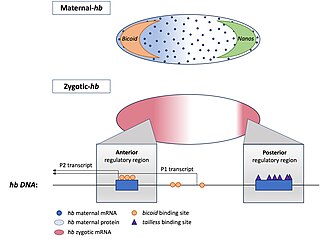
Hunchback is a maternal effect and zygotic gene expressed in the embryos of the fruit fly Drosophila melanogaster. In maternal effect genes, the RNA or protein from the mother’s gene is deposited into the oocyte or embryo before the embryo can express its own zygotic genes.

References

  1. Carroll, Sean B. "The Origins of Form". Natural History. Retrieved 12 October 2016.
  2. Russell, P. J. (2009). iGenetics, p. 564
  3. Bejsovec A, Wieschaus E (1993). "Segment polarity gene interactions modulate epidermal patterning in Drosophila embryos". Development. 119 (2): 501–517. doi:10.1242/dev.119.2.501. PMID   8287799.
  4. 1 2 Russel, Peter (2010). iGenetics: a molecular approach. San Francisco: Pearson Education. pp. 564–571. ISBN   978-0-321-56976-9.
  5. Rivera-Pomar R; Jackle H. (1996). "From gradients to stripes in Drosophila embryogenesis: Filling in the gaps". Trends Genet. 12 (11): 478–483. doi:10.1016/0168-9525(96)10044-5. PMID   8973159.
  6. 1 2 Wolpert, Lewis (2002). Principles of Development. Oxford University Press. pp. 151–161.

Sources