Related Research Articles

Bacterial vaginosis (BV) is an infection of the vagina caused by excessive growth of bacteria. Common symptoms include increased vaginal discharge that often smells like fish. The discharge is usually white or gray in color. Burning with urination may occur. Itching is uncommon. Occasionally, there may be no symptoms. Having BV approximately doubles the risk of infection by a number of sexually transmitted infections, including HIV/AIDS. It also increases the risk of early delivery among pregnant women.
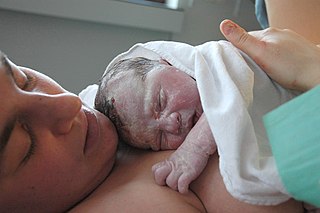
Childbirth, also known as labour, parturition and delivery, is the completion of pregnancy where one or more babies exits the internal environment of the mother via vaginal delivery or caesarean section. In 2019, there were about 140.11 million human births globally. In the developed countries, most deliveries occur in hospitals, while in the developing countries most are home births.

Amniocentesis is a medical procedure used primarily in the prenatal diagnosis of genetic conditions. It has other uses such as in the assessment of infection and fetal lung maturity. Prenatal diagnostic testing, which includes amniocentesis, is necessary to conclusively diagnose the majority of genetic disorders, with amniocentesis being the gold-standard procedure after 15 weeks' gestation.

Preterm birth, also known as premature birth, is the birth of a baby at fewer than 37 weeks gestational age, as opposed to full-term delivery at approximately 40 weeks. Extreme preterm is less than 28 weeks, very early preterm birth is between 28 and 32 weeks, early preterm birth occurs between 32 and 34 weeks, late preterm birth is between 34 and 36 weeks' gestation. These babies are also known as premature babies or colloquially preemies or premmies. Symptoms of preterm labor include uterine contractions which occur more often than every ten minutes and/or the leaking of fluid from the vagina before 37 weeks. Premature infants are at greater risk for cerebral palsy, delays in development, hearing problems and problems with their vision. The earlier a baby is born, the greater these risks will be.

Misoprostol is a synthetic prostaglandin medication used to prevent and treat stomach and duodenal ulcers, induce labor, cause an abortion, and treat postpartum bleeding due to poor contraction of the uterus. It is taken by mouth when used to prevent gastric ulcers in people taking nonsteroidal anti-inflammatory drugs (NSAID). For abortions it is used by itself or in conjunction with mifepristone or methotrexate. By itself, effectiveness for abortion is between 66% and 90%. For labor induction or abortion, it is taken by mouth, dissolved in the mouth, or placed in the vagina. For postpartum bleeding it may also be used rectally.

Obstetric ultrasonography, or prenatal ultrasound, is the use of medical ultrasonography in pregnancy, in which sound waves are used to create real-time visual images of the developing embryo or fetus in the uterus (womb). The procedure is a standard part of prenatal care in many countries, as it can provide a variety of information about the health of the mother, the timing and progress of the pregnancy, and the health and development of the embryo or fetus.
Labor induction is the process or treatment that stimulates childbirth and delivery. Inducing (starting) labor can be accomplished with pharmaceutical or non-pharmaceutical methods. In Western countries, it is estimated that one-quarter of pregnant women have their labor medically induced with drug treatment. Inductions are most often performed either with prostaglandin drug treatment alone, or with a combination of prostaglandin and intravenous oxytocin treatment.
Rupture of membranes (ROM) or amniorrhexis is a term used during pregnancy to describe a rupture of the amniotic sac. Normally, it occurs spontaneously at full term either during or at the beginning of labor. Rupture of the membranes is known colloquially as "breaking the water" or as one's "water breaking". A premature rupture of membranes (PROM) is a rupture of the amnion that occurs at full term and prior to the onset of labor. In cases of PROM, options include expectant management without intervention, or interventions such as oxytocin or other methods of labor induction, and both are usually accompanied by close monitoring of maternal and fetal health. Preterm premature rupture of membranes (PPROM) is when water breaks both before the onset of labor and before the pregnancy's 37 week gestation. In the United States, more than 120,000 pregnancies per year are affected by a premature rupture of membranes, which is the cause of about one third of preterm deliveries.
Tocolytics are medications used to suppress premature labor. Preterm birth accounts for 70% of neonatal deaths. Therefore, tocolytic therapy is provided when delivery would result in premature birth, postponing delivery long enough for the administration of glucocorticoids, which accelerate fetal lung maturity but may require one to two days to take effect.
Bishop score, also Bishop's score or cervix score, is a pre-labor scoring system to assist in predicting whether induction of labor will be required. It has also been used to assess the likelihood of spontaneous preterm delivery. The Bishop score was developed by Professor Emeritus of Obstetrics and Gynecology, Dr. Edward Bishop, and was first published in August 1964.
Bloody show or show is the passage of a small amount of blood or blood-tinged mucus through the vagina near the end of pregnancy. It is caused by thinning and dilation of the cervix, leading to detachment of the cervical mucus plug that seals the cervix during pregnancy and tearing of small cervical blood vessels, and is one of the signs that labor may be imminent. The bloody show may be expelled from the vagina in pieces or altogether and often appears as a jelly-like piece of mucus stained with blood. Although the bloody show may be alarming at first, it is not a concern of patient health after 37 weeks gestation.

Prelabor rupture of membranes (PROM), previously known as premature rupture of membranes, is breakage of the amniotic sac before the onset of labor. Women usually experience a painless gush or a steady leakage of fluid from the vagina. Complications in the baby may include premature birth, cord compression, and infection. Complications in the mother may include placental abruption and postpartum endometritis.

Cervical effacement or cervical ripening refers to the thinning and shortening of the cervix. This process occurs during labor to prepare the cervix for dilation to allow the fetus to pass through the vagina. While this a normal, physiological process that occurs at the later end of pregnancy, it can also be induced through medications and procedures.

Cervical cerclage, also known as a cervical stitch, is a treatment for cervical weakness, when the cervix starts to shorten and open too early during a pregnancy causing either a late miscarriage or preterm birth. In women with a prior spontaneous preterm birth and who are pregnant with one baby, and have shortening of the cervical length less than 25 mm, a cerclage prevents a preterm birth and reduces death and illness in the baby.
Cervical weakness, also called cervical incompetence or cervical insufficiency, is a medical condition of pregnancy in which the cervix begins to dilate (widen) and efface (thin) before the pregnancy has reached term. Definitions of cervical weakness vary, but one that is frequently used is the inability of the uterine cervix to retain a pregnancy in the absence of the signs and symptoms of clinical contractions, or labor, or both in the second trimester. Cervical weakness may cause miscarriage or preterm birth during the second and third trimesters. It has been estimated that cervical insufficiency complicates about 1% of pregnancies, and that it is a cause in about 8% of women with second trimester recurrent miscarriages.

Chorioamnionitis, also known as intra-amniotic infection (IAI), is inflammation of the fetal membranes, usually due to bacterial infection. In 2015, a National Institute of Child Health and Human Development Workshop expert panel recommended use of the term "triple I" to address the heterogeneity of this disorder. The term triple I refers to intrauterine infection or inflammation or both and is defined by strict diagnostic criteria, but this terminology has not been commonly adopted although the criteria are used.

Velamentous cord insertion is a complication of pregnancy where the umbilical cord is inserted in the fetal membranes. It is a major cause of antepartum hemorrhage that leads to loss of fetal blood and associated with high perinatal mortality. In normal pregnancies, the umbilical cord inserts into the middle of the placental mass and is completely encased by the amniotic sac. The vessels are hence normally protected by Wharton's jelly, which prevents rupture during pregnancy and labor. In velamentous cord insertion, the vessels of the umbilical cord are improperly inserted in the chorioamniotic membrane, and hence the vessels traverse between the amnion and the chorion towards the placenta. Without Wharton's jelly protecting the vessels, the exposed vessels are susceptible to compression and rupture.
Placental alpha microglobulin-1 (PAMG-1) is a human protein that was first isolated in 1975 from amniotic fluid. PAMG-1 is an important biomarker for the detection of premature rupture of fetal membrane (PROM) The high concentration of PAMG-1 in amniotic fluid means it can be used to detect if this fluid is present in the cervico-vaginal discharge of pregnant women; the presence of PAMG-1 in the discharge suggests that amniotic fluid is present, and therefore suggests that PROM has occurred. PAMG-1 was originally referred to as specific alpha-1 globulin of placenta.

The fetal membranes are the four extraembryonic membranes, associated with the developing embryo, and fetus in humans and other mammals. They are the amnion, chorion, allantois, and yolk sac. The amnion and the chorion are the chorioamniotic membranes that make up the amniotic sac which surrounds and protects the embryo. The fetal membranes are four of six accessory organs developed by the conceptus that are not part of the embryo itself, the other two are the placenta, and the umbilical cord.
The fern test is a medical laboratory test used in obstetrics and gynecology. The name refers to the detection of a characteristic "fern like" pattern of vaginal secretions when a specimen is allowed to dry on a glass slide and is viewed under a low-power microscope. The fern test is most commonly used to provide evidence of the presence of amniotic fluid and is used in obstetrics to detect preterm premature rupture of membranes and/or the onset of labor. It also may provide indirect evidence of ovulation and fertility, although it does not predict the time of ovulation.
References
- 1 2 Berghella V, Saccone G (July 2019). "Fetal fibronectin testing for reducing the risk of preterm birth". The Cochrane Database of Systematic Reviews. 7 (7): CD006843. doi:10.1002/14651858.CD006843.pub3. PMC 6663121 . PMID 31356681.
- ↑ "Fetal fibronectin test". Mayo Clinic. Retrieved 19 March 2016.
- ↑ Patten J, Wang K (March 2021). "Fibronectin in development and wound healing". Advanced Drug Delivery Reviews. 170: 353–368 (357). doi: 10.1016/j.addr.2020.09.005 . PMID 32961203.
- ↑ Lockwood CJ, Senyei AE, Dische MR, Casal D, Shah KD, Thung SN, et al. (September 1991). "Fetal fibronectin in cervical and vaginal secretions as a predictor of preterm delivery". The New England Journal of Medicine. 325 (10): 669–674. doi: 10.1056/NEJM199109053251001 . PMID 1870640.
- 1 2 Cornelissen LG, van Oostrum NH, van der Woude DA, Rolf C, Porath MM, Oei SG, van Laar JO (March 2020). "The diagnostic value of fetal fibronectin testing in clinical practice". The Journal of Obstetrics and Gynaecology Research. 46 (3): 405–412. doi:10.1111/jog.14201. PMID 31955485. S2CID 210831801.
- ↑ "Prelabor Rupture of Membranes: ACOG Practice Bulletin Summary, Number 217". Obstetrics and Gynecology. 135 (3): 739–743. March 2020. doi:10.1097/AOG.0000000000003701. PMID 32080044. S2CID 211231184.
- ↑ Berghella V, Saccone G, et al. (Cochrane Pregnancy and Childbirth Group) (July 2019). "Fetal fibronectin testing for reducing the risk of preterm birth". The Cochrane Database of Systematic Reviews. 7 (7): CD006843. doi:10.1002/14651858.CD006843.pub3. PMC 6663121 . PMID 31356681.
- ↑ Kiefer DG, Vintzileos AM (2008). "The utility of fetal fibronectin in the prediction and prevention of spontaneous preterm birth". Reviews in Obstetrics & Gynecology. 1 (3): 106–112. PMC 2582650 . PMID 19015761.