Germ-band extension

Last updated
Schematic of Drosophila embryo at the start of germ-band elongation and 30 minutes later. The germ-band (grey) is posterior to the cephalic furrow (curved-line) and folds dorsally (red arrow) upon cell intercalation. Each rectangle represents a field of cells before (0 min) and after (30 min) convergent extension. Colored nuclei arbitrarily mark rows of cells in order to visualize tissue morphogenesis. Germ-band extension.png
Schematic of Drosophila embryo at the start of germ-band elongation and 30 minutes later. The germ-band (grey) is posterior to the cephalic furrow (curved-line) and folds dorsally (red arrow) upon cell intercalation. Each rectangle represents a field of cells before (0 min) and after (30 min) convergent extension. Colored nuclei arbitrarily mark rows of cells in order to visualize tissue morphogenesis.

Germ-band extension is a morphological process widely studied in Drosophila melanogaster in which the germ-band, which develops into the segmented trunk of the embryo, approximately doubles in length along the anterior-posterior axis while subsequently narrowing along the dorsal-ventral axis. [1]

Contents

Germ-band extension begins shortly after gastrulation and is divided into two phases. The fast phase, in which most of the extension occurs, takes about 25 minutes. The remaining extension continues during the slow phase and is completed in the following 70 minutes. [2] During this process the ventral germ-band extends around the posterior end of the embryo, effectively folding over onto the dorsal side of the egg. Multiple individual cells intercalating mediolateral to the anterior-posterior axis drive the resulting global elongation of the embryo. In addition, cell shape changes, and oriented cell divisions in the posterior germ-band are in part required for full elongation. [3] However, elongation of the body axis seems to be primarily linked to changes in cell neighbor relations.

This article describes axis elongation in Drosophila. However, the basis of germ-band elongation is applicable to many organisms including other invertebrates and vertebrates alike. [4]

Cellular basis

Schematic of neighbor exchange or an elementary T1 process involving four cells. T1-magenta cells are in direct contact. T2-all cells share a common vertex. T3-resolution results in green cells sharing a common boundary. T1Junction.png
Schematic of neighbor exchange or an elementary T1 process involving four cells. T1-magenta cells are in direct contact. T2-all cells share a common vertex. T3-resolution results in green cells sharing a common boundary.

In order for cells to intercalate between one another the adherens junctions that maintain the integrity of the epithelial tissue must be dynamically remodeled. Time-lapse microscopy has captured this process of cell neighbor exchange, which is schematically represented to the right. In the type 1 configuration, two cells contact each other along the anterior-posterior axis, whereas two dorsal-ventral cells do not directly contact. Next, the cell boundary between the two anterior-posterior neighbors selectively shrinks, resulting in an obligatory intermediate type 2 junction, where the four cells share a vertex. Upon resolution of the type 2 junction, a new type 3 junctions forms perpendicular to the original type 1 configuration. During this process the two dorsal-ventral cells have become neighbors. [5] When multiple clusters of cells intercalate in the dorsal-ventral axis, through junctional neighbor exchange, the outcome is an extension of germ-band in the anterior-posterior axis.

In addition to the simple neighbor exchange, higher-ordered rosette formations have been observed in which five or more cells meet at a vertex. Multicellular rosettes form and resolve in a directional fashion that promotes germ-band elongation. [6] Neighbor exchange and multicellular rosette formation involve oriented junctional remodeling, which indicates that the intercalating cells are intrinsically polarized within the plane of the epithelium.

Molecular basis

Asymmetric localization of proteins to reciprocal cell borders in the apical plane of a polarized epithelial. Planarpolarity.png
Asymmetric localization of proteins to reciprocal cell borders in the apical plane of a polarized epithelial.

The dorsal-ventral intercalation of cells during germ-band extension ultimately arises from the asymmetric localization of proteins within individual cells. Immunofluorescence reveals that non-muscle Myosin II localizes to the anterior-posterior boundaries of cells, destabilizing adherens junctions, whereas the Bazooka/Par-3 complex localizes to dorsal-ventral boundaries, stabilizing adherens junctions. Moreover, bazooka mutants are defective in germ-band extension, which supports the idea that polarized protein localization is critical for cell rearrangements. [7] One mechanism by which Myosin II might promote polarized cell remodeling is through contractile activity that creates tension orienting junctional disassembly. [8] However, the precise mechanism in which asymmetrically localized protein complexes encourage directed intercalation remains disputed.

The source that establishes planar polarity during germ-band extension remains elusive. Polarized intercalation is largely unaffected in mutant embryos that lack dorsal-ventral cell types. Yet, mutations that disrupt segmental patterning along the anterior-posterior axis, such as eve and runt , decrease cell intercalation and subsequent germ-band elongation. [1] Furthermore, ectopic expression of eve or runt is sufficient to locally reorient the polarity of nearby cells. This evidence argues that planar polarity is established by cell-cell interactions, and not by a long-range polarizing cue. Thus, polarizing information can spread from one cell to the next, downstream of an Eve-dependent signal that remains to be identified. [7]

Tensile forces

Researchers suggest that mesoderm invagination provides an anterior-posterior pulling force that contributes to germ-band extension through passive cell shape changes. Although anterior-posterior patterning mutants fail to fully extend their germ-bands, during the fast phase the elongation length is normal despite defects in polarized cell intercalation. Time-lapse analysis revealed that an increase in cell shape stretching in the anterior-posterior axis was compensating for aberrant cell intercalation, independent of anterior-posterior patterning. Furthermore, during wild type development, it has been suggested that intercalary cell behavior relaxes the stress imposed on the germ-band, allowing stretched cells to restore to isometric shapes. [3]

In addition, there is evidence that mechanical tension is necessary and sufficient for the cortical localization of Myosin II. Thus, not only can Myosin II generate tension but it may also be up-regulated by tensile forces, creating a positive feedback loop that allows cells to dynamically respond to fluctuations in their mechanical environment. [9]

Related Research Articles

<span class="mw-page-title-main">Gastrulation</span> Stage in embryonic development in which germ layers form

Gastrulation is the stage in the early embryonic development of most animals, during which the blastula, or in mammals the blastocyst is reorganized into a multilayered structure known as the gastrula. Before gastrulation, the embryo is a continuous epithelial sheet of cells; by the end of gastrulation, the embryo has begun differentiation to establish distinct cell lineages, set up the basic axes of the body, and internalized one or more cell types including the prospective gut.

A coenocyte is a multinucleate cell which can result from multiple nuclear divisions without their accompanying cytokinesis, in contrast to a syncytium, which results from cellular aggregation followed by dissolution of the cell membranes inside the mass. The word syncytium in animal embryology is used to refer to the coenocytic blastoderm of invertebrates. A coenocytic colony is referred to as a coenobium, and most coenobia are composed of a distinct number of cells, often as a multiple of two.

<i>Drosophila</i> embryogenesis Embryogenesis of the fruit fly Drosophila, a popular model system

Drosophila embryogenesis, the process by which Drosophila embryos form, is a favorite model system for genetics and developmental biology. The study of its embryogenesis unlocked the century-long puzzle of how development was controlled, creating the field of evolutionary developmental biology. The small size, short generation time, and large brood size make it ideal for genetic studies. Transparent embryos facilitate developmental studies. Drosophila melanogaster was introduced into the field of genetic experiments by Thomas Hunt Morgan in 1909.

Compartments can be simply defined as separate, different, adjacent cell populations, which upon juxtaposition, create a lineage boundary. This boundary prevents cell movement from cells from different lineages across this barrier, restricting them to their compartment. Subdivisions are established by morphogen gradients and maintained by local cell-cell interactions, providing functional units with domains of different regulatory genes, which give rise to distinct fates. Compartment boundaries are found across species. In the hindbrain of vertebrate embryos, rhombomeres are compartments of common lineage outlined by expression of Hox genes. In invertebrates, the wing imaginal disc of Drosophila provides an excellent model for the study of compartments. Although other tissues, such as the abdomen, and even other imaginal discs are compartmentalized, much of our understanding of key concepts and molecular mechanisms involved in compartment boundaries has been derived from experimentation in the wing disc of the fruit fly.

<span class="mw-page-title-main">Intermediate mesoderm</span> Layer of cells in mammalian embryos

Intermediate mesoderm or intermediate mesenchyme is a narrow section of the mesoderm located between the paraxial mesoderm and the lateral plate of the developing embryo. The intermediate mesoderm develops into vital parts of the urogenital system.

<span class="mw-page-title-main">Epiblast</span> Embryonic inner cell mass tissue that forms the embryo itself, through the three germ layers

In amniote embryonic development, the epiblast is one of two distinct cell layers arising from the inner cell mass in the mammalian blastocyst, or from the blastula in reptiles and birds, the other layer is the hypoblast. It drives the embryo proper through its differentiation into the three primary germ layers, ectoderm, mesoderm and endoderm, during gastrulation. The amniotic ectoderm and extraembryonic mesoderm also originate from the epiblast.

<span class="mw-page-title-main">Apical ectodermal ridge</span>

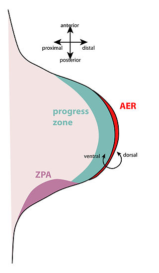
The apical ectodermal ridge (AER) is a structure that forms from the ectodermal cells at the distal end of each limb bud and acts as a major signaling center to ensure proper development of a limb. After the limb bud induces AER formation, the AER and limb mesenchyme—including the zone of polarizing activity (ZPA)—continue to communicate with each other to direct further limb development.

An asymmetric cell division produces two daughter cells with different cellular fates. This is in contrast to symmetric cell divisions which give rise to daughter cells of equivalent fates. Notably, stem cells divide asymmetrically to give rise to two distinct daughter cells: one copy of the original stem cell as well as a second daughter programmed to differentiate into a non-stem cell fate.

Decapentaplegic (Dpp) is a key morphogen involved in the development of the fruit fly Drosophila melanogaster and is the first validated secreted morphogen. It is known to be necessary for the correct patterning and development of the early Drosophila embryo and the fifteen imaginal discs, which are tissues that will become limbs and other organs and structures in the adult fly. It has also been suggested that Dpp plays a role in regulating the growth and size of tissues. Flies with mutations in decapentaplegic fail to form these structures correctly, hence the name. Dpp is the Drosophila homolog of the vertebrate bone morphogenetic proteins (BMPs), which are members of the TGF-β superfamily, a class of proteins that are often associated with their own specific signaling pathway. Studies of Dpp in Drosophila have led to greater understanding of the function and importance of their homologs in vertebrates like humans.

The heart is the first functional organ in a vertebrate embryo. There are 5 stages to heart development.

In the field of developmental biology, regional differentiation is the process by which different areas are identified in the development of the early embryo. The process by which the cells become specified differs between organisms.

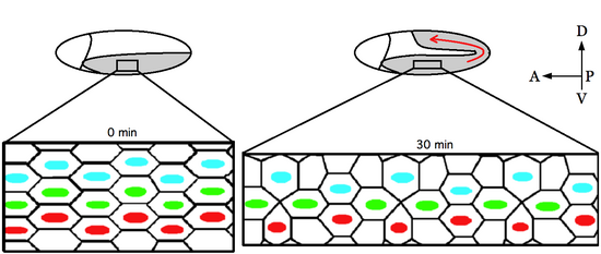
Convergent extension (CE), sometimes called convergence and extension (C&E), is the process by which the tissue of an embryo is restructured to converge (narrow) along one axis and extend (elongate) along a perpendicular axis by cellular movement.

<span class="mw-page-title-main">Fish development</span>

The development of fishes is unique in some specific aspects compared to the development of other animals.

<span class="mw-page-title-main">Stress fiber</span> Contractile actin bundles found in non-muscle cells

Stress fibers are contractile actin bundles found in non-muscle cells. They are composed of actin (microfilaments) and non-muscle myosin II (NMMII), and also contain various crosslinking proteins, such as α-actinin, to form a highly regulated actomyosin structure within non-muscle cells. Stress fibers have been shown to play an important role in cellular contractility, providing force for a number of functions such as cell adhesion, migration and morphogenesis.

<span class="mw-page-title-main">Planar cell polarity</span>

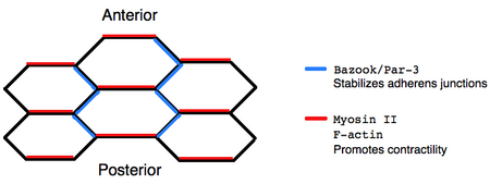
Planar cell polarity (PCP) is the protein-mediated signaling that coordinates the orientation of cells in a layer of epithelial tissue. In vertebrates, examples of mature PCP oriented tissue are the stereo-cilia bundles in the inner ear, motile cilia of the epithelium, and cell motility in epidermal wound healing. Additionally, PCP is known to be crucial to major developmental time points including coordinating convergent extension during gastrulation and coordinating cell behavior for neural tube closure. Cells orient themselves and their neighbors by establishing asymmetric expression of PCP components on opposing cell members within cells to establish and maintain the directionality of the cells. Some of these PCP components are transmembrane proteins which can proliferate the orientation signal to the surrounding cells.

<span class="mw-page-title-main">Cell polarity</span> Polar morphology of a cell, a specific orientation of the cell structure

Cell polarity refers to spatial differences in shape, structure, and function within a cell. Almost all cell types exhibit some form of polarity, which enables them to carry out specialized functions. Classical examples of polarized cells are described below, including epithelial cells with apical-basal polarity, neurons in which signals propagate in one direction from dendrites to axons, and migrating cells. Furthermore, cell polarity is important during many types of asymmetric cell division to set up functional asymmetries between daughter cells.

Thomas Lecuit, born 4 October 1971 in Saumur, is a French biologist specializing in the emergence of forms or morphogenesis. He is a professor at the Collège de France, holding the Dynamics of Life Chair. He leads a research team at the Institut de Biologie du Développement de Marseille (IBDM), and the Turing Centre for Living Systems, an interdisciplinary centre dedicated to the study of living organisms.

Barry James Thompson is an Australian and British developmental biologist and cancer biologist. He is a professor of the John Curtin School of Medical Research at the Australian National University in Canberra. Thompson is known for identifying genes, proteins and mechanisms involved in epithelial polarity, morphogenesis and cell signaling via the Wnt and Hippo signaling pathways, which have key roles in human cancer.

<span class="mw-page-title-main">Primordial germ cell migration</span>

Primordial germ cell (PGC) migration is the process of distribution of primordial germ cells throughout the embryo during embryogenesis.

References

  1. 1 2 Irvine, K., and Wieschaus, E. (1994). Cell intercalation during Drosophila germ-band extension and its regulation by pair-rule segmentation genes. Development 120, 827–841.
  2. da Silva, S. M. & Vincent, J. P. (2007). Oriented cell divisions in the extending germband of Drosophila. Development 134, 3049–3054.
  3. 1 2 Butler, L.C., et al. (2009). Cell shape changes indicate a role for extrinsic tensile forces in Drosophila germ-band extension. Nature Cell Biology 11, 859-864.
  4. Keller, R. (2006). Mechanisms of elongation in embryogenesis. Development 133, 2291–2302.
  5. Bertet, C., Sulak, L. & Lecuit, T. (2004). Myosin-dependent junction remodelling controls planar cell intercalation and axis elongation. Nature 429, 667–671.
  6. Blankenship, J. T., Backovic, S. T., Sanny, J. S., Weitz, O. and Zallen, J. A. (2006). Multicellular rosette formation links planar cell polarity to tissue morphogenesis. Dev. Cell 11, 459-470.
  7. 1 2 Zallen, J., and Wieschaus, E. (2004). Patterned gene expression directs bipolar planar polarity in Drosophila. Dev. Cell 6, 343–355.
  8. Baum B. (2004). Animal development: crowd control. Curr Biol. 7;14, 716-718.
  9. Fernandez-Gonzalez R, Simoes Sde M, Röper JC, Eaton S, Zallen JA. (2009). Myosin II dynamics are regulated by tension in intercalating cells. Dev Cell. 17, 736–743.