Halteria

Last updated

Halteria
Halteria 107300954.jpg
Scientific classification OOjs UI icon edit-ltr.svg
Domain: Eukaryota
Clade: Diaphoretickes
Clade: SAR
Clade: Alveolata
Phylum: Ciliophora
Class: Oligotrichea
Order: Halteriida
Family: Halteriidae
Genus: Halteria
Species

H. grandinella
H. bifurcate
H. chorelligera
H. cirrifera
H. decemsulcata
H. geleiana
H. máxima
H. mínima
H. minuta
H. oblonga
H. ovata
H. verrucosa
H. viridis
H. vorax

Contents

Halteria, sometimes referred to as the jumping oligotrich, is a genus of common planktonic ciliates that are found in many freshwater environments. Halteria are easy to locate due to their abundance and distinctive behaviour with observations of Halteria potentially dating back to the 17th century and the discovery of microorganisms. [1] Over time more has been established about their morphology and behavior, which has led to many changes in terms of classification.

Species of Halteria can exist in both a trophic and an encysted form but are most commonly described in the trophic form. [2] Species of Halteria can be identified by their unique jumping movement which is enabled by an equatorial row of stiff cirri that beat in unison, allowing the organism to move very quickly backwards. [3]


Members of the genus Halteria are heterotrophic and serve as important bacterivores in the habitats they occupy as well as being preyed upon primarily by metazoans. One recent paper identified Halteria sp. as the first identified "virovore", an organism that can feed on virus. [4] The cells of Halteria are roughly dome shaped and in addition to the equatorial cirri, they possess a collar of cilia around the buccal opening used for feeding and locomotion. [3] The important ecological role played by Halteria as well as its unique locomotion strategy, makes Halteria a genus of interest in different areas of protistology research.

History of knowledge

The genus Halteria is abundant in many freshwater environments. [5] The ubiquity of this genus is likely why observations date back hundreds of years. The original description of the genus is not clearly established, but it is possible that observations of Halteria date back to Antony van Leewenhoek’s observations in 1675 as the fourth animalcule observed in an earthen pot full of rainwater. The organism he observed was small, swift, and seen to stand still before quickly changing direction and travelling straight, which is consistent with the characteristic movement of Halteria. [1]

The name Halteria is credited to Félix Dujardin in 1840, who reclassified Trichodina grandinella and Trichodina vorax, which had been previously classified by Müller and Ehrenberg respectively, as H. grandinella and H. vorax. [6] Creating the new genus, Halteria, when the two species were found not to fit the subfamily Vorticellina, under which the genus Trichodina fell. [7] Descriptions of Halteria at this time were still rather vague, focusing on the quick jumping movement that results from the beating of its cirri and the presence of oral cilia. [6]

In 1858, Édouard Claparède and Johannes Lachmann described Halteria grandinella in greater detail. Noting explicitly for the first time, that the cirri are only found in an equatorial belt around the cell. New details relating to the buccal cavity were also discovered; Claparède and Lachmann observed that there was an indentation in a portion of the buccal apparatus and that at this site no oral cilia are present. This means that the oral cilia form an incomplete circle around the buccal cavity, and do not surround it completely as was previously assumed. [8]

Questions on the classification of Halteria have arisen again in more recent years. Halteria have been most commonly classified as a member of the oligotrich group of ciliates, because they possess the group’s characteristic prominent oral cilia arranged in an incomplete circle. However, recent deep sequencing and RNA analysis of Halteria indicate that Halteria may be more closely related to oxytrichids than oligotrichs, suggesting the similarity in oral apparatus with oligotrichs is the result of convergent evolution. [9]

Description

Halteria can exist in a trophic, ciliated stage or an encysted stage and the morphology of the cells varies significantly between stages. [2]

Trophic stage

In the trophic stage, Halteria cells are globular and between 15 and 35 μm in size. [2] Cells possess both oral cilia and rigid equatorial cirri. [2] A collar of prominent oral cilia can be found at the anterior end of Halteria cells, partially surrounding the buccal cavity. [8] This oral apparatus consists of fifteen membranelles that encircle the peristome and seven membranelles inside the buccal cavity. [2]

The rigid cirri of Halteria, sometimes referred to as jumping bristles, are each 15-25 μm long. [2] The cirri are organized equatorially around the cells in 7-10 longitudinal rows. [2] Each row is in turn organized into four groups of cirri. When species of Halteria beat these cirri in unison, they generate a characteristic jumping motion sufficiently distinct to Halteria that observation of this movement has been considered sufficient for visual identification of Halteria [3]

The cortex of Halteria is composed of four membranes. [2] Two of these membranes, the inner and outer alveolar membranes, cover the flat alveoli which lie entirely beneath the two remaining membranes. [2] The cell membrane sits directly above the outer alveolar membrane and covers the entire cell including the cilia. [2] The perilemma is the fragile outermost membrane seen covering only small portions of the cell. [2] The fragility of the perilemma may be the cause of this distribution as it would be difficult to preserve. [2] Just beneath the membranes of the cortex, the body shape of Halteria is stabilized by microtubules in a basket configuration. [2]

Within Halteria cells, a contractile vacuole is located approximately midway between the anterior and posterior ends of the cell. [2] The mitochondria of Halteria are usually spherical with tubular cristae. [10] Within the mitochondria of H. geleiana, microorganisms have been observed within the matrix. [10] The microorganisms were rod shaped and observed with various lengths and in different numbers. [10] No function or origin is currently known for these microorganisms or whether they are parasitic or symbiotic. Halteria have one micronucleus and a macronucleus with large band-like nucleoli. [11] [12] The macronucleus is oblong in shape while the micronucleus is more globular. [2]

Encystment

As Halteria cells transition from the trophic to the encysted stage, initially their globular bodies elongate, primarily at the anterior end, until the length of the cell has nearly doubled. [2] Owing to the uneven elongation, the buccal cavity is flattened, the membranelles of the oral apparatus move closer to the centre of the cell and the rows of cirri move closer to the posterior end of the cell. [2] While the cell stretches, the cytoplasm develops 5 μm long conical structures. [2] After this stage of elongation, the cells become more rounded, and a mucous envelope is extruded. Also during this next stage of encystment, the conical structures formed in the cytoplasm attach to the outer layer of the developing cyst, called the ectocyst. [2] Once attached to the ectocyst, the conical structures are called lepidosomes. [2] After encystment, cysts use the mucous envelope to firmly attach to any available substrate. [2]

Habitat and ecology

The genus Halteria consists of freshwater ciliates that typically live a planktonic lifestyle. The species Halteria grandinella is considered cosmopolitan, meaning that it is found in habitats across the world. [13] Other species are less common and so they are less well defined, however frequent descriptions of Halteria grandinella have provided insight into the genus as a whole. Halteria are heterotrophic and unlike many closely related genera like Pelagohalteria, they have no photosynthetic endosymbionts. Halteria do frequently eat green algae which, when observed in food vacuoles, has led to misclassifications in the past when mistaken for endosymbionts. [14]

Species of Halteria play a particularly large role in many freshwater habitats as bacteriovores. In a study that used fluorescently labelled bacteria in fishponds to observe protistan bacterivory, ciliate grazing accounted for 56% of total protistan grazing and Halteria, along with two other ciliate genera, Pelagohalteria and Rimostrombidium were responsible approximately 71% of the total ciliate bacterivory. [15] Halteria also act as prey for many metazoan predators. [16] It has been proposed that the characteristic jumping behavior of Halteria was evolved as an escape strategy to avoid such predation. [5] Halteria are also able to act as virovores and can consume viruses, such as chloroviruses, to fuel growth and division. [17] [18]

Much of the research related to Halteria is focused on their movement and their ecological roles. Halteria acts as a model organism for the study of their jumping movement through ciliary beating. It can be found in abundance in diverse freshwater habitats interacting with other organisms as both predators and prey. [16] [15]

Halteria spend most of the time either stationary or moving smoothly through water propelled by the cilia at their anterior end. [5] The halting jumping movement most associated with Halteria is the result of external stimulus such as currents, which is known because jumping in Halteria has been induced in a laboratory setting. [16] Jumping behavior in Halteria requires 41% of the organism’s total metabolic rate, [16] and so employing it too frequently would be an inefficient use of energy.

Reproduction

Asexual reproduction

Halteria can reproduce asexually by transverse binary fission. During this replication the majority of the ciliature that will be present on the daughter cells is formed de novo. [19] The only exception to this is the oral ciliature of the parent cell which is inherited by the proter daughter cell. [19] The parental cirri are resorbed by the cell during division and the cirri of both daughter cells are produced de novo from cirral anlagen and the oral ciliature of the opisthe daughter cell is generated de novo through the formation of an oral primordium at the posterior end of the cell. [19] Both the macronucleus and micronucleus divide during the process resulting in two daughter cells that are genetically identical to the parent cell. [19]

Conjugation

Halteria cells can reproduce sexually through a process that has been studied specifically in H. grandinella. [12] During sexual reproduction, the ventral sides of two Halteria cells fuse. Various changes in morphology then occur through maturation divisions including a decrease in the number of cirri in both cells and the loss of buccal membranelles in one of the pair and the entire oral apparatus disappears in the other. [12] The remaining membranelles are shared between the cells at the anterior end. [12] On a nuclear level, during conjugation the original macronuclei fragment and the micronuclei mature and divide three times, with only one derivative of the first two divisions continuing to divide, forming two pronuclei in the third division. [12] A pronuclei from each cell is exchanged and the two that end up in each cell fuse to form the synkaryon. [12] The synkaryon divides twice with one derivative from each of the second divisions degenerating and the remaining derivatives becoming the new micronucleus and the macronucleus analge. [12] After synkaryon division is complete, conjugates separate, now generating two cells with genetics distinct from the parent cells and from each other. [12]

Related Research Articles

<span class="mw-page-title-main">Spirotrich</span> Class of single-celled organisms

The spirotrichs are a large and diverse group of ciliate protozoa. They typically have prominent oral cilia in the form of a series of polykinetids, called the adoral zone of membranelles, beginning anterior to the oral cavity and running down to the left side of the mouth. There may also be one or two paroral membranes on its right side. The body cilia are fused to form polykinetids called cirri in some, and are sparse to absent in others.

<i>Paramecium</i> Genus of unicellular ciliates, commonly studied as a representative of the ciliate group

Paramecium is a genus of eukaryotic, unicellular ciliates, commonly studied as a model organism of the ciliate group. Paramecium are widespread in freshwater, brackish, and marine environments and are often abundant in stagnant basins and ponds. Because some species are readily cultivated and easily induced to conjugate and divide, they have been widely used in classrooms and laboratories to study biological processes. The usefulness of Paramecium as a model organism has caused one ciliate researcher to characterize it as the "white rat" of the phylum Ciliophora.

<span class="mw-page-title-main">Colpodea</span> Class of protists in the ciliates phylum

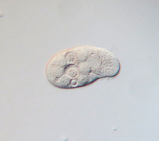
The Colpodea are a class of ciliates, of about 200 species common in freshwater and soil habitats. The body cilia are typically uniform, and are supported by dikinetids of characteristic structure, with cilia on both kinetosomes. The mouth may be apical or ventral, with more or less prominent associated polykinetids. Many are asymmetrical, the cells twisting sideways and then untwisting again prior to division, which often takes place within cysts. Colpoda, a kidney-shaped ciliate common in organic rich conditions, is representative.

<span class="mw-page-title-main">Hypotrich</span> Subclass of single-celled organisms


The hypotrichs are a group of ciliated protozoa, common in fresh water, salt water, soil and moss. Hypotrichs possess compound ciliary organelles called "cirri," which are made up of thick tufts of cilia, sparsely distributed on the ventral surface of the cell. The multiple fused cilia which form a cirrus function together as a unit, enabling the organism to crawl along solid substrates such as submerged debris or sediments. Hypotrichs typically possess a large oral aperture, bordered on one side by a wreath or collar of membranelles, forming an "adoral zone of membranelles," or AZM.

<i>Stylonychia</i> Genus of single-celled organisms

Stylonychia is a genus of ciliates, in the subclass Hypotrichia. Species of Stylonychia are very common in fresh water and soil, and may be found on filamentous algae, surface films, and among particles of sediment. Like other Hypotrichs, Stylonychia has cilia grouped into membranelles alongside the mouth and cirri over the body. It is distinguished partly by long cirri at the posterior, usually a cluster of three. The largest can just be seen at a 25x magnification, and the smallest can just be seen at a 450x magnification.

<i>Vorticella</i> Genus of single-celled organisms

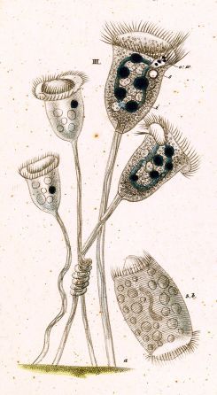
Vorticella is a genus of bell-shaped ciliates that have stalks to attach themselves to substrates. The stalks have contractile myonemes, allowing them to pull the cell body against substrates. The formation of the stalk happens after the free-swimming stage.

<i>Spirostomum</i> Genus of ciliated protists

Spirostomum is a genus of ciliated protists in the class Heterotrichea. It is known for being very contractile. Having been first identified by Christian Gottfried Ehrenberg in 1834, further research has identified eight additional true morphospecies. This bacterivore genus mainly lives in the sediment deposits at the bottom of various aquatic habitats, and members possess rquA genes that could be responsible for their ability to survive in these hypoxic and anoxic environments. They are identifiable by their relatively large tubular/flat vermiform bodies. Their life cycle consists of a growth stage, in which they mature, and asexual and sexual reproduction stages. Some species are model organisms for studies on human pathogenic bacteria, while others are sensitive and accurate bioindicators for toxic substances.

<i>Paramecium caudatum</i> Species of single-celled organism

Paramecium caudatum is a species of unicellular protist in the phylum Ciliophora. They can reach 0.33 mm in length and are covered with minute hair-like organelles called cilia. The cilia are used in locomotion and feeding. The species is very common, and widespread in marine, brackish and freshwater environments.

Karyorelictea is a class of ciliates in the subphylum Postciliodesmatophora. Most species are members of the microbenthos community, that is, microscopic organisms found in the marine interstitial habitat, though one genus, Loxodes, is found in freshwater.

<span class="mw-page-title-main">Ciliate</span> Taxon of protozoans with hair-like organelles called cilia

The ciliates are a group of alveolates characterized by the presence of hair-like organelles called cilia, which are identical in structure to eukaryotic flagella, but are in general shorter and present in much larger numbers, with a different undulating pattern than flagella. Cilia occur in all members of the group and are variously used in swimming, crawling, attachment, feeding, and sensation.

<i>Euplotes</i> Genus of single-celled organisms

Euplotes is a genus of ciliates in the subclass Euplotia. Species are widely distributed in marine and freshwater environments, as well as soil and moss. Most members of the genus are free-living, but two species have been recorded as commensal organisms in the digestive tracts of sea urchins.

<i>Climacostomum</i> Genus of single-celled organisms

Climacostomum is a genus of unicellular ciliates, belonging to the class Heterotrichea.

<i>Pseudomonilicaryon anser</i> Species of single-celled organism

Pseudomonilicaryon anser is a species of unicellular ciliates in the family Dileptidae, also known under the names Dileptus anser and Dileptus cygnus. The species is common in fresh water ponds, stagnant pools, mosses and soils.

<i>Colpidium colpoda</i> Species of protozoan

Colpidium colpoda are free-living ciliates commonly found in many freshwater environments including streams, rivers, lakes and ponds across the world. Colpidium colpoda is also frequently found inhabiting wastewater treatment plants. This species is used as an indicator of water quality and waste treatment plant performance.

<i>Colpoda inflata</i> Species of single-celled organism

Colpoda inflata is a unicellular organism, belonging to the genus Colpoda. Colpodeans are eucaryotic protozoans, that mainly feed on bacteria (bacteriophagous), vary a lot in size and have a funnel-shaped vestibule.

<span class="mw-page-title-main">Condylostoma</span> Genus of protists belonging to the ciliates phylum

Condylostoma is a genus of unicellular ciliate protists, belonging to the class Heterotrichea.

<i>Licnophora</i> Genus of single-celled organisms

Licnophora is a genus of ciliates in the family Licnophoridae. They typically have an hourglass-like shape and live as ectocommensals on marine animals.

Miamiensis avidus is a species of unicellular marine eukaryote that is a parasite of many different types of fish. It is one of several organisms known to cause the fish disease scuticociliatosis and is considered an economically significant pathogen of farmed fish. M. avidus is believed to be the cause of a 2017 die-off of fish and sharks in the San Francisco Bay.

<span class="mw-page-title-main">Stentor roeselii</span> Species of single-celled organism

Stentor roeselii is a free-living ciliate species of the genus Stentor, in the class Heterotrichea. It is a common and widespread protozoan, found throughout the world in freshwater ponds, lakes, rivers and ditches.

Parablepharismea is a class of free-living marine and brackish anaerobic ciliates that form a major clade of obligate anaerobes within the SAL group, together with the classes Muranotrichea and Armophorea.

References

  1. 1 2 van Leeuwenhoek, A.P. (1677). "Observations, communicated to the publisher by Mr. Antony van Leewenhoeck, in a dutch letter of the 9th Octob. 1676. here English'd: concerning little animals by him observed in rain-well-sea-and snow water; as also in water wherein pepper had lain infused". Philosophical Transactions of the Royal Society of London. 12 (133): 821–831. doi:10.1098/rstl.1677.0003.
  2. 1 2 3 4 5 6 7 8 9 10 11 12 13 14 15 16 17 18 19 20 21 Foissner, W.; Müller, H.; Agatha, S. (2007). "A comparative fine structural and phylogenetic analysis of resting cysts in oligotrich and hypotrich Spirotrichea (Ciliophora)". European Journal of Protistology. 43 (4): 295–314. doi:10.1016/j.ejop.2007.06.001. PMC   2848329 . PMID   17766095.
  3. 1 2 3 Patterson, D.J.; Hedley, S. (1996). Freeliving Freshwater Protozoa. CRC Press. ISBN   9781840765847.
  4. DeLong, John P.; Van Etten, James L.; Al-Ameeli, Zeina; Agarkova, Irina V.; Dunigan, David D. (2023-01-03). "The consumption of viruses returns energy to food chains". Proceedings of the National Academy of Sciences. 120 (1): e2215000120. Bibcode:2023PNAS..12015000D. doi: 10.1073/pnas.2215000120 . ISSN   0027-8424. PMID   36574690. S2CID   255219850.
  5. 1 2 3 Archbold, J.H.; Berger, J. (1985). "A qualitative assessment of some metazoan predators of Halteria grandinella, a common freshwater ciliate". Hydrobiologia. 126 (2): 97–102. doi:10.1007/BF00008675. S2CID   28208345.
  6. 1 2 Dujardin, F. (1841). "Histoire naturelle des zoophytes. Infusoires: comprenant la physiologie et la classification de ces animaux et la manière de les étudier à l'aide du microscope.". Librairie Encyclopèdique de Roret. Paris, France. OCLC   910488425.
  7. A., Pritchard (1861). "A History of Infusoria, Including the Desmidiaceae and Diatomaceae, British and Foreign: Enlarged and Revised by JT Anlidge, W". The British and Foreign Medico-Chirurgical Review. 27 (54). Whittaker and Company: 445–446. doi:10.5962/bhl.title.101827. OCLC   969523285. PMC   5182355 .
  8. 1 2 Claparède, R.É. (1858). Études sur les infusoires et les rhizopodes. Vol. 1. Geneva, Switzerland: Vaney. OCLC   1253409690.
  9. Lynn, D.H.; Kolisko, M. (2017). "Molecules illuminate morphology: phylogenomics confirms convergent evolution among 'oligotrichous' ciliates". International Journal of Systematic and Evolutionary Microbiology. 67 (9): 3676–82. doi: 10.1099/ijsem.0.002060 . PMID   28829032.
  10. 1 2 3 Yamataka, S.; Hayashi, R. (1970). "Electron microscopic studies on the mitochondria and intramitochondrial microorganisms of Halteria geleiana". Journal of Electron Microscopy. 19 (1): 50–62. PMID   4990783.
  11. Petz, W.; Foissner, W. (1992). "Morphology and Morphogenesis of Strobilidium caudatum (Fromentel), Meseres corlissi N. Sp., Halteria grandinella (Müller), and Strombidium rehwaldi N. Sp., and a Proposed Phylogenetic System for Oligotrich Ciliates (Protozoa, Ciliophora) 1". The Journal of Protozoology. 39 (1): 159–176. doi:10.1111/j.1550-7408.1992.tb01296.x.
  12. 1 2 3 4 5 6 7 8 Agatha, S.; Foissner, W. (2009). "Conjugation in the spirotrich ciliate Halteria grandinella (Müller, 1773) Dujardin, 1841 (Protozoa, Ciliophora) and its phylogenetic implications". European Journal of Protistology. 45 (1): 51–63. doi:10.1016/j.ejop.2008.07.004. PMC   2847824 . PMID   18929469.
  13. Foissner, W.; Chao, A.; Katz, L.A. (2009). "Diversity and geographic distribution of ciliates (Protista: Ciliophora)". Protist diversity and geographical distribution. Springer. pp. 111–129. ISBN   978-90-481-2801-3.
  14. Foissner, W. (1994). "Progress in taxonomy of planktonic freshwater ciliates". Marine Microbial. Food Webs. 8 (1–2): 9–35.
  15. 1 2
  16. 1 2 3 4 Gilbert, J.J. (1994). "Jumping behavior in the oligotrich ciliates Strobilidium velox and Halteria grandinella, and its significance as a defense against rotifer predators". Microbial Ecology. 27 (2): 189–200. doi:10.1007/BF00165817. PMID   24190275. S2CID   35676499.
  17. DeLong, John P.; Van Etten, James L.; Al-Ameeli, Zeina; Agarkova, Irina V.; Dunigan, David D. (2023-01-03). "The consumption of viruses returns energy to food chains". Proceedings of the National Academy of Sciences. 120 (1): e2215000120. Bibcode:2023PNAS..12015000D. doi: 10.1073/pnas.2215000120 . ISSN   0027-8424. PMID   36574690. S2CID   255219850.
  18. Irving, Michael (28 December 2022). "First "virovore" discovered: An organism that eats viruses". New Atlas. Archived from the original on 29 December 2022. Retrieved 29 December 2022.
  19. 1 2 3 4 Song, W. (1993). "Studies on the cortical morphogenesis during cell division in Halteria grandinella (Muller, 1773) (Ciliophora, Oligotrichida)". Chinese Journal of Oceanology and Limnology. 11 (2): 122–9. doi:10.1007/BF02850862. S2CID   84111747.