Hemolytic anemia

Last updated
Hemolytic anemia
Other namesHaemolytic anaemia
Modified sickle cell 01.jpg
Figure shows normal red blood cells flowing freely in a blood vessel. The inset image shows a cross-section of a normal red blood cell with normal hemoglobin. [1]
Specialty Hematology
Causes Hemolysis

Hemolytic anemia or haemolytic anaemia is a form of anemia due to hemolysis, the abnormal breakdown of red blood cells (RBCs), either in the blood vessels (intravascular hemolysis) or elsewhere in the human body (extravascular). [2] This most commonly occurs within the spleen, but also can occur in the reticuloendothelial system or mechanically (prosthetic valve damage). [2] Hemolytic anemia accounts for 5% of all existing anemias. [2] It has numerous possible consequences, ranging from general symptoms to life-threatening systemic effects. [2] The general classification of hemolytic anemia is either intrinsic or extrinsic. [3] Treatment depends on the type and cause of the hemolytic anemia. [2]

Contents

Symptoms of hemolytic anemia are similar to other forms of anemia (fatigue and shortness of breath), but in addition, the breakdown of red cells leads to jaundice and increases the risk of particular long-term complications, such as gallstones [4] and pulmonary hypertension. [5]

Signs and symptoms

Symptoms of hemolytic anemia are similar to the general signs of anemia. [2] General signs and symptoms include fatigue, pallor, shortness of breath, and tachycardia. [2] In small children, failure to thrive may occur in any form of anemia. [6] [7] In addition, symptoms related to hemolysis may be present such as chills, jaundice, dark urine, and an enlarged spleen. [2] Certain aspects of the medical history can suggest a cause for hemolysis, such as drugs, medication side effects, autoimmune disorders, blood transfusion reactions, the presence of prosthetic heart valve, or other medical illness. [2]

Chronic hemolysis leads to an increased excretion of bilirubin into the biliary tract, which in turn may lead to gallstones. [8] The continuous release of free hemoglobin has been linked with the development of pulmonary hypertension (increased pressure over the pulmonary artery); this, in turn, leads to episodes of syncope (fainting), chest pain, and progressive breathlessness. [9] Pulmonary hypertension eventually causes right ventricular heart failure, the symptoms of which are peripheral edema (fluid accumulation in the skin of the legs) and ascites (fluid accumulation in the abdominal cavity). [9]

Causes

They may be classified according to the means of hemolysis, being either intrinsic in cases where the cause is related to the red blood cell (RBC) itself, or extrinsic in cases where factors external to the RBC dominate. [10] Intrinsic effects may include problems with RBC proteins or oxidative stress handling, whereas external factors include immune attack and microvascular angiopathies (RBCs are mechanically damaged in circulation). [2] [3]

Intrinsic causes

Hereditary (inherited) hemolytic anemia can be due to :

Extrinsic causes

Acquired hemolytic anemia may be caused by immune-mediated causes, drugs, and other miscellaneous causes. [2]

Mechanism

In hemolytic anemia, there are two principal mechanisms of hemolysis; intravascular and extravascular. [25]

Intravascular hemolysis

Intravascular hemolysis describes hemolysis that happens mainly inside the vasculature. [26] As a result, the contents of the red blood cell are released into the general circulation, leading to hemoglobinemia [27] and increasing the risk of ensuing hyperbilirubinemia. [28]

Intravascular hemolysis may occur when red blood cells are targeted by autoantibodies, leading to complement fixation, or by damage by parasites such as Babesia. [29]

Extravascular hemolysis

Extravascular hemolysis refers to hemolysis taking place in the liver, spleen, bone marrow, and lymph nodes. [26] In this case little hemoglobin escapes into blood plasma. [28] The macrophages of the reticuloendothelial system in these organs engulf and destroy structurally-defective red blood cells, or those with antibodies attached, and release unconjugated bilirubin into the blood plasma circulation. [30] [31] Typically, the spleen destroys mildly abnormal red blood cells or those coated with IgG-type antibodies, [32] [33] while severely abnormal red blood cells or those coated with IgM-type antibodies are destroyed in the circulation or in the liver. [32]

If extravascular hemolysis is extensive, hemosiderin can be deposited in the spleen, bone marrow, kidney, liver, and other organs, resulting in hemosiderosis. [28]

In a healthy person, a red blood cell survives 90 to 120 days in the circulation, so about 1% of human red blood cells break down each day. [34] [ unreliable medical source? ] The spleen (part of the reticulo-endothelial system) is the main organ that removes old and damaged RBCs from the circulation. [2] In healthy individuals, the breakdown and removal of RBCs from the circulation is matched by the production of new RBCs in the bone marrow. [2]

In conditions where the rate of RBC breakdown is increased, the body initially compensates by producing more RBCs; however, breakdown of RBCs can exceed the rate that the body can make RBCs, and so anemia can develop. [34] Bilirubin, a breakdown product of hemoglobin, can accumulate in the blood, causing jaundice. [29]

In general, hemolytic anemia occurs as a modification of the RBC life cycle. [35] [ unreliable medical source? ] That is, instead of being collected at the end of its useful life and disposed of normally, the RBC disintegrates in a manner allowing free iron-containing molecules to reach the blood. [35] With their complete lack of mitochondria, RBCs rely on pentose phosphate pathway (PPP) for the materials needed to reduce oxidative damage. Any limitations of PPP can result in more susceptibility to oxidative damage and a short or abnormal lifecycle. [36] [ unreliable medical source? ] If the cell is unable to signal to the reticuloendothelial phagocytes by externalizing phosphatidylserine, it is likely to lyse through uncontrolled means. [37] [38] [39]

The distinguishing feature of intravascular hemolysis is the release of RBC contents into the blood stream. The metabolism and elimination of these products, largely iron-containing compounds capable of doing damage through Fenton reactions, is an important part of the condition. Several reference texts exist on the elimination pathways, for example. [40] [41] [42] Free hemoglobin can bind to haptoglobin, and the complex is cleared from the circulation; thus, a decrease in haptoglobin can support a diagnosis of hemolytic anemia. Alternatively, hemoglobin may oxidize and release the heme group that is able to bind to either albumin or hemopexin. The heme is ultimately converted to bilirubin and removed in stool and urine. [40] Hemoglobin may be cleared directly by the kidneys resulting in fast clearance of free hemoglobin but causing the continued loss of hemosiderin loaded renal tubular cells for many days.

Additional effects of free hemoglobin seem to be due to specific reactions with NO. [43]

Diagnosis

The diagnosis of hemolytic anemia can be suspected on the basis of a constellation of symptoms and is largely based on the presence of anemia, an increased proportion of immature red cells (reticulocytes) and a decrease in the level of haptoglobin, a protein that binds free hemoglobin. Examination of a peripheral blood smear and some other laboratory studies can contribute to the diagnosis. Symptoms of hemolytic anemia include those that can occur in all anemias as well as the specific consequences of hemolysis. All anemias can cause fatigue, shortness of breath, decreased ability to exercise when severe. Symptoms specifically related to hemolysis include jaundice and dark colored urine due to the presence of hemoglobin (hemoglobinuria). When restricted to the morning hemoglobinuria may suggest paroxysmal nocturnal haemoglobinuria. Direct examination of blood under a microscope in a peripheral blood smear may demonstrate red blood cell fragments called schistocytes, red blood cells that look like spheres (spherocytes), and/or red blood cells missing small pieces (bite cells). An increased number of newly made red blood cells (reticulocytes) may also be a sign of bone marrow compensation for anemia. Laboratory studies commonly used to investigate hemolytic anemia include blood tests for breakdown products of red blood cells, bilirubin and lactate dehydrogenase, a test for the free hemoglobin binding protein haptoglobin, and the direct Coombs test (also called direct antiglobulin test or DAT) to evaluate complement factors and/or antibodies binding to red blood cells: [44]

Algorithm for the main diagnoses in a positive DAT, using C3 to represent complement factors, and IgG as antibody type. Algorithm in positive direct antiglobulin test (DAT, or direct Coombs test).png
Algorithm for the main diagnoses in a positive DAT, using C3 to represent complement factors, and IgG as antibody type.

Treatment

Definitive therapy depends on the cause:

Mitapivat was approved for medical use in the United States in February 2022. [46]

Other animals

Hemolytic anemia affects nonhuman species as well as humans. It has been found, in a number of animal species, to result from specific triggers. [47]

Some notable cases include hemolytic anemia found in black rhinos kept in captivity, with the disease, in one instance, affecting 20% of captive rhinos at a specific facility. [48] [49] [50] The disease is also found in wild rhinos. [51]

Dogs and cats differ slightly from humans in some details of their RBC composition and have altered susceptibility to damage, notably, increased susceptibility to oxidative damage from consumption of onion. Garlic is less toxic to dogs than onion. [52]

Related Research Articles

<span class="mw-page-title-main">Hemolysis</span> Rupturing of red blood cells and release of their contents

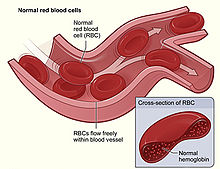
Hemolysis or haemolysis, also known by several other names, is the rupturing (lysis) of red blood cells (erythrocytes) and the release of their contents (cytoplasm) into surrounding fluid. Hemolysis may occur in vivo or in vitro.

<span class="mw-page-title-main">Anemia</span> Medical condition

Anemia or anaemia is a blood disorder in which the blood has a reduced ability to carry oxygen due to a lower than normal number of red blood cells, a reduction in the amount of hemoglobin or hemoglobin abnormalities. The name is derived from Ancient Greek: ἀναιμία anaimia, meaning 'lack of blood', from ἀν- an-, 'not' and αἷμα haima, 'blood'. When anemia comes on slowly, the symptoms are often vague, such as tiredness, weakness, shortness of breath, headaches, and a reduced ability to exercise. When anemia is acute, symptoms may include confusion, feeling like one is going to pass out, loss of consciousness, and increased thirst. Anemia must be significant before a person becomes noticeably pale. Symptoms of anemia depend on how quickly hemoglobin decreases. Additional symptoms may occur depending on the underlying cause. Preoperative anemia can increase the risk of needing a blood transfusion following surgery. Anemia can be temporary or long term and can range from mild to severe.

<span class="mw-page-title-main">Spherocytosis</span> Medical condition

Spherocytosis is the presence of spherocytes in the blood, i.e. erythrocytes that are sphere-shaped rather than bi-concave disk shaped as normal. Spherocytes are found in all hemolytic anemias to some degree. Hereditary spherocytosis and autoimmune hemolytic anemia are characterized by having only spherocytes.

<span class="mw-page-title-main">Haptoglobin</span> Mammalian protein found in Homo sapiens

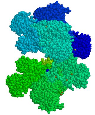
Haptoglobin is the protein that in humans is encoded by the HP gene. In blood plasma, haptoglobin binds with high affinity to free hemoglobin released from erythrocytes, and thereby inhibits its deleterious oxidative activity. Compared to Hp, hemopexin binds to free heme. The haptoglobin-hemoglobin complex will then be removed by the reticuloendothelial system.

<span class="mw-page-title-main">Glucose-6-phosphate dehydrogenase deficiency</span> Medical condition

Glucose-6-phosphate dehydrogenase deficiency (G6PDD), which is the most common enzyme deficiency worldwide, is an inborn error of metabolism that predisposes to red blood cell breakdown. Most of the time, those who are affected have no symptoms. Following a specific trigger, symptoms such as yellowish skin, dark urine, shortness of breath, and feeling tired may develop. Complications can include anemia and newborn jaundice. Some people never have symptoms.

Microangiopathic hemolytic anemia (MAHA) is a microangiopathic subgroup of hemolytic anemia caused by factors in the small blood vessels. It is identified by the finding of anemia and schistocytes on microscopy of the blood film.

<span class="mw-page-title-main">Hereditary spherocytosis</span> Medical condition

Hereditary spherocytosis (HS) is a congenital hemolytic disorder, wherein a genetic mutation coding for a structural membrane protein phenotype leads to a spherical shaping of erythrocytic cellular morphology. As erythrocytes are sphere-shaped (spherocytosis), rather than the normal biconcave disk-shaped, their morphology interferes with these cells' abilities to be flexible during circulation throughout the entirety of the body - arteries, arterioles, capillaries, venules, veins, and organs. This difference in shape also makes the red blood cells more prone to rupture under osmotic and/or mechanical stress. Cells with these dysfunctional proteins are degraded in the spleen, which leads to a shortage of erythrocytes resulting in hemolytic anemia.

<span class="mw-page-title-main">Paroxysmal nocturnal hemoglobinuria</span> Medical condition

Paroxysmal nocturnal hemoglobinuria (PNH) is a rare, acquired, life-threatening disease of the blood characterized by destruction of red blood cells by the complement system, a part of the body's innate immune system. This destructive process occurs due to deficiency of the red blood cell surface protein DAF, which normally inhibits such immune reactions. Since the complement cascade attacks the red blood cells within the blood vessels of the circulatory system, the red blood cell destruction (hemolysis) is considered an intravascular hemolytic anemia. There is ongoing research into other key features of the disease, such as the high incidence of venous blood clot formation. Research suggests that PNH thrombosis is caused by both the absence of GPI-anchored complement regulatory proteins on PNH platelets and the excessive consumption of nitric oxide (NO).

<span class="mw-page-title-main">Hemolytic disease of the newborn</span> Fetal and neonatal alloimmune blood condition

Hemolytic disease of the newborn, also known as hemolytic disease of the fetus and newborn, HDN, HDFN, or erythroblastosis fetalis, is an alloimmune condition that develops in a fetus at or around birth, when the IgG molecules produced by the mother pass through the placenta. Among these antibodies are some which attack antigens on the red blood cells in the fetal circulation, breaking down and destroying the cells. The fetus can develop reticulocytosis and anemia. The intensity of this fetal disease ranges from mild to very severe, and fetal death from heart failure can occur. When the disease is moderate or severe, many erythroblasts are present in the fetal blood, earning these forms of the disease the name erythroblastosis fetalis.

Autoimmune hemolytic anemia (AIHA) occurs when antibodies directed against the person's own red blood cells (RBCs) cause them to burst (lyse), leading to an insufficient number of oxygen-carrying red blood cells in the circulation. The lifetime of the RBCs is reduced from the normal 100–120 days to just a few days in serious cases. The intracellular components of the RBCs are released into the circulating blood and into tissues, leading to some of the characteristic symptoms of this condition. The antibodies are usually directed against high-incidence antigens, therefore they also commonly act on allogenic RBCs. AIHA is a relatively rare condition, with an incidence of 5–10 cases per 1 million persons per year in the warm-antibody type and 0.45 to 1.9 cases per 1 million persons per year in the cold antibody type. Autoimmune hemolysis might be a precursor of later onset systemic lupus erythematosus.

Paroxysmal cold hemoglobinuria (PCH) or Donath–Landsteiner hemolytic anemia (DLHA) is an autoimmune hemolytic anemia featured by complement-mediated intravascular hemolysis after cold exposure. It can present as an acute non-recurrent postinfectious event in children, or chronic relapsing episodes in adults with hematological malignancies or tertiary syphilis. Described by Julius Donath (1870–1950) and Karl Landsteiner (1868–1943) in 1904, PCH is one of the first clinical entities recognized as an autoimmune disorder.

In ABO hemolytic disease of the newborn maternal IgG antibodies with specificity for the ABO blood group system pass through the placenta to the fetal circulation where they can cause hemolysis of fetal red blood cells which can lead to fetal anemia and HDN. In contrast to Rh disease, about half of the cases of ABO HDN occur in a firstborn baby and ABO HDN does not become more severe after further pregnancies.

Cold agglutinin disease (CAD) is a rare autoimmune disease characterized by the presence of high concentrations of circulating cold sensitive antibodies, usually IgM and autoantibodies that are also active at temperatures below 30 °C (86 °F), directed against red blood cells, causing them to agglutinate and undergo lysis. It is a form of autoimmune hemolytic anemia, specifically one in which antibodies bind red blood cells only at low body temperatures, typically 28–31 °C.

<span class="mw-page-title-main">Hemoglobinemia</span> Abnormally increased hemoglobin in blood plasma

Hemoglobinemia is a medical condition in which there is an excess of hemoglobin in the blood plasma. This is an effect of intravascular hemolysis, in which hemoglobin separates from red blood cells, a form of anemia.

Congenital hemolytic anemia (CHA) is a diverse group of rare hereditary conditions marked by decreased life expectancy and premature removal of erythrocytes from blood flow. Defects in erythrocyte membrane proteins and red cell enzyme metabolism, as well as changes at the level of erythrocyte precursors, lead to impaired bone marrow erythropoiesis. CAH is distinguished by variable anemia, chronic extravascular hemolysis, decreased erythrocyte life span, splenomegaly, jaundice, biliary lithiasis, and iron overload. Immune-mediated mechanisms may play a role in the pathogenesis of these uncommon diseases, despite the paucity of data regarding the immune system's involvement in CHAs.

<span class="mw-page-title-main">Drug-induced autoimmune hemolytic anemia</span> Medical condition

Drug-induced autoimmune hemolytic anemia also known as Drug-induced immune hemolytic anemia (DIIHA) is a rare cause of hemolytic anemia. It is difficult to differentiate from other forms of anemia which can lead to delays in diagnosis and treatment. Many different types of antibiotics can cause DIIHA and discontinuing the offending medication is the first line of treatment. DIIHA has is estimated to affect one to two people per million worldwide.

This page is currently under construction.

Intravascular hemolysis describes hemolysis that happens mainly inside the vasculature. As a result, the contents of the red blood cell are released into the general circulation, leading to hemoglobinemia and increasing the risk of ensuing hyperbilirubinemia.

Cold sensitive antibodies (CSA) are antibodies sensitive to cold temperature. Some cold sensitive antibodies are pathological and can lead to blood disorder. These pathological cold sensitive antibodies include cold agglutinins, Donath–Landsteiner antibodies, and cryoglobulins which are the culprits of cold agglutinin disease, paroxysmal cold hemoglobinuria in the process of Donath–Landsteiner hemolytic anemia, and vasculitis, respectively.

Hemolytic jaundice, also known as prehepatic jaundice, is a type of jaundice arising from hemolysis or excessive destruction of red blood cells, when the byproduct bilirubin is not excreted by the hepatic cells quickly enough. Unless the patient is concurrently affected by hepatic dysfunctions or is experiencing hepatocellular damage, the liver does not contribute to this type of jaundice.

References

  1. "Red Blood Cells". US National Library of Medicine. Archived from the original on 2017-01-01.
  2. 1 2 3 4 5 6 7 8 9 10 11 12 13 14 15 16 Capriotti, Theresa (2016). Pathophysiology : introductory concepts and clinical perspectives. Frizzell, Joan Parker. Philadelphia. ISBN   978-0-8036-1571-7. OCLC   900626405.{{cite book}}: CS1 maint: location missing publisher (link)
  3. 1 2 Philadelphia, The Children's Hospital of (2014-03-30). "Hemolytic Anemia". chop.edu. Retrieved 2020-02-25.
  4. Trotman, BW (1991). "Pigment gallstone disease". Gastroenterology Clinics of North America. 20 (1): 111–26. doi:10.1016/S0889-8553(21)00536-7. ISSN   0889-8553. PMID   2022417.
  5. Machado, Roberto F.; Gladwin, Mark T. (2010). "Pulmonary Hypertension in Hemolytic Disorders". Chest. Elsevier BV. 137 (6): 30S–38S. doi:10.1378/chest.09-3057. ISSN   0012-3692. PMC   2882115 . PMID   20522578.
  6. Kahre, Tiina; Teder, Maris; Panov, Maarja; Metspalu, Andres (2004). "Severe CF manifestation with anaemia and failure to thrive in a 394delTT homozygous patient". Journal of Cystic Fibrosis. Elsevier BV. 3 (1): 58–60. doi: 10.1016/j.jcf.2003.12.009 . ISSN   1569-1993. PMID   15463888.
  7. Hypoproteinemia, Anemia, and Failure to Thrive in an Infant
  8. Levitt, Robert E.; Ostrow, Donald J. (1980). "Hemolytic Jaundice and Gallstones". Gastroenterology. 78 (4): 821–830. doi: 10.1016/0016-5085(80)90690-3 . PMID   7353766.
  9. 1 2 Schrier, Robert W.; Bansal, Shweta (2008). "Pulmonary Hypertension, Right Ventricular Failure, and Kidney". Clinical Journal of the American Society of Nephrology. 3 (5): 1232–1237. doi:10.2215/CJN.01960408. PMC   4571151 . PMID   18614776.
  10. McPhee, Stephen J.; Papadakis, Maxine A. (2008-10-31). CURRENT Medical Diagnosis and Treatment 2009. McGraw Hill Professional. ISBN   978-0-07-164137-1.
  11. Eisa, Mahmoud S.; Mohamed, Shehab F.; Ibrahim, Firyal; Shariff, Khalid; Sadik, Nagham; Nashwan, Abdulqadir; Yassin, Mohamed A. (2019-11-01). "Paroxysmal Nocturnal Hemoglobinuria with Glucose-6-Phosphate Dehydrogenase Deficiency: A Case Report and Review of the Literature". Case Reports in Oncology. 12 (3): 838–844. doi:10.1159/000503817. ISSN   1662-6575. PMC   6873095 . PMID   31762758.
  12. Grace, Rachael F.; Bianchi, Paola; van Beers, Eduard J.; Eber, Stefan W.; Glader, Bertil; Yaish, Hassan M.; Despotovic, Jenny M.; Rothman, Jennifer A.; Sharma, Mukta; McNaull, Melissa M.; Fermo, Elisa (2018-05-17). "Clinical spectrum of pyruvate kinase deficiency: data from the Pyruvate Kinase Deficiency Natural History Study". Blood. 131 (20): 2183–2192. doi: 10.1182/blood-2017-10-810796 . ISSN   0006-4971. PMID   29549173.
  13. Sharma, Sunita; Toppo, Anupa; Rath, B.; Harbhajanka, Aparna; Lalita Jyotsna, P. (September 2010). "Hemolytic Anemia as a Presenting Feature of Wilson's Disease: A Case Report". Indian Journal of Hematology and Blood Transfusion. 26 (3): 101–102. doi:10.1007/s12288-010-0034-2. PMC   3002091 . PMID   21886393.
  14. Khoury, Tawfik; Abu Rmeileh, Ayman; Kornspan, Jonathan David; Abel, Roy; Mizrahi, Meir; Nir-Paz, Ran (2015-02-19). "Mycoplasma pneumoniae Pneumonia Associated With Methemoglobinemia and Anemia: An Overlooked Association?". Open Forum Infectious Diseases. 2 (1): ofv022. doi:10.1093/ofid/ofv022. ISSN   2328-8957. PMC   4438901 . PMID   26034771.
  15. 1 2 Hill, Anita; Hill, Quentin A. (2018-11-30). "Autoimmune hemolytic anemia". Hematology. 2018 (1): 382–389. doi:10.1182/asheducation-2018.1.382. ISSN   1520-4391. PMC   6246027 . PMID   30504336.
  16. Privitera, G., & Meli, G. (2016). An unusual cause of anemia in cirrhosis: spur cell anemia, a case report with review of literature. Gastroenterology and hepatology from bed to bench, 9(4), 335–339.
  17. Li, Hao; Guan, Dongyao; Xu, Junqiang; Jin, Enhao; Sun, Shu (January 2020). "Atraumatic splenic rupture was attributed to intra-cystic haemorrhage and hypersplenism in a patient with cirrhosis and portal hypertension: A case report". SAGE Open Medical Case Reports. 8: 2050313X2090190. doi:10.1177/2050313X20901900. ISSN   2050-313X. PMC   6984417 . PMID   32047630.
  18. Zahid, Mohammad Faizan; Alsammak, Mohamed S. (2018). "Spurious thrombocytosis in the setting of hemolytic anemia and microcytosis secondary to extensive burn injury". Turkish Journal of Hematology. 35 (3): 205–206. doi:10.4274/tjh.2017.0466. PMC   6110454 . PMID   29391327.
  19. Brodsky, Robert A. (2014-10-30). "Paroxysmal nocturnal hemoglobinuria". Blood. 124 (18): 2804–2811. doi:10.1182/blood-2014-02-522128. ISSN   0006-4971. PMC   4215311 . PMID   25237200.
  20. Valentine, W N; Paglia, D E; Fink, K; Madokoro, G (October 1976). "Lead poisoning: association with hemolytic anemia, basophilic stippling, erythrocyte pyrimidine 5'-nucleotidase deficiency, and intraerythrocytic accumulation of pyrimidines". Journal of Clinical Investigation. 58 (4): 926–932. doi:10.1172/JCI108545. ISSN   0021-9738. PMC   333255 . PMID   965496.
  21. Correia, Nuno; Carvalho, Catarina; Friões, Fernando; Araújo, José P; Almeida, Jorge; Azevedo, Ana (2009-08-11). "Haemolytic anaemia secondary to arsenic poisoning: a case report". Cases Journal. 2: 7768. doi:10.4076/1757-1626-2-7768. ISSN   1757-1626. PMC   2769370 . PMID   19918480.
  22. Telford RD, Sly GJ, Hahn AG, Cunningham RB, Bryant C, Smith JA (January 2003). "Footstrike is the major cause of hemolysis during running". J. Appl. Physiol. 94 (1): 38–42. doi:10.1152/japplphysiol.00631.2001. PMID   12391035. S2CID   5750453.
  23. Lippi G, Schena F, Salvagno GL, Aloe R, Banfi G, Guidi GC (July 2012). "Foot-strike haemolysis after a 60-km ultramarathon". Blood Transfus. 10 (3): 377–383. doi:10.2450/2012.0167-11. PMC   3417738 . PMID   22682343.
  24. Wise, Donald Lee (2000). Biomaterials Engineering and Devices: Orthopedic, dental, and bone graft applications. Humana Press. ISBN   978-0-89603-859-2.[ permanent dead link ]
  25. Dhaliwal G, Cornett PA, Tierney LM., Jr Hemolytic anemia. Am Fam Physician. 2004;69:2599–606. [PubMed] [Google Scholar]
  26. 1 2 Stanley L Schrier, MD. William C Mentzer, MD, Jennifer S Tirnauer, MD (eds.). "Diagnosis of hemolytic anemia in the adult". UpToDate. Archived from the original on 2017-12-26. Retrieved 2019-05-04.
  27. "Intravascular hemolysis". eClinpath. Retrieved 2019-05-08.
  28. 1 2 3 Muller, Andre; Jacobsen, Helene; Healy, Edel; McMickan, Sinead; Istace, Fréderique; Blaude, Marie-Noëlle; Howden, Peter; Fleig, Helmut; Schulte, Agnes (2006). "Hazard classification of chemicals inducing haemolytic anaemia: An EU regulatory perspective" (PDF). Regulatory Toxicology and Pharmacology. Elsevier BV. 45 (3): 229–241. doi:10.1016/j.yrtph.2006.04.004. hdl:10029/5596. ISSN 0273-2300. PMID 16793184. Retrieved 2019-05-04
  29. 1 2 "Bilirubin and hemolytic anemia". eClinpath. Retrieved 2019-05-08.
  30. Rhodes, Carl E.; Varacallo, Matthew (2019-03-04). "Physiology, Oxygen Transport". NCBI Bookshelf. PMID 30855920. Retrieved 2019-05-04.
  31. Sokol RJ, Hewitt S, Stamps BK (June 1981). "Autoimmune haemolysis: an 18-year study of 865 cases referred to a regional transfusion centre". Br Med J (Clin Res Ed). 282(6281): 2023–7. doi:10.1136/bmj.282.6281.2023. PMC 1505955. PMID 6788179.
  32. 1 2 BRAUNSTEIN.EVAN (2019-05-03). "Overview of Hemolytic Anemia – Hematology and Oncology". Merck Manuals Professional Edition (in Latin). Retrieved 2019-05-05.
  33. "Hypersplenism: MedlinePlus Medical Encyclopedia". MedlinePlus. 2019-04-30. Retrieved 2019-05-08.
  34. 1 2 Bosman, Giel J. C. G. M. (2013). "Survival of red blood cells after transfusion: processes and consequences". Frontiers in Physiology. 4: 376. doi: 10.3389/fphys.2013.00376 . ISSN   1664-042X. PMC   3866658 . PMID   24391593.
  35. 1 2 Alaarg, Amr; Schiffelers, Raymond M.; van Solinge, Wouter W.; van Wijk, Richard (2013). "Red blood cell vesiculation in hereditary hemolytic anemia". Frontiers in Physiology. 4: 365. doi: 10.3389/fphys.2013.00365 . ISSN   1664-042X. PMC   3862113 . PMID   24379786.
  36. Kosenko, Elena A.; Tikhonova, Lyudmila A.; Montoliu, Carmina; Barreto, George E.; Aliev, Gjumrakch; Kaminsky, Yury G. (2018-01-05). "Metabolic Abnormalities of Erythrocytes as a Risk Factor for Alzheimer's Disease". Frontiers in Neuroscience. 11: 728. doi: 10.3389/fnins.2017.00728 . ISSN   1662-453X. PMC   5760569 . PMID   29354027.
  37. Kolb S, Vranckx R, Huisse MG, Michel JB, Meilhac O (July 2007). "The phosphatidylserine receptor mediates phagocytosis by vascular smooth muscle cells". The Journal of Pathology. 212 (3): 249–59. doi:10.1002/path.2190. PMID   17534843. S2CID   22923550.
  38. Bosman GJ, Willekens FL, Werre JM (2005). "Erythrocyte aging: a more than superficial resemblance to apoptosis?" (PDF). Cellular Physiology and Biochemistry. 16 (1–3): 1–8. doi: 10.1159/000087725 . hdl:2066/47441. PMID   16121027. S2CID   188974.
  39. Bratosin D, Mazurier J, Tissier JP, et al. (February 1998). "Cellular and molecular mechanisms of senescent erythrocyte phagocytosis by macrophages. A review". Biochimie. 80 (2): 173–95. doi:10.1016/S0300-9084(98)80024-2. PMID   9587675.
  40. 1 2 Hillman, Robert S.; Ault, Kenneth A.; Rinder, Henry (June 2005). Hematology in Clinical Practice. McGraw Hill Professional. ISBN   978-0-07-144035-6.
  41. Greer, John P. (2008). Wintrobe's Clinical Hematology. Lippincott Williams & Wilkins. ISBN   978-0-7817-6507-7.
  42. Bradencarter (21 January 2017). "What is Hemolytic Anemia?". thestackerz.com. Retrieved 21 January 2017.
  43. Boretti FS, Buehler PW, D'Agnillo F, et al. (August 2009). "Sequestration of extracellular hemoglobin within a haptoglobin complex decreases its hypertensive and oxidative effects in dogs and guinea pigs" (PDF). The Journal of Clinical Investigation. 119 (8): 2271–80. doi:10.1172/JCI39115. PMC   2719941 . PMID   19620788. Archived from the original (PDF) on 2022-02-11. Retrieved 2018-10-31.
  44. 1 2 Image by Mikael Häggström, MD. Source for conditions: Joe Chaffin (2017-02-27). "028: Who DAT? with Sue Johnson". Citing: C Feldman & J O'Connor.
  45. "Hemolytic Anemias, F. Spherocytosis". MedicalAssistantOnlinePrograms.org/. Archived from the original on 8 January 2015. Retrieved 6 November 2013.
  46. "Agios Announces FDA Approval of Pyrukynd (mitapivat) as First Disease-Modifying Therapy for Hemolytic Anemia in Adults with Pyruvate Kinase Deficiency" (Press release). Agios Pharmaceuticals. 17 February 2022. Retrieved 19 February 2022 via GlobeNewswire.
  47. Mary Anna Thrall, Dale C. Baker, E. Duane Lassen, Veterinary hematology and clinical chemistry, ISBN   0-7817-6850-0, 2004.
  48. Edward F. Gibbons, Barbara Susan Durrant, Jack Demarest, Conservation of endangered species in captivity: an interdisciplinary approach, page 324, 2005, ISBN   0-7914-1911-8
  49. Oliver A. Ryder, Zoological Society of San Diego, Rhinoceros biology and conservation, Zoological Society of San Diego, 1993, page 312, 335.
  50. Texas Monthly, Oct 1992, Vol. 20, No. 10, ISSN 0148-7736, page 98-100.
  51. Jutta Meister, ed. Catharine E. Bell, Encyclopedia of the world's zoos, Volume 3, page 1008, ISBN   1-57958-174-9, 2001.
  52. Kovalkovičová N, Sutiaková I, Pistl J, Sutiak V (2009). "Some food toxic for pets". Interdisciplinary Toxicology. 2 (3): 169–76. doi:10.2478/v10102-009-0012-4. PMC   2984110 . PMID   21217849.