Drug-induced autoimmune hemolytic anemia

Last updated
Drug-induced autoimmune heamolytic anemia
Other namesDrug-induced immune hemolytic anemia, DIIHA.
Warm autoimmune hemolytic anemia blood smear.jpg
Blood smear from a patient with warm autoimmune hemolytic anemia showing spherocytes, marked polychromasia and a nucleated red blood cell.
Specialty Hematology
Symptoms Fatigue, shortness of breath, dizziness, bloody urine, jaundice, weakness, and palpitations [1]
Complications Hemolysis, shock, ischemia, acute respiratory distress syndrome, disseminated intravascular coagulation, [1] and acute renal failure. [2]
Usual onsetHours to months of the initial drug exposure. [1]
Causes Antimicrobials, nonsteroidal anti-inflammatory drugs, antineoplastic drugs, and other drugs. [3]
Diagnostic method Blood tests, blood smear, and Direct antiglobulin testing [4]
Differential diagnosis Warm antibody autoimmune hemolytic anemia. [4]
TreatmentStopping the offending drug, blood transfusions, and thromboprophylaxis. [4]
FrequencyOne to two people per million worldwide. [1]

Drug-induced autoimmune hemolytic anemia also known as Drug-induced immune hemolytic anemia (DIIHA) is a rare cause of hemolytic anemia. It is difficult to differentiate from other forms of anemia which can lead to delays in diagnosis and treatment. Many different types of antibiotics can cause DIIHA and discontinuing the offending medication is the first line of treatment. DIIHA has is estimated to affect one to two people per million worldwide. [1]

Contents

In some cases, a drug can cause the immune system to mistakenly think the body's own red blood cells are dangerous, foreign substances. Antibodies then develop against the red blood cells. The antibodies attach to red blood cells and cause them to break down too early. It is known that more than 150 drugs can cause this type of hemolytic anemia. [5] The list includes:

Signs and symptoms

Initial symptoms of drug-induced autoimmune hemolytic anemia are typically vague and reflect mild, moderate, or severe anemia. Symptoms of DIIHA can manifest within hours to months of the initial drug exposure. [1] DIIHA ranges in severity from severe intravascular hemolysis to milder presentations of extravascular hemolysis. [7] Common symptoms of DIIHA are fatigue, shortness of breath, dizziness, bloody or dark urine, [1] weakness, and palpitations. DIIHA will occasionally present as hemoglobinuria with chills, however this is quite rare. [2] Patients with DIIHA may appear pale and have jaundice. Hepatomegaly, splenomegaly, and adenopathy have also been observed. [1]

When DIIHA is not recognized promptly it can have life-threatening complications such as hemolysis leading to shock, ischemia, acute respiratory distress syndrome, disseminated intravascular coagulation, [1] and acute renal failure. [2]

Causes

As of 2020 over 130 drugs have been reported to cause DIIHA. That number will continue to rise as new drugs are discovered. [1] Antimicrobials are responsible for 42% of DIIHA cases, making them the most common cause. Nonsteroid anti-inflammatory drugs cause about 15% of cases and antineoplastic drugs cause around 11%. [3]

Mechanism

The main mechanism of DIIHA is the development of antibodies. Drug-induced antibodies can be classified into two groups, drug-dependent antibodies and drug-independent autoantibodies. Drug-dependent antibodies are common in DIIHA. They require the offending drug to be present in order to bind and lyse cells. [8]

Drug-independent autoantibodies are a less common factor in DIIHA. Drug-independent autoantibodies are found in Drug-induced autoimmune hemolytic anemia because of beta-lactamase inhibitors and platinum-based chemotherapeutics. These autoantibodies can sometimes bind and react to red blood cells even in the absence of whatever drug triggered the anemia. [8]

Diagnosis

Drug-induced autoimmune hemolytic anemia causes a significant drop in hemoglobin and hematocrit. Occasionally DIIHA can present with mild leukocytosis. In its earlier stages patients with DIIHA will have low reticulocytes. As HIIHA progresses reticulocytes increase leading to an elevated mean corpuscular volume. Indirect bilirubin and Lactate dehydrogenase become elevated. LFTs occasionally become elevated. In some cases, a peripheral blood smear may show schistocytes, anisocytosis, polychromasia, or poikilocytosis. [7]

Direct antiglobulin testing is the only way to confirm DIIHA. Direct antiglobulin testing can determine if complement C3 antibody and/or immunoglobulin G is bound to the red blood cell membrane. [4] A positive direct antiglobulin test differentiates immune-mediated hemolytic anemia from a nonimmune-mediated cause. Other situations such as liver disease, post-transfusion or immunoglobulin administration, renal disease, and malignancy can cause a positive direct antiglobulin test. [1] If both complement C3 Antibodies and immunoglobulin G are positive or if only immunoglobulin G is positive then warm antibody autoimmune hemolytic anemia must be considered as a differential diagnosis. [4]

Treatment

An appropriate course of treatment for drug-induced autoimmune hemolytic anemia hasn't yet been established. Once DIIHA has been recognized, the patient must stop whatever drug caused the anemia in order to provide proper treatment. Patients should be given blood transfusions as needed. The use of thromboprophylaxis is encouraged because despite being anemic, patients are often hypercoagulable. [4] Although corticosteroids have been used to treat DIIHA it is difficult to differentiate how much effects corticosteroids actually have on DIIHA. [9]

If drug-independent autoantibodies are involved and stopping the offending agents results in no response then intravenous immunoglobulins and immunosuppressants such as rituximab, azathioprine, cyclophosphamide, cyclosporine, danazol, and mycophenolate can be used. [8] [10] Improvement is typically seen within a few weeks of cessation of the offending drug. [1]

See also

Related Research Articles

<span class="mw-page-title-main">Hemolysis</span> Rupturing of red blood cells and release of their contents

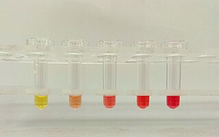
Hemolysis or haemolysis, also known by several other names, is the rupturing (lysis) of red blood cells (erythrocytes) and the release of their contents (cytoplasm) into surrounding fluid. Hemolysis may occur in vivo or in vitro.

<span class="mw-page-title-main">Hemolytic anemia</span> Medical condition

Hemolytic anemia or haemolytic anaemia is a form of anemia due to hemolysis, the abnormal breakdown of red blood cells (RBCs), either in the blood vessels or elsewhere in the human body (extravascular). This most commonly occurs within the spleen, but also can occur in the reticuloendothelial system or mechanically. Hemolytic anemia accounts for 5% of all existing anemias. It has numerous possible consequences, ranging from general symptoms to life-threatening systemic effects. The general classification of hemolytic anemia is either intrinsic or extrinsic. Treatment depends on the type and cause of the hemolytic anemia.

Serology is the scientific study of serum and other body fluids. In practice, the term usually refers to the diagnostic identification of antibodies in the serum. Such antibodies are typically formed in response to an infection, against other foreign proteins, or to one's own proteins. In either case, the procedure is simple.

<span class="mw-page-title-main">Hemolytic disease of the newborn</span> Fetal and neonatal alloimmune blood condition

Hemolytic disease of the newborn, also known as hemolytic disease of the fetus and newborn, HDN, HDFN, or erythroblastosis fetalis, is an alloimmune condition that develops in a fetus at or around birth, when the IgG molecules produced by the mother pass through the placenta. Among these antibodies are some which attack antigens on the red blood cells in the fetal circulation, breaking down and destroying the cells. The fetus can develop reticulocytosis and anemia. The intensity of this fetal disease ranges from mild to very severe, and fetal death from heart failure can occur. When the disease is moderate or severe, many erythroblasts are present in the fetal blood, earning these forms of the disease the name erythroblastosis fetalis.

The direct and indirect Coombs tests, also known as antiglobulin test (AGT), are blood tests used in immunohematology. The direct Coombs test detects antibodies that are stuck to the surface of the red blood cells. Since these antibodies sometimes destroy red blood cells they can cause anemia; this test can help clarify the condition. The indirect Coombs test detects antibodies that are floating freely in the blood. These antibodies could act against certain red blood cells; the test can be carried out to diagnose reactions to a blood transfusion.

Warm antibody autoimmune hemolytic anemia (WAIHA) is the most common form of autoimmune haemolytic anemia. About half of the cases are of unknown cause, with the other half attributable to a predisposing condition or medications being taken. Contrary to cold autoimmune hemolytic anemia which happens in cold temperature (28–31 °C), WAIHA happens at body temperature.

Autoimmune hemolytic anemia (AIHA) occurs when antibodies directed against the person's own red blood cells (RBCs) cause them to burst (lyse), leading to an insufficient number of oxygen-carrying red blood cells in the circulation. The lifetime of the RBCs is reduced from the normal 100–120 days to just a few days in serious cases. The intracellular components of the RBCs are released into the circulating blood and into tissues, leading to some of the characteristic symptoms of this condition. The antibodies are usually directed against high-incidence antigens, therefore they also commonly act on allogenic RBCs. AIHA is a relatively rare condition, with an incidence of 5–10 cases per 1 million persons per year in the warm-antibody type and 0.45 to 1.9 cases per 1 million persons per year in the cold antibody type. Autoimmune hemolysis might be a precursor of later onset systemic lupus erythematosus.

Paroxysmal cold hemoglobinuria (PCH) or Donath–Landsteiner hemolytic anemia (DLHA) is an autoimmune hemolytic anemia featured by complement-mediated intravascular hemolysis after cold exposure. It can present as an acute non-recurrent postinfectious event in children, or chronic relapsing episodes in adults with hematological malignancies or tertiary syphilis. Described by Julius Donath (1870–1950) and Karl Landsteiner (1868–1943) in 1904, PCH is one of the first clinical entities recognized as an autoimmune disorder.

In ABO hemolytic disease of the newborn maternal IgG antibodies with specificity for the ABO blood group system pass through the placenta to the fetal circulation where they can cause hemolysis of fetal red blood cells which can lead to fetal anemia and HDN. In contrast to Rh disease, about half of the cases of ABO HDN occur in a firstborn baby and ABO HDN does not become more severe after further pregnancies.

Cold agglutinin disease (CAD) is a rare autoimmune disease characterized by the presence of high concentrations of circulating cold sensitive antibodies, usually IgM and autoantibodies that are also active at temperatures below 30 °C (86 °F), directed against red blood cells, causing them to agglutinate and undergo lysis. It is a form of autoimmune hemolytic anemia, specifically one in which antibodies bind red blood cells only at low body temperatures, typically 28–31 °C.

Hemolytic disease of the newborn (anti-Kell1) is the second most common cause of severe hemolytic disease of the newborn (HDN) after Rh disease. Anti-Kell1 is becoming relatively more important as prevention of Rh disease is also becoming more effective.

Hemolytic disease of the newborn (anti-Rhc) can range from a mild to a severe disease. It is the third most common cause of severe HDN. Rh disease is the most common and hemolytic disease of the newborn (anti-Kell) is the second most common cause of severe HDN. It occurs more commonly in women who are Rh D negative.

Hemolytic disease of the newborn (anti-RhE) is caused by the anti-RhE antibody of the Rh blood group system. The anti-RhE antibody can be naturally occurring, or arise following immune sensitization after a blood transfusion or pregnancy.

Acquired hemolytic anemia can be divided into immune and non-immune mediated forms of hemolytic anemia.

This page is currently under construction.

<span class="mw-page-title-main">Red cell agglutination</span> Clumping of red blood cells

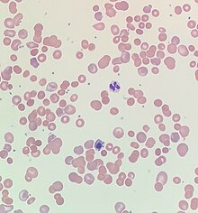
In hematology, red cell agglutination or autoagglutination is a phenomenon in which red blood cells clump together, forming aggregates. It is caused by the surface of the red cells being coated with antibodies. This often occurs in cold agglutinin disease, a type of autoimmune hemolytic anemia in which people produce antibodies that bind to their red blood cells at cold temperatures and destroy them. People may develop cold agglutinins from lymphoproliferative disorders, from infection with Mycoplasma pneumoniae or Epstein–Barr virus, or idiopathically. Red cell agglutination can also occur in paroxysmal nocturnal hemoglobinuria and warm autoimmune hemolytic anemia. In cases of red cell agglutination, the direct antiglobulin test can be used to demonstrate the presence of antibodies bound to the red cells.

Cold autoimmune hemolytic anemia caused by cold-reacting antibodies. Autoantibodies that bind to the erythrocyte membrane leading to premature erythrocyte destruction (hemolysis) characterize autoimmune hemolytic anemia.

Cold sensitive antibodies (CSA) are antibodies sensitive to cold temperature. Some cold sensitive antibodies are pathological and can lead to blood disorder. These pathological cold sensitive antibodies include cold agglutinins, Donath–Landsteiner antibodies, and cryoglobulins which are the culprits of cold agglutinin disease, paroxysmal cold hemoglobinuria in the process of Donath–Landsteiner hemolytic anemia, and vasculitis, respectively.

Mixed autoimmune hemolytic anemia (MAIHA) is a type of autoimmune hemolytic anemia which combines the features of cold sensitive antibody-induced diseases and warm autoimmune hemolytic anemia. The work-up for diagnosis is complex and the condition can be over-diagnosed.

Hemolytic jaundice, also known as prehepatic jaundice, is a type of jaundice arising from hemolysis or excessive destruction of red blood cells, when the byproduct bilirubin is not excreted by the hepatic cells quickly enough. Unless the patient is concurrently affected by hepatic dysfunctions or is experiencing hepatocellular damage, the liver does not contribute to this type of jaundice.

References

  1. 1 2 3 4 5 6 7 8 9 10 11 12 Chan Gomez, Janet; Saleem, Tabinda; Snyder, Samantha; Joseph, Maria; Kanderi, Tejaswi (June 17, 2020). "Drug-Induced Immune Hemolytic Anemia due to Amoxicillin-Clavulanate: A Case Report and Review". Cureus. 12 (6). Cureus, Inc.: e8666. doi: 10.7759/cureus.8666 . ISSN   2168-8184. PMC   7370667 . PMID   32699666.
  2. 1 2 3 Chen, Fei; Zhan, Zhuying (2014). "Severe drug-induced immune haemolytic anaemia due to ceftazidime". Blood Transfusion. 12 (3). Edizioni SIMTI: 435–437. doi: 10.2450/2014.0237-13 . PMC   4111830 . PMID   24887228.
  3. 1 2 Garratty, George (January 1, 2009). "Drug-induced immune hemolytic anemia". Hematology. 2009 (1). American Society of Hematology: 73–79. doi: 10.1182/asheducation-2009.1.73 . ISSN   1520-4391. PMID   20008184 . Retrieved November 21, 2023.
  4. 1 2 3 4 5 6 Hill, Quentin A.; Stamps, Robert; Massey, Edwin; Grainger, John D.; Provan, Drew; Hill, Anita (2017). "Guidelines on the management of drug-induced immune and secondary autoimmune, haemolytic anaemia". British Journal of Haematology. 177 (2). Wiley: 208–220. doi: 10.1111/bjh.14654 . ISSN   0007-1048. PMID   28369704.
  5. Barcellini, Wilma (2015-10-01). "Immune Hemolysis: Diagnosis and Treatment Recommendations". Seminars in Hematology. Anemia in Clinical Practice. 52 (4): 304–312. doi:10.1053/j.seminhematol.2015.05.001. ISSN   0037-1963. PMID   26404442.
  6. MedlinePlus Encyclopedia : Drug-induced immune hemolytic anemia
  7. 1 2 Karunathilaka, H. G. C. S.; Chandrasiri, D. P.; Ranasinghe, P.; Ratnamalala, V.; Fernando, A. H. N. (March 31, 2020). "Co-Amoxiclav Induced Immune Haemolytic Anaemia: A Case Report". Case Reports in Hematology. 2020. Hindawi Limited: 1–3. doi: 10.1155/2020/9841097 . ISSN   2090-6560. PMC   7150724 . PMID   32292611.
  8. 1 2 3 Leicht, Hans Benno; Weinig, Elke; Mayer, Beate; Viebahn, Johannes; Geier, Andreas; Rau, Monika (October 25, 2018). "Ceftriaxone-induced hemolytic anemia with severe renal failure: a case report and review of literature". BMC Pharmacology and Toxicology. 19 (1). Springer Science and Business Media LLC: 67. doi: 10.1186/s40360-018-0257-7 . ISSN   2050-6511. PMC   6203207 . PMID   30359322.
  9. Garbe, Edeltraut; Andersohn, Frank; Bronder, Elisabeth; Klimpel, Andreas; Thomae, Michael; Schrezenmeier, Hubert; Hildebrandt, Martin; Späth-Schwalbe, Ernst; Grüneisen, Andreas; Mayer, Beate; Salama, Abdulgabar; Kurtal, Hanife (July 12, 2011). "Drug induced immune haemolytic anaemia in the Berlin Case-Control Surveillance Study". British Journal of Haematology. 154 (5). Wiley: 644–653. doi: 10.1111/j.1365-2141.2011.08784.x . ISSN   0007-1048. PMID   21749359.
  10. Wu, Yuanjun; Wu, Yong; Ji, Yanli; Liang, Jiajie; He, Ziyi; Liu, Yanhui; Tang, Li; Guo, Ganping (August 12, 2021). "Case Report: Drug-Induced Immune Haemolytic Anaemia Caused by Cefoperazone-Tazobactam/ Sulbactam Combination Therapy". Frontiers in Medicine. 8. Frontiers Media SA. doi: 10.3389/fmed.2021.697192 . ISSN   2296-858X. PMC   8415779 . PMID   34485334.

Further reading