Holocentric chromosome

Last updated

Holocentric chromosomes are chromosomes that possess multiple kinetochores along their length rather than the single centromere typical of other chromosomes. [1] They were first described in cytogenetic experiments in 1935. Since this first observation, the term holocentric chromosome has referred to chromosomes that: i) lack the primary constriction corresponding to the centromere observed in monocentric chromosomes; [2] and ii) possess multiple kinetochores dispersed along the entire chromosomal axis, such that microtubules bind to the chromosome along its entire length and move broadside to the pole from the metaphase plate. [3] Holocentric chromosomes are also termed holokinetic, because, during cell division, the sister chromatids move apart in parallel and do not form the classical V-shaped figures typical of monocentric chromosomes. [4] [5] [6]

Contents

Holocentric chromosomes have evolved several times during both animal and plant evolution, and are currently reported in about eight hundred diverse species, including plants, insects, arachnids, and nematodes. [7] [8] As a consequence of their diffuse kinetochores, holocentric chromosomes may stabilize chromosomal fragments created by accidental double-strand breaks, preventing loss of the fragments and favouring karyotype rearrangements. [9] [10] However, holocentric chromosomes may also present limitations to crossing over, causing a restriction of the number of chiasma in bivalents, [11] and may cause a restructuring of meiotic divisions resulting in an "inverted" meiosis. [12]

Evolution

Holocentric chromosomes were described for the first time by Franz Schrader in 1935, who identified chromosomes with a diffuse kinetochore (or with a diffuse kinetochore activity), making these chromosomes able to bind to microtubules along their entire length rather than at a single primary constriction, as is typical of chromosomes with centromeres. In recent decades, several studies determined that this behaviour during mitosis can be observed in holocentric/holokinetic chromosomes but also in polykinetic chromosomes, which contain numerous (but discrete) microtubule-binding sites; even so, the term “holocentric/holokinetic” is still commonly used to refer to both processes. [1] [5] [7]

Top box: In monocentric chromosomes (left), kinetochores (red circles) are located at a single chromosomal primary constriction (centromere) during metaphase (M). During anaphase (A), sister chromatids move towards the poles after their attachment to microtubules, and V-shaped structures can be observed resulting from the passive movement of the chromosomal arms. In holocentric chromosomes (right), a chromosome-wide kinetochore is present (red lines) and no primary constriction is present during metaphase (M). During anaphase (A), holocentric chromatids move towards poles as linear bars, in parallel.

Bottom box: If a chromosomal breakage occurs in a monocentric chromosome, acentric chromosome fragments cannot be attached to microtubules during metaphase (M) and they are subsequently lost during anaphase (A). On the contrary, chromosomal breakage of a holocentric chromosome results in chromosomal fragments that retain kinetic activity due to the chromosome-wide centromere extension and can be properly inherited. Figure holocentric chromosomes.jpg
Top box: In monocentric chromosomes (left), kinetochores (red circles) are located at a single chromosomal primary constriction (centromere) during metaphase (M). During anaphase (A), sister chromatids move towards the poles after their attachment to microtubules, and V-shaped structures can be observed resulting from the passive movement of the chromosomal arms. In holocentric chromosomes (right), a chromosome-wide kinetochore is present (red lines) and no primary constriction is present during metaphase (M). During anaphase (A), holocentric chromatids move towards poles as linear bars, in parallel.

Bottom box: If a chromosomal breakage occurs in a monocentric chromosome, acentric chromosome fragments cannot be attached to microtubules during metaphase (M) and they are subsequently lost during anaphase (A). On the contrary, chromosomal breakage of a holocentric chromosome results in chromosomal fragments that retain kinetic activity due to the chromosome-wide centromere extension and can be properly inherited.

Before molecular methods became available, the presence of holocentric chromosomes was evaluated mostly using cytology and, considering that many species are difficult to study cytologially, it can be surmised that the true presence of holocentrism may be underestimated. In addition, there are several taxa whose chromosomes are still uncharacterized, though their phylogenetic position suggests that they should have holocentric chromosomes. [7] [13] The presence of holocentric chromosomes has been up till now assessed in about 800 species, including insects, plants, arachnids, and nematodes, [1] [5] [7] suggesting that, in general, holocentric chromosomes probably originated by convergent evolution from ancestors possessing monocentric chromosomes. Interesting exceptions are represented by insects belonging to the Oligoneoptera and Neoptera, whose monocentric chromosomes probably evolved from an holocentric ancestor in two different and independent events. [7] Evidence of convergent evolution suggests that holocentrism is adaptive, but the specific conditions under which holocentrism provides a selective advantage seem to be diverse for different taxa. [7] [14] Indeed, in phytophagous insects (such as aphids and lepidopterans), holocentrism could be an evolved defense against the production by plants of compounds able to induce chromosomal breakages (clastogens), whereas in other cases, holocentrism may constitute a defense against DNA damage resulting from desiccation and/or other chromosome-breaking factors. [14]

Despite these differences, holocentric chromosomes present intrinsic benefits since chromosomal mutations, such as fissions and fusions, are potentially neutral in holocentric chromosomes with respect to monocentric ones. However, the hypothesis of holocentrism as an anticlastogenic adaptation needs more systematic testing, including both controlled laboratory experiments and field studies across clastogenic gradients and large-scale phylogenetic analyses. [8] At the same time, Nagaki et al. [15] proposed that holocentrism can be easily acquired during plant and animal evolution by a slight difference in the kinetochore origin. In particular, they hypothesized that if the direction of kinetochore origin turns by 90° and occurs along the chromosome axes up to the telomeric regions, it is possible to “generate” holocentric chromosomes without any further step.

Structure

A detailed molecular analysis of the structure of holocentric chromosomes is currently available only for the nematode Caenorhabditis elegans , [16] [17] whereas the presence of true holokinetic nature has also been confirmed in other taxa by the evidence that experimentally induced chromosome fragments continue to attach to the spindle and segregate correctly. [3] For most of the species, data about holocentrism are related to the analysis of the behaviour of chromosomes during anaphase migration since holocentric sister chromatids migrate in parallel to the spindle poles, in contrast to monocentric ones in which pulling forces are exerted on a single chromosomal point and chromosome arms trail behind. As a consequence, chromatids of holocentric chromosomes move apart in parallel and do not form the classical V-shaped figures typical of monocentric ones. [4] Moreover, if a holocentric chromosome is fragmented (for instance by X-ray irradiation), each fragment retains centromere activity and can segregate properly to the poles.


In different organisms

Arthropods

Among arthropods, the presence of holocentric chromosome has been reported in different species belonging to insects (Odonata, Zoraptera, Dermaptera, Psocoptera, Phthiraptera, Thysanoptera, Hemiptera, Trichoptera and Lepidoptera), scorpions (Buthoidea), mites and ticks of the superorder Acariformes and genus Rhipicephalus (Ixodidae), spiders (Dysderidae and Segestridae), [7] [14] millipedes [18] and centipedes. [18] Despite this widespread occurrence, most of the currently available data on holocentrism is related to aphid and lepidopteran species. [7] [5] In aphids, holocentric chromosomes have been deeply studied and their ability to stabilize chromosomal fragments has been associated to their phytophagous life style. Indeed, several plants produce chemicals able to induce DNA damage to pest insects. Nicotine, for instance, is a naturally occurring alkaloid found primarily in members of the solanaceous plant family (including Nicotiana tabacum ) that can cause replication fork stress resulting in various forms of DNA damage, including chromosomal fragmentations. [19] [20] Similar effects have been also reported by other plant-produced molecules, such as caffeine and ethanol. [19] [20] In view of their ability to favour the inheritance of chromosomal fragments, holocentrism has been associated to recurrent changes in the karyotypes of some aphid species and in particular in the peach potato aphid Myzus persicae , where both inter- and intra-individual rearranged karyotypes have been also observed. [21] [22] Aphids also possess a constitutive expression of the telomerase coding gene so that they can initiate a de novo synthesis of telomere sequences at internal breakpoints, resulting in the stabilization of chromosomal fragments. [23] [24] Among non-polyploid animals, Lepidoptera exhibit the highest variance in chromosome number between species within a genus and notable levels of interspecific and intraspecific karyotype variability. [12] [25] [26] Lepidoptera indeed tolerate chromosomal variations in view of their holokinetic chromosomes, which facilitate the successful inheritance of novel fission or fusion fragments. As a consequence, Lepidoptera can avoid the deleterious consequences of large-scale chromosomal fission and fusion. [12] [25] [26] Nevertheless, they can sometimes tolerate heterozygosity for multiple rearrangements in hybrids between population with differences in their karyotype, raising questions about additional mechanisms that rescue fertility in chromosomal hybrids. In Lepidoptera, therefore, chromosome evolution is believed to play a role in reinforcing speciation. [12] Comparing the genomes of lepidopteran species it has been also possible to analyse the effect of holocentrism in terms of rate of fixed chromosomal rearrangements. This approach evidenced in Lepidoptera two chromosome breaks per megabase of DNA per Million of years: a rate that is much higher than what observed in Drosophila and it is a direct consequence of the holocentric nature of the lepidopteran genomes. [27] [28] At a structural level, insect holocentric chromosomes have not been studied in details, but it is interesting to underline the absence of homologues of CENP-C and CENP-A, previously considered essential for kinetochore functioning in eukaryotes. [29]

Nematodes

The best known group of holocentric species can be found in the Secernentea class of the nematodes, which includes C. elegans. [16] [17] other nematodes are usually described as holocentric because of their phylogenetic relationship to C. elegans, but real karyotypic evidences are scarce or controversial. [30] [31] [32] Nematode development is typically characterized by fixed lineages and a single inappropriate cell death, therefore, it has been suggested that holocentrism could avoid the disastrous consequences of unrepaired chromosome breakage events. [33] The availability of several molecular and genomic resources allowed a detailed characterization of C. elegans holocentric chromosomes and in particular the structure of the kinetochore has been molecularly dissected. [34] [35] Current data suggest that C. elegans kinetochores form paired lines or plates on opposite faces of condensed mitotic chromosomes, [35] where each line represents the diffuse kinetochore of a single chromatid. transmission electron microscopy of C. elegans chromosomes revealed that the kinetochore has a trilaminar structure very similar to that observed in monocentric chromosomes [35] [36] More than 30 different proteins have been identified as components of the C. elegans kinetochore and half of them was already known as functioning in the kinetochores of monocentric chromosomes. Among these, highly studied proteins include homologues of CENP-C and CENP-A, which are highly conserved structural component of the kinetochore in eukaryotes. [36] [37] Contrarily to what generally observed in monocentric chromosomes, in holocentric ones the preferential localization of centromeres within heterochromatic areas is missing together with the presence of specific DNA sequences that in C. elegans are not required for the assembly of a functional kinetochore. [36] [37] In this regard, it has been observed that holocentric chromosomes of nematodes are unique because they have a large number of satellites scattered throughout their genome, whereas no scattered satellites are found in the monocentric chromosomes of the nematode trichinella spiralis. [38] Interestingly, these satellite DNAs are not conserved in their sequences among species suggesting that highly repetitive DNAs may facilitate the formation of kinetochores in view of their repetitiveness rather than for their specific sequence. [38] The absence of a localized centromere prompted several studies to identify proteins that are involved in the sister chromatid cohesion assessing that it is accomplished by a separate complex of conserved proteins, termed cohesin, that is composed of the core subunits Scc3, Smc1, Smc3 and Scc1. Interestingly, they play the same function in organisms with monocentric chromosomes with an exception related to the subunit Scc1, whose gene in addition to the Scc1 orthologue present three additional paralogous genes. [36] [37]

Plants

In plants, holocentric chromosomes have been found in zygnematophycean algae, [39] in the genera Myristica (Myristicaceae), Chionographis (Melanthiaceae), Cuscuta (Convolvulaceae) and Droseraceae, [40] [41] [42] in the species Trithuria submersa (Hydatellaceae), Prionium serratum (Thurniaceae), [43] [44] and, among higher plants, in many genera belonging to the families Cyperaceae and Juncaceae, including the snowy woodrush Luzula nivea (Juncaceae), the most well-studied holocentric plant. [45] [46] In Luzula spp., the centromeric activity is localized simultaneously at several evenly spaced sites along each chromosome and chromosomes can be fragmented naturally or by irradiation into smaller (but viable) chromosomes. [47] [48] The presence of rearranged karyotypes does not affect fitness, as assessed by studies reporting that Luzula hybrids with parents possessing smaller and larger chromosomes showed the smaller chromosomes aligned and paired with the larger ones. [47] Similarly, in plants belonging to the genus Carex , differentiation of the karyotype has been demonstrated to correlate with genetic divergence within species, [49] among populations within species, [50] and within populations, [51] suggesting that, as previously reported in the Lepidoptera, [12] holocentric chromosome rearrangements contribute to genetic differentiation at different evolutionary scales in Carex evolution and speciation. In plants it has also been suggested that the diffuse kinetochore of holocentric chromosomes may suppress the meiotic drive of centromeric repeats and its negative consequences. [46] In particular, the expansions (or contractions) of centromeric repeats may lead to a larger (or smaller) kinetochore, which attracts more (or fewer) microtubules during meiosis. [17] [46] This hypothesis, which correlates the presence of holocentric chromosomes with centromere drive suppression, is very intriguing but it only explains the evolution of chromosomal holocentrism in meiosis and not in mitosis, a fact which is not trivial considering that some species with holocentric chromosomes may present a restriction of kinetochore activity during meiosis. [17] [52] Similarly to what was previously reported for C. elegans, in L. elegans centromeres are not made by centromere-associated retrotransposons nor centromere-associated satellite DNAs, but cenH3 proteins seem to be associated with a centromere-specific chromatin folding rather than with specific centromeric DNA sequences. [53] Conservation of elements between mono- and holocentric chromosomes is not limited to centromeric proteins, but it is also extended to epigenetic marks. Indeed, the cell cycle-dependent phosphorylation of serine 10 or serine 28 of H3 (that is typically enriched in pericentric regions of monocentric plant chromosomes) occurs uniformly along the Luzula chromosomes. [53] As previously described in aphids, L. elegans possesses a rapid and efficient de novo telomere formation based on a telomerase-mediated healing process that is active immediately after chromosomal damage by irradiation of chromosomes. [54] Newly formed telomere repeats were cytologially detectable 21 days after irradiation in about 50% of cases, with a complete healing of telomeres after three months favouring the fragment stabilization and karyotype fixation. [54]

Meiosis

In the late 19th century, van Beneden (1883) and Boveri (1890) described meiosis for the first time through a careful observation of germ cell formation in the nematode Ascaris . These observations, together with several further analyses, evidenced that canonical meiosis consists of a first division (called reductional division) that involves the segregation of chromosomal homologs resulting in the reduction of chromosome number, followed by a second division (called equational division) that involves the segregation of sister chromatids. A general rule for meiosis is therefore: first homologs, then sisters.

Schematic comparison of the chromosomal separation occurring during the first meiotic division in standard and inverted meiosis Inverted meiosis.jpg
Schematic comparison of the chromosomal separation occurring during the first meiotic division in standard and inverted meiosis

However, the understanding of the reductional division in meiosis of Ascaris spp. has been obtained by studying the holocentric chromosomes which, in many other taxa, follow a reverse order of meiotic division. [12] Indeed, as reported in several nematodes, in insects belonging to the Hemiptera and Lepidoptera, [55] [56] in mites, [57] and in some flowering plants, [8] species with holocentric chromosomes generally present an inverted meiotic sequence, in which segregation of homologs is postponed until the second meiotic division.

Furthermore, in most cases of inverted meiosis the absence of a canonical kinetochore structure has been observed, together with a restriction of the kinetic activity to the chromosomal ends. [12] [55] [56] These changes are related to the peculiar cohesion occurring in tetrads of the holocentric homologous chromosomes during meiosis that impose obstacles to the releases of chromosomes involved in multiple crossing over events. [55] [56] [57] In the holocentric chromosomes of C. elegans female meiosis, [58] this problem is circumvented by restricting crossing over to form only a single chiasma per bivalent and triggering the redistribution of kinetochore proteins along the bivalent axis, forming meiosis-specific cup-like structures that uniformly coat each half-bivalent but are excluded from the mid-bivalent region. [58] During anaphase I, C. elegans homologous chromosomes are segregated to the poles by microtubule-pushing from the mid-bivalent regions towards the poles. [58]

In contrast to C. elegans, other organisms with holocentric chromosomes, including both plants and insects, [12] [55] [56] circumvent this problem by segregating sister chromatids during meiosis I, leading to the term inverted meiosis, in which the order of reductional and equational division is inverted with respect to canonical meiosis. In this case therefore the separation of homologous chromosomes follows rather than precedes the segregation of sister chromatids. However, in order to have a successful inverted meiosis, it is necessary that a bipolar orientation of sister kinetochores occurs, together with their attachment to microtubules from opposite spindle poles in meiosis I. This allows the segregation of sister chromatids to opposite poles in anaphase I (equational division), but requires a mechanism to align and pair homologous chromosomes during the second meiotic division. [55] [56] [57] Interestingly, the presence of inverted meiosis can also facilitate proper chromosome segregation in hybrids from parental species with differences in their karyotypes or derived by populations with rearranged karyotypes, allowing rescue of the fertility and viability of hybrids and promoting a fast karyotype evolution and possibly chromosomal speciation, as reported in the Lepidoptera. [12]

Related Research Articles

<span class="mw-page-title-main">Chromosome</span> DNA molecule containing genetic material of a cell

A chromosome is a package of DNA with part or all of the genetic material of an organism. In most chromosomes, the very long thin DNA fibers are coated with nucleosome forming packaging proteins; in eukaryotic cells the most important of these proteins are the histones. These proteins, aided by chaperone proteins, bind to and condense the DNA molecule to maintain its integrity. These chromosomes display a complex three-dimensional structure, which plays a significant role in transcriptional regulation.

<span class="mw-page-title-main">Centromere</span> Specialized DNA sequence of a chromosome that links a pair of sister chromatids

The centromere links a pair of sister chromatids together during cell division. This constricted region of chromosome connects the sister chromatids, creating a short arm (p) and a long arm (q) on the chromatids. During mitosis, spindle fibers attach to the centromere via the kinetochore.

<span class="mw-page-title-main">Meiosis</span> Cell division producing haploid gametes

Meiosis is a special type of cell division of germ cells and apicomplexans in sexually-reproducing organisms that produces the gametes, such as sperm or egg cells. It involves two rounds of division that ultimately result in four cells with only one copy of each chromosome (haploid). Additionally, prior to the division, genetic material from the paternal and maternal copies of each chromosome is crossed over, creating new combinations of code on each chromosome. Later on, during fertilisation, the haploid cells produced by meiosis from a male and a female will fuse to create a cell with two copies of each chromosome again, the zygote.

<span class="mw-page-title-main">Mitosis</span> Process in which chromosomes are replicated and separated into two new identical nuclei

Mitosis is a part of the cell cycle in which replicated chromosomes are separated into two new nuclei. Cell division by mitosis is an equational division which gives rise to genetically identical cells in which the total number of chromosomes is maintained. Mitosis is preceded by the S phase of interphase and is followed by telophase and cytokinesis; which divides the cytoplasm, organelles and cell membrane of one cell into two new cells containing roughly equal shares of these cellular components. The different stages of mitosis altogether define the mitotic phase of a cell cycle—the division of the mother cell into two daughter cells genetically identical to each other.

<span class="mw-page-title-main">Cell division</span> Process by which living cells divide

Cell division is the process by which a parent cell divides into two daughter cells. Cell division usually occurs as part of a larger cell cycle in which the cell grows and replicates its chromosome(s) before dividing. In eukaryotes, there are two distinct types of cell division: a vegetative division (mitosis), producing daughter cells genetically identical to the parent cell, and a cell division that produces haploid gametes for sexual reproduction (meiosis), reducing the number of chromosomes from two of each type in the diploid parent cell to one of each type in the daughter cells. In Mitosis is a part of the cell cycle, in which, replicated chromosomes are separated into two new nuclei. Cell division gives rise to genetically identical cells in which the total number of chromosomes is maintained. In general, mitosis is preceded by the S stage of interphase and is followed by telophase and cytokinesis; which divides the cytoplasm, organelles, and cell membrane of one cell into two new cells containing roughly equal shares of these cellular components. The different stages of mitosis all together define the M phase of an animal cell cycle—the division of the mother cell into two genetically identical daughter cells. To ensure proper progression through the cell cycle, DNA damage is detected and repaired at various checkpoints throughout the cycle. These checkpoints can halt progression through the cell cycle by inhibiting certain cyclin-CDK complexes. Meiosis undergoes two divisions resulting in four haploid daughter cells. Homologous chromosomes are separated in the first division of meiosis, such that each daughter cell has one copy of each chromosome. These chromosomes have already been replicated and have two sister chromatids which are then separated during the second division of meiosis. Both of these cell division cycles are used in the process of sexual reproduction at some point in their life cycle. Both are believed to be present in the last eukaryotic common ancestor.

<span class="mw-page-title-main">Prophase</span> First phase of cell division in both mitosis and meiosis

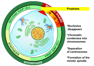
Prophase is the first stage of cell division in both mitosis and meiosis. Beginning after interphase, DNA has already been replicated when the cell enters prophase. The main occurrences in prophase are the condensation of the chromatin reticulum and the disappearance of the nucleolus.

<span class="mw-page-title-main">Homologous chromosome</span> Chromosomes that pair in fertilization

A couple of homologous chromosomes, or homologs, are a set of one maternal and one paternal chromosome that pair up with each other inside a cell during fertilization. Homologs have the same genes in the same loci, where they provide points along each chromosome that enable a pair of chromosomes to align correctly with each other before separating during meiosis. This is the basis for Mendelian inheritance, which characterizes inheritance patterns of genetic material from an organism to its offspring parent developmental cell at the given time and area.

<span class="mw-page-title-main">Nondisjunction</span> Failure to separate properly during cell division

Nondisjunction is the failure of homologous chromosomes or sister chromatids to separate properly during cell division (mitosis/meiosis). There are three forms of nondisjunction: failure of a pair of homologous chromosomes to separate in meiosis I, failure of sister chromatids to separate during meiosis II, and failure of sister chromatids to separate during mitosis. Nondisjunction results in daughter cells with abnormal chromosome numbers (aneuploidy).

<span class="mw-page-title-main">Kinetochore</span> Protein complex that allows microtubules to attach to chromosomes during cell division

A kinetochore is a disc-shaped protein structure associated with duplicated chromatids in eukaryotic cells where the spindle fibers attach during cell division to pull sister chromatids apart. The kinetochore assembles on the centromere and links the chromosome to microtubule polymers from the mitotic spindle during mitosis and meiosis. The term kinetochore was first used in a footnote in a 1934 Cytology book by Lester W. Sharp and commonly accepted in 1936. Sharp's footnote reads: "The convenient term kinetochore has been suggested to the author by J. A. Moore", likely referring to John Alexander Moore who had joined Columbia University as a freshman in 1932.

<span class="mw-page-title-main">Chromosomal inversion</span> Chromosome rearrangement in which a segment of a chromosome is reversed

An inversion is a chromosome rearrangement in which a segment of a chromosome becomes inverted within its original position. An inversion occurs when a chromosome undergoes a two breaks within the chromosomal arm, and the segment between the two breaks inserts itself in the opposite direction in the same chromosome arm. The breakpoints of inversions often happen in regions of repetitive nucleotides, and the regions may be reused in other inversions. Chromosomal segments in inversions can be as small as 100 kilobases or as large as 100 megabases. The number of genes captured by an inversion can range from a handful of genes to hundreds of genes. Inversions can happen either through ectopic recombination, chromosomal breakage and repair, or non-homologous end joining.

<span class="mw-page-title-main">Isochromosome</span>

An isochromosome is an unbalanced structural abnormality in which the arms of the chromosome are mirror images of each other. The chromosome consists of two copies of either the long (q) arm or the short (p) arm because isochromosome formation is equivalent to a simultaneous duplication and deletion of genetic material. Consequently, there is partial trisomy of the genes present in the isochromosome and partial monosomy of the genes in the lost arm.

<span class="mw-page-title-main">Synapsis</span> Biological phenomenon in meiosis

Synapsis is the pairing of two chromosomes that occurs during meiosis. It allows matching-up of homologous pairs prior to their segregation, and possible chromosomal crossover between them. Synapsis takes place during prophase I of meiosis. When homologous chromosomes synapse, their ends are first attached to the nuclear envelope. These end-membrane complexes then migrate, assisted by the extranuclear cytoskeleton, until matching ends have been paired. Then the intervening regions of the chromosome are brought together, and may be connected by a protein-RNA complex called the synaptonemal complex. During synapsis, autosomes are held together by the synaptonemal complex along their whole length, whereas for sex chromosomes, this only takes place at one end of each chromosome.

<span class="mw-page-title-main">Cohesin</span> Protein complex that regulates the separation of sister chromatids during cell division

Cohesin is a protein complex that mediates sister chromatid cohesion, homologous recombination, and DNA looping. Cohesin is formed of SMC3, SMC1, SCC1 and SCC3. Cohesin holds sister chromatids together after DNA replication until anaphase when removal of cohesin leads to separation of sister chromatids. The complex forms a ring-like structure and it is believed that sister chromatids are held together by entrapment inside the cohesin ring. Cohesin is a member of the SMC family of protein complexes which includes Condensin, MukBEF and SMC-ScpAB.

<span class="mw-page-title-main">Bivalent (genetics)</span>

A bivalent is one pair of chromosomes in a tetrad. A tetrad is the association of a pair of homologous chromosomes physically held together by at least one DNA crossover. This physical attachment allows for alignment and segregation of the homologous chromosomes in the first meiotic division. In most organisms, each replicated chromosome elicits formation of DNA double-strand breaks during the leptotene phase. These breaks are repaired by homologous recombination, that uses the homologous chromosome as a template for repair. The search for the homologous target, helped by numerous proteins collectively referred as the synaptonemal complex, cause the two homologs to pair, between the leptotene and the pachytene phases of meiosis I.

<span class="mw-page-title-main">Aurora kinase B</span> Protein

Aurora kinase B is a protein that functions in the attachment of the mitotic spindle to the centromere.

Chromosome segregation is the process in eukaryotes by which two sister chromatids formed as a consequence of DNA replication, or paired homologous chromosomes, separate from each other and migrate to opposite poles of the nucleus. This segregation process occurs during both mitosis and meiosis. Chromosome segregation also occurs in prokaryotes. However, in contrast to eukaryotic chromosome segregation, replication and segregation are not temporally separated. Instead segregation occurs progressively following replication.

<span class="mw-page-title-main">Chiasma (genetics)</span>

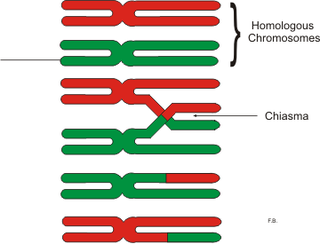
In genetics, a chiasma is the point of contact, the physical link, between two (non-sister) chromatids belonging to homologous chromosomes. At a given chiasma, an exchange of genetic material can occur between both chromatids, what is called a chromosomal crossover, but this is much more frequent during meiosis than mitosis. In meiosis, absence of a chiasma generally results in improper chromosomal segregation and aneuploidy.

<span class="mw-page-title-main">Neocentromere</span>

Neocentromeres are new centromeres that form at a place on the chromosome that is usually not centromeric. They typically arise due to disruption of the normal centromere. These neocentromeres should not be confused with “knobs”, which were also described as “neocentromeres” in maize in the 1950s. Unlike most normal centromeres, neocentromeres do not contain satellite sequences that are highly repetitive but instead consist of unique sequences. Despite this, most neocentromeres are still able to carry out the functions of normal centromeres in regulating chromosome segregation and inheritance. This raises many questions on what is necessary versus what is sufficient for constituting a centromere.

Achiasmate Meiosis refers to meiosis without chiasmata, which are structures that are necessary for recombination to occur and that usually aid in the segregation of non-sister homologs. The pachytene stage of prophase I typically results in the formation of chiasmata between homologous non-sister chromatids in the tetrad chromosomes that form. The formation of a chiasma is also referred to as crossing over. When two homologous chromatids cross over, they form a chiasma at the point of their intersection. However, it has been found that there are cases where one or more pairs of homologous chromosomes do not form chiasmata during pachynema. Without a chiasma, no recombination between homologs can occur.

<span class="mw-page-title-main">Monocentric chromosome</span> Chromosome that has only one centromere in a chromosome and forms a narrow constriction.

The monocentric chromosome is a chromosome that has only one centromere in a chromosome and forms a narrow constriction.

References

Open Access logo PLoS transparent.svg This article was adapted from the following source under a CC BY 4.0 license (2020) (reviewer reports): Mauro Mandrioli; Gian Carlo Manicardi (2020). "Holocentric chromosomes". PLOS Genetics . 16 (7): e1008918. doi:10.1371/JOURNAL.PGEN.1008918. ISSN   1553-7390. PMC   7392213 . PMID   32730246. Wikidata   Q97932961.{{cite journal}}: CS1 maint: unflagged free DOI (link)

  1. 1 2 3 Schrader F (1935). "Notes an the Mitotic Behavior of Long Chromosomes". Cytologia. 6 (4): 422–430. doi: 10.1508/cytologia.6.422 . ISSN   0011-4545.
  2. Mandrioli M, Manicardi GC (2003). "Analysis of insect holocentric chromosomes by atomic force microscopy". Hereditas. 138 (2): 129–32. doi:10.1034/j.1601-5223.2003.01661.x. hdl: 11380/5054 . PMID   12921164.
  3. 1 2 Hughes-Schrader S; Schrader F (January 1961). "The kinetochore of the Hemiptera". Chromosoma. 12 (1): 327–50. doi:10.1007/bf00328928. PMID   13716663. S2CID   5169204.
  4. 1 2 Wrensch DL, Kethley JB, Norton RA (1994). "Cytogenetics of Holokinetic Chromosomes and Inverted Meiosis: Keys to the Evolutionary Success of Mites, with Generalizations on Eukaryotes". Mites. Springer US. pp. 282–343. doi:10.1007/978-1-4615-2389-5_11. ISBN   978-1-4613-6012-4.
  5. 1 2 3 4 White MJ (1973). Animal cytology and evolution (3d ed.). Cambridge [England]: University Press. ISBN   0-521-07071-6. OCLC   674359.
  6. Mandrioli M, Manicardi GC (August 2012). "Unlocking holocentric chromosomes: new perspectives from comparative and functional genomics?". Current Genomics. 13 (5): 343–9. doi:10.2174/138920212801619250. PMC   3401891 . PMID   23372420.
  7. 1 2 3 4 5 6 7 8 Melters DP, Paliulis LV, Korf IF, Chan SW (July 2012). "Holocentric chromosomes: convergent evolution, meiotic adaptations, and genomic analysis". Chromosome Research. 20 (5): 579–93. doi: 10.1007/s10577-012-9292-1 . PMID   22766638. S2CID   3351527.
  8. 1 2 3 Benavente R (May 1982). "Holocentric chromosomes of arachnids: Presence of kinetochore plates during meiotic divisions". Genetica. 59 (1): 23–27. doi:10.1007/bf00130811. ISSN   0016-6707. S2CID   21290751.
  9. Monti V, Lombardo G, Loxdale HD, Manicardi GC, Mandrioli M (March 2012). "Continuous occurrence of intra-individual chromosome rearrangements in the peach potato aphid, Myzus persicae (Sulzer) (Hemiptera: Aphididae)". Genetica. 140 (1–3): 93–103. doi:10.1007/s10709-012-9661-x. hdl: 11380/739277 . PMID   22644285. S2CID   15715405.
  10. Manicardi GC, Nardelli A, Mandrioli M (2015-08-01). "Fast chromosomal evolution and karyotype instability: recurrent chromosomal rearrangements in the peach potato aphidMyzus persicae(Hemiptera: Aphididae)". Biological Journal of the Linnean Society. 116 (3): 519–529. doi: 10.1111/bij.12621 . ISSN   0024-4066.
  11. Nokkala S, Kuznetsova VG, Maryanska-Nadachowska A, Nokkala C (2004). "Holocentric chromosomes in meiosis. I. Restriction of the number of chiasmata in bivalents". Chromosome Research. 12 (7): 733–9. doi:10.1023/b:chro.0000045797.74375.70. PMID   15505408. S2CID   22226624.
  12. 1 2 3 4 5 6 7 8 9 Lukhtanov VA, Dincă V, Friberg M, Šíchová J, Olofsson M, Vila R, et al. (October 2018). "Versatility of multivalent orientation, inverted meiosis, and rescued fitness in holocentric chromosomal hybrids". Proceedings of the National Academy of Sciences of the United States of America. 115 (41): E9610–E9619. Bibcode:2018PNAS..115E9610L. doi: 10.1073/pnas.1802610115 . PMC   6187165 . PMID   30266792.
  13. Bureš P, Zedek F, Marková M (2012-08-23). "Holocentric Chromosomes". Plant Genome Diversity Volume 2. Vol. 2. Springer Vienna. pp. 187–208. doi:10.1007/978-3-7091-1160-4_12. ISBN   978-3-7091-1159-8.
  14. 1 2 3 Escudero M, Márquez-Corro JI, Hipp AL (2016-09-01). "The Phylogenetic Origins and Evolutionary History of Holocentric Chromosomes". Systematic Botany. 41 (3): 580–585. doi:10.1600/036364416x692442. ISSN   0363-6445. S2CID   89161837.
  15. Nagaki K, Kashihara K, Murata M (July 2005). "Visualization of diffuse centromeres with centromere-specific histone H3 in the holocentric plant Luzula nivea". The Plant Cell. 17 (7): 1886–93. doi:10.1105/tpc.105.032961. PMC   1167539 . PMID   15937225.
  16. 1 2 Howe M, McDonald KL, Albertson DG, Meyer BJ (June 2001). "HIM-10 is required for kinetochore structure and function on Caenorhabditis elegans holocentric chromosomes". The Journal of Cell Biology. 153 (6): 1227–38. doi:10.1083/jcb.153.6.1227. PMC   2192032 . PMID   11402066.
  17. 1 2 3 4 Zedek F, Bureš P (2012-01-23). "Evidence for centromere drive in the holocentric chromosomes of Caenorhabditis". PLOS ONE. 7 (1): e30496. Bibcode:2012PLoSO...730496Z. doi: 10.1371/journal.pone.0030496 . PMC   3264583 . PMID   22291967.
  18. 1 2 Ogawa K (1953). "Chromosome Studies in the Myriapoda". Japanese Journal of Genetics (遺伝學雑誌). 28 (1): 12–18. doi: 10.1266/jjg.28.12 .
  19. 1 2 Richards RI (June 2001). "Fragile and unstable chromosomes in cancer: causes and consequences". Trends in Genetics. 17 (6): 339–45. doi:10.1016/s0168-9525(01)02303-4. PMID   11377796.
  20. 1 2 Freudenreich CH (September 2007). "Chromosome fragility: molecular mechanisms and cellular consequences". Frontiers in Bioscience. 12 (12): 4911–24. doi: 10.2741/2437 . PMID   17569619.
  21. Manicardi GC, Mandrioli M, Blackman RL (February 2015). "The cytogenetic architecture of the aphid genome". Biological Reviews of the Cambridge Philosophical Society. 90 (1): 112–25. doi:10.1111/brv.12096. hdl: 11380/1062551 . PMID   24593177. S2CID   21793030.
  22. Monti V, Mandrioli M, Rivi M, Manicardi GC (2011-12-16). "The vanishing clone: karyotypic evidence for extensive intraclonal genetic variation in the peach potato aphid, Myzus persicae (Hemiptera: Aphididae)". Biological Journal of the Linnean Society. 105 (2): 350–358. doi:10.1111/j.1095-8312.2011.01812.x. ISSN   0024-4066. S2CID   82310768.
  23. Monti V, Giusti M, Bizzaro D, Manicardi GC, Mandrioli M (July 2011). "Presence of a functional (TTAGG)(n) telomere-telomerase system in aphids". Chromosome Research. 19 (5): 625–33. doi:10.1007/s10577-011-9222-7. hdl: 11380/659235 . PMID   21667174. S2CID   25127536.
  24. Mandrioli M, Borsatti F (2007-11-17). "Analysis of heterochromatic epigenetic markers in the holocentric chromosomes of the aphid Acyrthosiphon pisum". Chromosome Research. 15 (8): 1015–22. doi:10.1007/s10577-007-1176-4. hdl: 11380/594885 . PMID   18004669. S2CID   20329150.
  25. 1 2 Marec F, Tothova A, Sahara K, Traut W (December 2001). "Meiotic pairing of sex chromosome fragments and its relation to atypical transmission of a sex-linked marker in Ephestia kuehniella (Insecta: Lepidoptera)". Heredity. 87 (Pt 6): 659–71. doi: 10.1046/j.1365-2540.2001.00958.x . PMID   11903561. S2CID   415409.
  26. 1 2 Hill J, Rastas P, Hornett EA, Neethiraj R, Clark N, Morehouse N, et al. (June 2019). "Unprecedented reorganization of holocentric chromosomes provides insights into the enigma of lepidopteran chromosome evolution". Science Advances. 5 (6): eaau3648. Bibcode:2019SciA....5.3648H. doi:10.1126/sciadv.aau3648. PMC   6561736 . PMID   31206013.
  27. d'Alençon E, Sezutsu H, Legeai F, Permal E, Bernard-Samain S, Gimenez S, et al. (April 2010). "Extensive synteny conservation of holocentric chromosomes in Lepidoptera despite high rates of local genome rearrangements". Proceedings of the National Academy of Sciences of the United States of America. 107 (17): 7680–5. Bibcode:2010PNAS..107.7680D. doi: 10.1073/pnas.0910413107 . PMC   2867904 . PMID   20388903.
  28. Lukhtanov VA, Dincă V, Talavera G, Vila R (April 2011). "Unprecedented within-species chromosome number cline in the Wood White butterfly Leptidea sinapis and its significance for karyotype evolution and speciation". BMC Evolutionary Biology. 11 (1): 109. Bibcode:2011BMCEE..11..109L. doi: 10.1186/1471-2148-11-109 . PMC   3113740 . PMID   21507222.
  29. Drinnenberg IA, deYoung D, Henikoff S, Malik HS (2014-08-18). "Author response: Recurrent loss of CenH3 is associated with independent transitions to holocentricity in insects". eLife. doi: 10.7554/elife.03676.020 .
  30. Mutafova T, Dimitrova Y, Komandarev S (1982). "The karyotype of four Trichinella species". Zeitschrift für Parasitenkunde. 67 (1): 115–20. doi:10.1007/bf00929519. PMID   7072318. S2CID   22178390.
  31. Spakulová M, Králová I, Cutillas C (March 1994). "Studies on the karyotype and gametogenesis in Trichuris muris". Journal of Helminthology. 68 (1): 67–72. doi:10.1017/s0022149x0001350x. PMID   8006389. S2CID   35922875.
  32. Post R (November 2005). "The chromosomes of the Filariae". Filaria Journal. 4 (1): 10. doi: 10.1186/1475-2883-4-10 . PMC   1282586 . PMID   16266430.
  33. Pimpinelli S, Goday C (September 1989). "Unusual kinetochores and chromatin diminution in Parascaris". Trends in Genetics. 5 (9): 310–5. doi:10.1016/0168-9525(89)90114-5. PMID   2686123.
  34. Dernburg AF (June 2001). "Here, there, and everywhere: kinetochore function on holocentric chromosomes". The Journal of Cell Biology. 153 (6): F33-8. doi:10.1083/jcb.153.6.f33. PMC   2192025 . PMID   11402076.
  35. 1 2 3 Maddox PS, Oegema K, Desai A, Cheeseman IM (2004). ""Holo"er than thou: chromosome segregation and kinetochore function in C. elegans". Chromosome Research. 12 (6): 641–53. doi:10.1023/b:chro.0000036588.42225.2f. PMID   15289669. S2CID   38989489.
  36. 1 2 3 4 Albertson DG, Thomson JN (October 1982). "The kinetochores of Caenorhabditis elegans". Chromosoma. 86 (3): 409–28. doi:10.1007/bf00292267. PMID   7172865. S2CID   24941882.
  37. 1 2 3 Monen J, Maddox PS, Hyndman F, Oegema K, Desai A (December 2005). "Differential role of CENP-A in the segregation of holocentric C. elegans chromosomes during meiosis and mitosis". Nature Cell Biology. 7 (12): 1248–55. doi:10.1038/ncb1331. PMID   16273096. S2CID   37436777.
  38. 1 2 Subirana JA, Messeguer X (2013-04-24). "A satellite explosion in the genome of holocentric nematodes". PLOS ONE. 8 (4): e62221. Bibcode:2013PLoSO...862221S. doi: 10.1371/journal.pone.0062221 . PMC   3634726 . PMID   23638010.
  39. Godward MB (1966). Chromosomes of the Algae. Edward Arnold. pp. 24–51. ISBN   9780713120585.
  40. Kolodin P, Cempírková H, Bureš P, Horová L, Veleba A, Francová J, Adamec L, Zedek F (December 2018). "Holocentric chromosomes may be an apomorphy of Droseraceae". Plant Systematics and Evolution. 304 (10): 1289–96. Bibcode:2018PSyEv.304.1289K. doi:10.1007/s00606-018-1546-8. S2CID   52882155.
  41. Luceño M, Vanzela AL, Guerra M (1998-03-01). "Cytotaxonomic studies in Brazilian Rhynchospora (Cyperaceae), a genus exhibiting holocentric chromosomes". Canadian Journal of Botany. 76 (3): 440–449. doi:10.1139/b98-013. ISSN   0008-4026.
  42. Kuta E, Bohanec B, Dubas E, Vizintin L, Przywara L (April 2004). "Chromosome and nuclear DNA study on Luzula - a genus with holokinetic chromosomes". Genome. 47 (2): 246–56. doi:10.1139/g03-121. PMID   15060577.
  43. Kynast RG, Joseph JA, Pellicer J, Ramsay MM, Rudall PJ (September 2014). "Chromosome behavior at the base of the angiosperm radiation: karyology of Trithuria submersa (Hydatellaceae, Nymphaeales)". American Journal of Botany. 101 (9): 1447–55. doi: 10.3732/ajb.1400050 . PMID   25253705.
  44. Zedek F, Veselý P, Horová L, Bureš P (June 2016). "Flow cytometry may allow microscope-independent detection of holocentric chromosomes in plants". Scientific Reports. 6 (1): 27161. Bibcode:2016NatSR...627161Z. doi:10.1038/srep27161. PMC   4891681 . PMID   27255216.
  45. Haizel T, Lim YK, Leitch AR, Moore G (2005). "Molecular analysis of holocentric centromeres of Luzula species". Cytogenetic and Genome Research. 109 (1–3): 134–43. doi:10.1159/000082392. PMID   15753569. S2CID   45200884.
  46. 1 2 3 Zedek F, Bureš P (December 2016). "Absence of positive selection on CenH3 in Luzula suggests that holokinetic chromosomes may suppress centromere drive". Annals of Botany. 118 (7): 1347–1352. doi:10.1093/aob/mcw186. PMC   5155603 . PMID   27616209.
  47. 1 2 Madej A, Kuta E (2001). "Holokinetic chromosomes of Luzula luzuloides [Juncaceae] in callus culture". Acta Biologica Cracoviensia. Series Botanica (43). ISSN   0001-5296.
  48. Nordenskiöld, Hedda (1961). "Tetrad Analysis and the Course of Meiosis in Three Hybrids of Luzula Campestris". Hereditas. 47 (2): 203–238. doi:10.1111/j.1601-5223.1961.tb01771.x. ISSN   1601-5223.
  49. Escudero M, Vargas P, Arens P, Ouborg NJ, Luceño M (January 2010). "The east-west-north colonization history of the Mediterranean and Europe by the coastal plant Carex extensa (Cyperaceae)". Molecular Ecology. 19 (2): 352–70. Bibcode:2010MolEc..19..352E. doi:10.1111/j.1365-294X.2009.04449.x. PMID   20002603. S2CID   3577580.
  50. Hipp AL, Rothrock PE, Roalson EH (2008-12-05). "The Evolution of Chromosome Arrangements in Carex (Cyperaceae)". The Botanical Review. 75 (1): 96–109. doi:10.1007/s12229-008-9022-8. ISSN   0006-8101. S2CID   4489708.
  51. Escudero M, Weber JA, Hipp AL (August 2013). "Species coherence in the face of karyotype diversification in holocentric organisms: the case of a cytogenetically variable sedge (Carex scoparia, Cyperaceae)". Annals of Botany. 112 (3): 515–26. doi:10.1093/aob/mct119. PMC   3718211 . PMID   23723260.
  52. Wanner G, Schroeder-Reiter E, Ma W, Houben A, Schubert V (December 2015). "The ultrastructure of mono- and holocentric plant centromeres: an immunological investigation by structured illumination microscopy and scanning electron microscopy". Chromosoma. 124 (4): 503–17. doi:10.1007/s00412-015-0521-1. PMID   26048589. S2CID   14145859.
  53. 1 2 Cuacos M, H Franklin FC, Heckmann S (2015-10-26). "Atypical centromeres in plants-what they can tell us". Frontiers in Plant Science. 6: 913. doi: 10.3389/fpls.2015.00913 . PMC   4620154 . PMID   26579160.
  54. 1 2 Jankowska M, Fuchs J, Klocke E, Fojtová M, Polanská P, Fajkus J, et al. (December 2015). "Holokinetic centromeres and efficient telomere healing enable rapid karyotype evolution". Chromosoma. 124 (4): 519–28. doi:10.1007/s00412-015-0524-y. PMID   26062516. S2CID   2530401.
  55. 1 2 3 4 5 Bogdanov YF (May 2016). "[Inverted meiosis and its place in the evolution of sexual reproduction pathways]". Genetika. 52 (5): 541–60. doi:10.1134/s1022795416050033. PMID   29368476. S2CID   18427008.
  56. 1 2 3 4 5 Viera A, Page J, Rufas JS (2009). "Inverted meiosis: the true bugs as a model to study". Genome Dynamics. Karger. 5: 137–156. doi:10.1159/000166639. ISBN   978-3-8055-8967-3. PMID   18948713.
  57. 1 2 3 Vanzela AL, Cuadrado A, Guerra M (2003). "Localization of 45S rDNA and telomeric sites on holocentric chromosomes of Rhynchospora tenuis Link (Cyperaceae)". Genetics and Molecular Biology. 26 (2): 199–201. doi: 10.1590/S1415-47572003000200014 .
  58. 1 2 3 Martinez-Perez E, Schvarzstein M, Barroso C, Lightfoot J, Dernburg AF, Villeneuve AM (October 2008). "Crossovers trigger a remodeling of meiotic chromosome axis composition that is linked to two-step loss of sister chromatid cohesion". Genes & Development. 22 (20): 2886–901. doi:10.1101/gad.1694108. PMC   2569886 . PMID   18923085.