Trichinella spiralis

Last updated

Trichinella spiralis
Trichinella larv1 DPDx.JPG
Scientific classification OOjs UI icon edit-ltr.svg
Domain: Eukaryota
Kingdom: Animalia
Phylum: Nematoda
Class: Enoplea
Order: Trichocephalida
Family: Trichinellidae
Genus: Trichinella
Species:
T. spiralis
Binomial name
Trichinella spiralis
(Owen, 1835)

Trichinella spiralis is a viviparous [1] nematode parasite, occurring in rodents, pigs, bears, hyenas and humans, and is responsible for the disease trichinosis. It is sometimes referred to as the "pork worm" due to it being typically encountered in undercooked pork products. It should not be confused with the distantly related pork tapeworm.

Contents

Description

Trichinella species, the smallest nematode parasite of humans, has an unusual life cycle, and are one of the most widespread and clinically important parasites in the world. [2] The small adult worms mature in the small intestine of a definitive host, such as a pig. Each adult female produces batches of live larvae, which bore through the intestinal wall, enters the blood (to feed on it) and lymphatic system, and are carried to striated muscle. Once in the muscle, they encyst, or become enclosed in a capsule. Humans can become infected by eating infected pork, horsemeat, or wild carnivores such as fox, cat, hyena or bear. [2]

Morphology

Males of T. spiralis measure between 1.4 and 1.6 mm long, and are more flat anteriorly than posteriorly. The anus can be found in the terminal end, and they have a large copulatory pseudobursa on each side. [2] The females of T. spiralis are about twice the size of the males, and have an anus found terminally. The vulva is located near the esophagus. The single uterus of the female is filled with developing eggs in the posterior portion, while the anterior portion contains the fully developed juveniles. [2]

Life cycle

Trichinella spiralis can live the majority of its adult life in the intestines of humans. To begin its life cycle, T. spiralis adults invade the intestinal wall of a pig, and produce larvae that invade the pig's muscles. The larval forms are encapsulated as a small cystic structure within a muscle cell of the infected host. When another animal (perhaps a human) eats the infected meat, the larvae are released from the nurse cells in the meat (due to stomach pH), and migrate to the intestine, where they burrow into the intestinal mucosa, mature, and reproduce. [3] Juveniles within nurse cells have an anaerobic or facultative anaerobic metabolism, but when they become activated, they adopt the aerobic metabolism characteristics of the adult. [2]

Trichinella spiralis life cycle Trichinella life cycle.jpg
Trichinella spiralis life cycle

Female Trichinella worms live for about six weeks, and in that time can produce up to 1,500 larvae; when a spent female dies, she passes out of the host. The larvae gain access to the circulation and migrate around the body of the host, in search of a muscle cell in which to encyst. [3] The migration and encystment of larvae can cause fever and pain, brought on by the host inflammatory response. In some cases, accidental migration to specific organ tissues can cause myocarditis and encephalitis that can result in death.

Animal tissue infected with the parasite that causes the disease trichinosis: Most parasites are shown in cross section, but some randomly appear in long section. Trichinella Spiralis x.s. & l.s. 40x.png
Animal tissue infected with the parasite that causes the disease trichinosis: Most parasites are shown in cross section, but some randomly appear in long section.

Nurse cell formation

This nematode is a multicellular parasite that lives within a single muscle cell, which it extensively modifies according to its own requirements. [4]

T. spiralis larvae within the diaphragm muscle of a pig Trichinella spiralis larvae within muscle.jpg
T. spiralis larvae within the diaphragm muscle of a pig

Nurse cell formation in skeletal muscle tissue is mediated by the hypoxic environment surrounding the new vessel formation. [5] The hypoxic environment stimulates muscle cells in the surrounding tissue to upregulate and secrete angiogenic cytokines, such as vascular endothelial growth factor (VEGF). This allows the migrating T. spiralis larva to enter the myocyte and induce the transformation into the nurse cell. VEGF expression is detected surrounding the nurse cell immediately after nurse cell formation, and the continued secretion of VEGF can maintain the constant state of hypoxia. [6] [7]

Symptoms

The first symptoms may appear between 12 hours and two days after ingestion of infected meat. The migration of adult worms in the intestinal epithelium can cause traumatic damage to the host tissue, and the waste products they excrete can provoke an immunological reaction. [2] The resulting inflammation can cause symptoms such as nausea, vomiting, sweating, and diarrhea. Five to seven days after the appearance of these symptoms, facial edema and fever may occur. Ten days following ingestion, intense muscular pain, difficulty breathing, weakening of pulse and blood pressure, heart damage, and various nervous disorders may occur, eventually leading to death due to heart failure, respiratory complications, or kidney malfunction, all due to larval migration. [2]

In pigs, infection is usually subclinical, but large worm burdens can be fatal in some cases. [8]

Diagnosis and treatment

Muscle biopsy may be used for trichinosis detection. Several immunodiagnostic tests are also available. Typically, patients are treated with either mebendazole or albendazole, but efficacy of such products is uncertain. Symptoms can be relieved by use of analgesics and corticosteroids. [2]

In pigs, ELISA testing is possible as a method of diagnosis. Anthelmintics can treat and prevent Trichinella infections. [8]

Prevention and control

Trichinosis (trichinellosis) is a disease caused by tissue-dwelling roundworms of the species Trichinella spiralis. In the United States, the national trichinellosis surveillance system has documented a steady decline in the reported incidence of this disease. During 1947 to 1951, a median of 393 human cases was reported annually, including 57 trichinellosis-related deaths. During 1997–2001, the incidence in the US decreased to a median of 12 cases annually, with no reported deaths. The decrease was largely due to improved compliance with standards and regulations by commercial pork producers. [9]

In the United States, Congress passed the Federal Swine Health Protection Act, restricting the use of uncooked garbage as feed stock for pigs, and creating a voluntary Trichinae Herd Certification Program. [9] The Trichinae Herd Certification Program is a voluntary pre-slaughter pork safety program that provides documentation of swine management practices to minimize Trichinella exposure. The goal of the program is to establish a system under which pork production facilities that follow good production practices might be certified as Trichinella-safe. [10] In addition to the reduction in Trichinella prevalence in commercial pork, processing methods also have contributed to the dramatic decline in human trichinellosis associated with pork products.[ citation needed ] Through the U.S. Code of Federal Regulations, the USDA has created guidelines for specific cooking temperatures and times, freezing temperatures and times, and curing methods for processed pork products to reduce the risk of human infection from Trichinella contaminated meat. [9] Pork products meeting these guidelines are designated certified pork.[ citation needed ]

It was reported in 2005 that the prevalence of human infections from Trichinella spiralis was low in the United States, despite nonexistent meat inspection with respect to trichinella. This was due to strict enforcement of the regulations applying to large meat production facilities: most cases have been from raw or undercooked meat from game animals. [11]

Also reported in 2005, the rate of infection from Trichinella spiralis was significantly higher in people living in parts of Europe, Asia, and Southeast Asia than in the United States. However, EU nations employ several strategies for detecting meat infected with Trichinella spiralis. If tests are consistently negative, then a trichinella-free designation is applied to a given meat supply. Rare outbreaks still occur despite this rigorous system: France, Italy, and Poland have reported outbreaks due to eating raw horsemeat. At that time, the parasite was considered endemic in Japan and China, while Korea had recently reported its first human cases of trichinosis. [11]

In most abattoirs, the diaphragms of pigs are routinely sampled to detect Trichinella infections. [8]

Post-slaughter human exposure is also preventable by educating consumers on simple steps that can be taken to kill any larvae that can potentially be in meat bought at the local supermarket. Freezing meat in an average household freezer for 20 days before consumption will kill some species of Trichinella. Cooking pork products to a minimum internal temperature of 160 °F (72 °C) will kill most species, and is the best way to ensure the meat is safe to eat. [12]

Economic impact

It was reported in 2009 that political and economic changes had caused an increase in the prevalence and incidence rates of this parasite in many former eastern European countries due to weakened veterinary control on susceptible animals. [13] This complicated the meat trade industry within European Union countries, and exportation of pork outside the EU. [13] As a result, the European Union and some associated countries implemented a Trichinella monitoring program for pigs, horses, wild boar, and other wildlife species; while the European Commission implemented a new regulation to control Trichinella in meat in order to improve food safety for European consumers [13]

Illegal pork importation from places with low safety standards allows the spread of the parasite from endemic to nonendemic countries. [13] Illegal importation and new food practices and dishes including raw meat have resulted in human trichinosis outbreaks in many European countries, including Denmark, Germany, Italy, Spain, and the United Kingdom. [13]

The economic cost of detecting trichinosis can be another cost burden. In 1998, a rough global cost estimate was $3.00 per pig to detect the parasite. [13] At the same time, in the 15 countries comprising the European Union in 1998, about 190 million pigs were killed in slaughterhouses annually, leading to an estimated economic impact of testing of about $570 million per year. [13] However, depending on the size of the specific slaughterhouse, the actual costs could be more than an order of magnitude smaller (i.e. less than $0.30 per pig). [13]

Genome

The Trichinella spiralis draft genome became available in March, 2011. [14] The genome size was 58.55 Mbp with an estimated 16,549 genes. [15] The T. spiralis genome is the only known nematode genome to be subject to DNA methylation, [16] an epigenetic mechanism that was not previously thought to exist in nematodes.

See also

Related Research Articles

<span class="mw-page-title-main">Trichuriasis</span> Infection by Trichuris trichiura (whipworm)

Trichuriasis, also known as whipworm infection, is an infection by the parasitic worm Trichuris trichiura (whipworm). If infection is only with a few worms, there are often no symptoms. In those who are infected with many worms, there may be abdominal pain, fatigue and diarrhea. The diarrhea sometimes contains blood. Infections in children may cause poor intellectual and physical development. Low red blood cell levels may occur due to loss of blood.

<span class="mw-page-title-main">Trichinosis</span> Parasitic disease due to invasion by Trichinella spp.

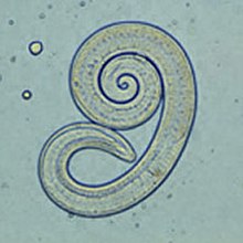
Trichinosis, also known as trichinellosis, is a parasitic disease caused by roundworms of the Trichinella type. During the initial infection, invasion of the intestines can result in diarrhea, abdominal pain, and vomiting. Migration of larvae to muscle, which occurs about a week after being infected, can cause swelling of the face, inflammation of the whites of the eyes, fever, muscle pains, and a rash. Minor infection may be without symptoms. Complications may include inflammation of heart muscle, central nervous system involvement, and inflammation of the lungs.

<span class="mw-page-title-main">Parasitology</span> Study of parasites, their hosts, and the relationship between them

Parasitology is the study of parasites, their hosts, and the relationship between them. As a biological discipline, the scope of parasitology is not determined by the organism or environment in question but by their way of life. This means it forms a synthesis of other disciplines, and draws on techniques from fields such as cell biology, bioinformatics, biochemistry, molecular biology, immunology, genetics, evolution and ecology.

Gnathostomiasis, also known as larva migrans profundus, is the human infection caused by the nematode Gnathostoma spinigerum and/or Gnathostoma hispidum, which infects vertebrates.

<span class="mw-page-title-main">Toxocaridae</span> Family of roundworms

The Toxocaridae are a zoonotic family of parasitic nematodes that infect canids and felids and which cause toxocariasis in humans. The worms are unable to reproduce in humans.

<i>Trichinella</i> Genus of worms

Trichinella is the genus of parasitic roundworms of the phylum Nematoda that cause trichinosis. Members of this genus are often called trichinella or trichina worms. A characteristic of Nematoda is the one-way digestive tract, with a pseudocoelom.

<span class="mw-page-title-main">Paragonimiasis</span> Medical condition

Paragonimiasis is a food-borne parasitic disease caused by several species of lung flukes belonging to genus Paragonimus. Infection is acquired by eating crustaceans such as crabs and crayfishes which host the infective forms called metacercariae, or by eating raw or undercooked meat of mammals harboring the metacercariae from crustaceans.

<span class="mw-page-title-main">Parasitic worm</span> Large type of parasitic organism

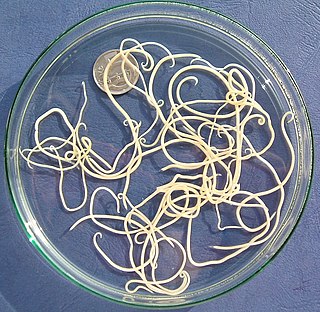
Parasitic worms, also known as helminths, are large macroparasites; adults can generally be seen with the naked eye. Many are intestinal worms that are soil-transmitted and infect the gastrointestinal tract. Other parasitic worms such as schistosomes reside in blood vessels.

Spirometra erinaceieuropaei is a parasitic tapeworm that infects domestic animals and humans. The medical term for this infection in humans and other animals is sparganosis. Morphologically, these worms are similar to other worms in the genus Spirometra. They have a long body consisting of three sections: the scolex, the neck, and the strobilia. They have a complex life cycle that consists of three hosts, and can live in varying environments and bodily tissues. Humans can contract this parasite in three main ways. Historically, humans are considered a paratenic host; however, the first case of an adult S. erinaceieuropaei infection in humans was reported in 2017. Spirometra tapeworms exist worldwide and infection is common in animals, but S. erinaceieuropaei infections are rare in humans. Treatment for infection typically includes surgical removal and anti-worm medication.

Sparganosis is a parasitic infection caused by the plerocercoid larvae of the genus Spirometra including S. mansoni, S. ranarum, S. mansonoides and S. erinacei. It was first described by Patrick Manson in 1882, and the first human case was reported by Charles Wardell Stiles from Florida in 1908. The infection is transmitted by ingestion of contaminated water, ingestion of a second intermediate host such as a frog or snake, or contact between a second intermediate host and an open wound or mucous membrane. Humans are the accidental hosts in the life cycle, while dogs, cats, and other mammals are definitive hosts. Copepods are the first intermediate hosts, and various amphibians and reptiles are second intermediate hosts.

<span class="mw-page-title-main">Dickson Despommier</span> American academic, microbiologist, ecologist

Dickson D. Despommier is an emeritus professor of microbiology and Public Health at Columbia University. From 1971 to 2009, he conducted research on intracellular parasitism and taught courses on parasitic diseases, medical ecology and ecology. Despommier has received media coverage for his ideas on vertical farming.

<span class="mw-page-title-main">Nematode</span> Phylum of worms with tubular digestive systems with openings at both ends

The nematodesroundworms or eelworms, constitute the phylum Nematoda. They are a diverse animal phylum inhabiting a broad range of environments. Most species are free-living, feeding on microorganisms, but there are many that are parasitic. The parasitic worms (helminths) are the cause of soil-transmitted helminthiases.

<span class="mw-page-title-main">Microfilaria</span> Early stage in the life cycle of certain parasitic nematodes in the family Onchocercidae

The microfilaria is an early stage in the life cycle of certain parasitic nematodes in the family Onchocercidae. In these species, the adults live in a tissue or the circulatory system of vertebrates. They release microfilariae into the bloodstream of the vertebrate host. The microfilariae are taken up by blood-feeding arthropod vectors. In the intermediate host the microfilariae develop into infective larvae that can be transmitted to a new vertebrate host.

<i>Trichinella britovi</i> Species of roundworm

Trichinella britovi is a nematode parasite responsible for a zoonotic disease called trichinellosis. Currently, eight species of Trichinella are known, only three of which cause trichinellosis, and Trichinella britovi is one of them. Numerous mammal species, as well as birds and crocodiles, can harbor the parasite worldwide, but the sylvatic cycle is mainly maintained by wild carnivores.

<i>Alaria</i> (trematode) Genus of flukes

Alaria is a genus of flatworms, or trematodes, in the family Diplostomidae.

Trichinella papuae is a nematode parasite responsible for a zoonotic disease called trichinellosis, predominantly in Thailand. Currently, eight species of Trichinella are known.

<span class="mw-page-title-main">Cysticercus</span> Larval tapeworm

Cysticercus is a scientific name given to the young tapeworms (larvae) belonging to the genus Taenia. It is a small, sac-like vesicle resembling a bladder; hence, it is also known as bladder worm. It is filled with fluid, in which the main body of the larva, called scolex, resides. It normally develops from the eggs, which are ingested by the intermediate hosts, such as pigs and cattle. The tissue infection is called cysticercosis. Inside such hosts, they settle in the muscles. When humans eat raw or undercooked pork or beef that is contaminated with cysticerci, the larvae grow into adult worms inside the intestine. Under certain circumstances, specifically for the pork tapeworm, the eggs can be accidentally eaten by humans through contaminated foodstuffs. In such case, the eggs hatch inside the body, generally moving to muscles as well as inside the brain. Such brain infection can lead to a serious medical condition called neurocysticercosis. This disease is the leading cause of acquired epilepsy.

<span class="mw-page-title-main">Gastropod-borne parasitic disease</span> Medical condition

Gastropod-borne parasitic diseases (GPDs) are a group of infectious diseases that require a gastropod species to serve as an intermediate host for a parasitic organism that can infect humans upon ingesting the parasite or coming into contact with contaminated water sources. These diseases can cause a range of symptoms, from mild discomfort to severe, life-threatening conditions, with them being prevalent in many parts of the world, particularly in developing regions. Preventive measures such as proper sanitation and hygiene practices, avoiding contact with infected gastropods and cooking or boiling food properly can help to reduce the risk of these diseases.

<span class="mw-page-title-main">Cat worm infections</span> Worm infections in cats

Cat worm infections, the infection of cats (Felidae) with parasitic worms, occur frequently. Most worm species occur worldwide in both domestic and other cats, but there are regional, species and lifestyle differences in the frequency of infestation. According to the classification of the corresponding parasites in the zoological system, infections can be divided into those caused by nematode and flatworms - in the case of the latter, mainly cestoda and trematoda - while other strains are of no veterinary significance. While threadworms usually do not require an intermediate host for their reproduction, the development cycle of flatworms always proceeds via alternate hosts.

<span class="mw-page-title-main">Nematode infection in dogs</span> Threadworm infections of dogs are frequent

Nematode infection in dogs - the infection of dogs with parasitic nemamotodes - are, along with tapeworm infections and infections with protozoa, frequent parasitoses in veterinary practice. Nematodes, as so-called endoparasites, colonize various internal organs - most of them the digestive tract - and the skin. To date, about 30 different species of nematode have been identified in domestic dogs; they are essentially also found in wild dog species. However, the majority of them often cause no or only minor symptoms of disease in adult animals. The infection therefore does not necessarily have to manifest itself in a worm disease (helminthosis). For most nematodes, an infection can be detected by examining the feces for eggs or larvae. Roundworm infection in dogs and the hookworm in dogs is of particular health significance in Central Europe, as they can also be transmitted to humans (zoonosis). Regular deworming can significantly reduce the frequency of infection and thus the risk of infection for humans and dogs.

References

  1. Xiaolei Liu,#1 Yanxia Song,#1,2 Ning Jiang,1 Jielin Wang,1 Bin Tang,1 Huijun Lu,1 Shuai Peng,1 Zhiguang Chang,1 Yizhi Tang,1 Jigang Yin,1 Mingyuan Liu,1 Yan Tan,2,* and Qijun Chen1,3,* (August 2012). "Global Gene Expression Analysis of the Zoonotic Parasite Trichinella spiralis Revealed Novel Genes in Host Parasite Interaction". PLOS Negl Trop Dis. 6 (8): e1794. doi:10.1371/journal.pntd.0001794. PMC   3429391 . PMID   22953016.{{cite journal}}: CS1 maint: multiple names: authors list (link)
  2. 1 2 3 4 5 6 7 8 Roberts, Larry S., John Janovay (2005). Foundations of Parasitology (7th ed.). New York: McGraw-Hill. pp. 405–407.{{cite book}}: CS1 maint: multiple names: authors list (link)
  3. 1 2 Crowley, Leonard (2009). An Introduction to Human Disease: Pathology and Pathophysiology Correlations (8th ed.). Jones and Bartlett.
  4. Combes, Claude (2005). The art of Being a Parasite (English translation ed.). The University of Chicago Press.
  5. Fong GH (2008). "Mechanism of adaptative angiogenesis to tissue hypoxia". Angiogenesis. 11 (2): 121–140. doi:10.1007/s10456-008-9107-3. PMID   18327686. S2CID   11528564.
  6. Capo VA, Despommier DD, Polvere RI (1998). "Trichinella spiralis: vascular endothelial growth factor is up-regulated within the nurse cell during the early phase of its formation". J. Parasitol. 84 (2): 209–214. doi:10.2307/3284472. JSTOR   3284472. PMID   9576489.
  7. Novo E, Cannito S, Zamara E, Valfre di Bonzo L, Caligiuri A, Cravanzola C, et al. (2007). "Proangiogenic cytokines as hypoxia-dependent factors stimulating migration of human hepatic stellate cells". Am. J. Pathol. 170 (6): 1942–1953. doi:10.2353/ajpath.2007.060887. PMC   1899450 . PMID   17525262.
  8. 1 2 3 "Trichinella". WikiVet . Retrieved 12 October 2011.
  9. 1 2 3 Sharon L. Roy, M.D.; Adriana S. Lopez, M.H.S.; Peter M. Schantz, V.M.D., Ph.D. (2003-07-25). "Trichinellosis Surveillance --- United States, 1997--2001". Division of Parasitic Diseases, National Center for Infectious Diseases. Retrieved 2020-04-11. The majority of the decline in reported trichinellosis cases is a result of improved observance of standards and regulations in the U.S. commercial pork industry, which has altered animal husbandry practices resulting in reduced Trichinella prevalence among swine.{{cite web}}: CS1 maint: multiple names: authors list (link)
  10. National Pork Board. Trichinae Herd Certification. Des Moines, Iowa: National Pork Producers Council, 2000. Available at http://www.aphis.usda.gov/vs/trichinella/.
  11. 1 2 Despommier; Gwadz; Hotez; Knirsch (2005). Parasitic Diseases (5th ed.). Apple Trees Productions LLC. ISBN   0-9700027-7-7. Archived from the original on 2018-09-06.
  12. Morbidity and Mortality Weekly Report: Surveillance Summaries (2003): 1–8. JSTOR. Centers for Disease Control & Prevention (CDC). Web. 1 Dec. 2014.
  13. 1 2 3 4 5 6 7 8 Gottstein, Bruno; Pozio, Edoardo; Nöckler, Karsten (2009-01-01). "Epidemiology, Diagnosis, Treatment, and Control of Trichinellosis". Clinical Microbiology Reviews. 22 (1): 127–145. doi:10.1128/CMR.00026-08. ISSN   0893-8512. PMC   2620635 . PMID   19136437.
  14. "Trichinella spiralis". Genome. NCBI. Retrieved 2012-04-19.
  15. Mitreva M, Jasmer DP, Zarlenga DS, Wang Z, Abubucker S, Martin J, Taylor CM, Yin Y, Fulton L, Minx P, Yang SP, Warren WC, Fulton RS, Bhonagiri V, Zhang X, Hallsworth-Pepin K, Clifton SW, McCarter JP, Appleton J, Mardis ER, Wilson RK (March 2011). "The draft genome of the parasitic nematode Trichinella spiralis". Nat. Genet. 43 (3): 228–235. doi:10.1038/ng.769. PMC   3057868 . PMID   21336279.
  16. Gao F, Liu X, Wu XP, Wang XL, Gong D, Lu H, Xia Y, Song Y, Wang J, Du J, Liu S, Han X, Tang Y, Yang H, Jin Q, Zhang X, Liu M (October 2012). "Differential DNA methylation in discrete developmental stages of the parasitic nematode Trichinella spiralis". Genome Biol. 13 (10): R100. doi:10.1186/gb-2012-13-10-r100. PMC   4053732 . PMID   23075480.

14 "Microbiology: An Introduction 9/e" (2006)

Further reading