Gnathostoma spinigerum

Last updated

Gnathostoma spinigerum
Scientific classification OOjs UI icon edit-ltr.svg
Domain: Eukaryota
Kingdom: Animalia
Phylum: Nematoda
Class: Chromadorea
Order: Rhabditida
Family: Gnathostomatidae
Genus: Gnathostoma
Species:
G. spinigerum
Binomial name
Gnathostoma spinigerum
Levinsen, 1889

Gnathostoma spinigerum is a parasitic nematode that causes gnathostomiasis in humans, also known as its clinical manifestations are creeping eruption , larva migrans, Yangtze edema, Choko-Fuschu Tua chid and wandering swelling. Gnathostomiasis in animals can be serious, and even fatal. The first described case of gnathostomiasis was in a young tiger that died in the London Zoo in 1835. The larval nematode is acquired by eating raw or undercooked fish and meat.

Contents

Gnathostoma spinigerum has a multi-host life history. The eggs hatch in fresh water and the larvae are eaten by water fleas of the genus Cyclops . The water fleas are in turn eaten by small fish. Eventually, the larvae end up in the stomachs of carnivores, usually cats and dogs. The larva bores through the stomach wall and migrates around in the host's body for about three months before returning to the stomach and attaching itself in the gastric mucosa. It then takes another six months to mature. The eggs are carried in the host's feces, and if they reach fresh water the cycle begins again. As humans are not a normal host for the larvae, they do not mature in humans, but can cause various degrees of damage, depending on where the larvae wander in the body.

Description

Video showing movement of G. spinigerum larvae in a copepod.

This species grows to a length of 11 to 54 mm (0.43 to 2.13 in).

Species within the genus Gnathostoma are recognized by a bulbous head with a pair of lateral lips surrounding a mouth on the longitudinal axis. The cephalic region is covered by transverse rows of cuticular spines. Internally, the head is divided into four glandular cervical sacs that attach near the esophagus, as well as four hollow spaces called ballonets, each being continuous with a cervical sac via a central canal. [1]

The body is typically pink and is also covered anteriorly with circles of flat spines, which become sparser and single-tipped further toward the end. Behind a bare region that constitutes roughly half of the body length, many small spines can be found on the posterior tip. the four species gnathostomiasis--G. doloresi, G. hispidum, G. nipponicum, and G. spinigerum can cause human –these species are differences in these external appearance and third larvae stage which is helpful for identification. [2]

As a nematode, Gnathostoma spinigerum has cylindrical, a cuticle layer with three main outer layers made of collagen and other compounds. The outer layers are non-cellular and are secreted by the epidermis. The cuticle layer protects the nematodes so they can invade the digestive tracts of animals. [3]

Nematodes have longitudinal muscles along the body wall. The muscles are obliquely arranged in bands. Dorsal, ventral and longitudinal nerve cords are connected to the main body of the muscle. As a nematode in the group Secernentea, Gnathostoma have specialized tubular excretory system with three canals. The canals are arranged to form an H. [3]

On average, female worms are larger than males by an estimated 4 mm in length and 0.65 mm in width. Respectively, their size ranges from 11 to 54 mm and 11 to 31 mm long. Females are different from males in that they possess only two large papillae around their slightly rounded posterior ends. From the side view, the dorsal outline is rounded, whereas the ventral side appears flat. Males have eight caudal papillae encompassing the anus on their bluntly rounded ends. Characteristic of males is the presence of blunt spicules that play the important reproductive role of holding open the vagina during sperm transfer. These male copulatory organs measure 1.1 mm and 0.4 mm long. [4]

Just before molting into an adult, fourth-stage larvae have eight transverse rows of cephalic hooklets. At this point of development males can easily be differentiated from females based on identifiable sexual organs. Advanced third-stage larvae bear four rows of hooklets on their head bulbs and measure 3 to 4 mm in length and 0.3 to 0.4 mm wide. The distinctive swollen head morphology, in addition to the four rows of hooklets, already becomes apparent in second-stage larvae. Eggs have a polar cap at only one end and are laid un-embryonated within pitted shells. Their dimensions are 65-70 µm by 38-40 µm. [2]

Geographic range

Although Gnathostoma spinigerum are considered endemic to Thailand, they are also found in many other countries of Southeast Asia. These nematode parasites have also been reported to be found in Japan, Australia, United States, and Mexico. However, the incidence of infection is rarer outside the Asian continent. [5]

Habitat

Gnathostoma spinigerum normally could be found in wet tropical environments. Larvae can infect many classes of animals. Canine and feline species, and possibly pigs, are determined as c the definitive hosts. In Japan, freshwater fishes, Ophicephalus argus and O. tadianus, are the most important vectors of human gnathostomiasis. In addition to freshwater fishes, domestic duck, Anas platyrhynchus and the domestic chicken Gallus gallus domesticus carry the parasite in Thailand. [6]

Prevention of gnathostomiasis

In a study done in Thailand, researchers have proposed a method of prevention of gnathostomiasis. It states that humans can become infected via eating fresh water fish. For example, a popular dish serving fermented fresh water fish is popular among the women of Thailand, which may explain their higher incidence of the infection. In order to avoid or kill the infective larva living in fish muscle, one must boil the fish first for a minimum of five minutes, placed in a solution of vinegar and 4% acetic acid for a period of 5.5 hours, or just to avoid raw or undercooked fish, always being sure to cook fresh water fish thoroughly. [7]

Treatment of gnathostomiasis

Laboratory testing on mice treated once or twice daily with albendazole were shown to have a significant reduction in worm count when compared to the untreated mice group. The effective dosage at which albendazole has been shown effective is 90 mg/kg twice a day for 21 straight days. On the other hand, there are other antihelminthic drugs available such as bithionol, thiabendazole, metronidazole, and others, that did not reveal any significant therapeutic effects in experimental G. spinigerum infections. [3]

See also

Related Research Articles

<i>Strongyloides stercoralis</i> Species of worm

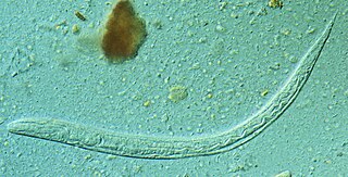
Strongyloides stercoralis is a human pathogenic parasitic roundworm causing the disease strongyloidiasis. Its common name in the US is threadworm. In the UK and Australia, however, the term threadworm can also refer to nematodes of the genus Enterobius, otherwise known as pinworms.

<span class="mw-page-title-main">Trichinosis</span> Parasitic disease due to invasion by Trichinella spp.

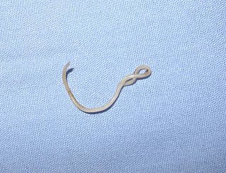
Trichinosis, also known as trichinellosis, is a parasitic disease caused by roundworms of the Trichinella type. During the initial infection, invasion of the intestines can result in diarrhea, abdominal pain, and vomiting. Migration of larvae to muscle, which occurs about a week after being infected, can cause swelling of the face, inflammation of the whites of the eyes, fever, muscle pains, and a rash. Minor infection may be without symptoms. Complications may include inflammation of heart muscle, central nervous system involvement, and inflammation of the lungs.

<i>Loa loa</i> Species of roundworm

Loa loa is a filarial (arthropod-borne) nematode (roundworm) that causes Loa loa filariasis. Loa loa actually means "worm worm", but is commonly known as the "eye worm", as it localizes to the conjunctiva of the eye. Loa loa is commonly found in Africa. It mainly inhabits rain forests in West Africa and has native origins in Ethiopia. The disease caused by Loa loa is called loiasis and is one of the neglected tropical diseases.

<span class="mw-page-title-main">Trematoda</span> Class of parasitic flatworms

Trematoda is a class of flatworms known as flukes or trematodes. They are obligate internal parasites with a complex life cycle requiring at least two hosts. The intermediate host, in which asexual reproduction occurs, is usually a snail. The definitive host, where the flukes sexually reproduce, is a vertebrate. Infection by trematodes can cause disease in all five traditional vertebrate classes: mammals, birds, amphibians, reptiles, and fish.

Gnathostomiasis, also known as larva migrans profundus, is the human infection caused by the nematode Gnathostoma spinigerum and/or Gnathostoma hispidum, which infects vertebrates.

<i>Anisakis</i> Genus of parasitic nematodes

Anisakis is a genus of parasitic nematodes that have life cycles involving fish and marine mammals. They are infective to humans and cause anisakiasis. People who produce immunoglobulin E in response to this parasite may subsequently have an allergic reaction, including anaphylaxis, after eating fish infected with Anisakis species.

Opisthorchis viverrini, common name Southeast Asian liver fluke, is a food-borne trematode parasite from the family Opisthorchiidae that infects the bile duct. People are infected after eating raw or undercooked fish. Infection with the parasite is called opisthorchiasis. O. viverrini infection also increases the risk of cholangiocarcinoma, a cancer of the bile ducts.

<span class="mw-page-title-main">Eucestoda</span> Subclass of flatworms

Eucestoda, commonly referred to as tapeworms, is the larger of the two subclasses of flatworms in the class Cestoda. Larvae have six posterior hooks on the scolex (head), in contrast to the ten-hooked Cestodaria. All tapeworms are endoparasites of vertebrates, living in the digestive tract or related ducts. Examples are the pork tapeworm with a human definitive host, and pigs as the secondary host, and Moniezia expansa, the definitive hosts of which are ruminants.

Spirometra erinaceieuropaei is a parasitic tapeworm that infects domestic animals and humans. The medical term for this infection in humans and other animals is sparganosis. Morphologically, these worms are similar to other worms in the genus Spirometra. They have a long body consisting of three sections: the scolex, the neck, and the strobilia. They have a complex life cycle that consists of three hosts, and can live in varying environments and bodily tissues. Humans can contract this parasite in three main ways. Historically, humans are considered a paratenic host; however, the first case of an adult S. erinaceieuropaei infection in humans was reported in 2017. Spirometra tapeworms exist worldwide and infection is common in animals, but S. erinaceieuropaei infections are rare in humans. Treatment for infection typically includes surgical removal and anti-worm medication.

<i>Toxocara canis</i> Species of roundworm

Toxocara canis is a worldwide-distributed helminth parasite that primarily infects dogs and other canids, but can also infect other animals including humans. The name is derived from the Greek word "toxon," meaning bow or quiver, and the Latin word "caro," meaning flesh. T. canis live in the small intestine of the definitive host. This parasite is very common in puppies and somewhat less common in adult dogs. In adult dogs, infection is usually asymptomatic but may be characterized by diarrhea. By contrast, untreated infection with Toxocara canis can be fatal in puppies, causing diarrhea, vomiting, pneumonia, enlarged abdomen, flatulence, poor growth rate, and other complications.

<span class="mw-page-title-main">Asian swamp eel</span> Species of fish

The Asian swamp eel, also known as rice eel, ricefield eel, rice paddy eel or white rice-field eel, is a commercially important, air-breathing species of fish in the family Synbranchidae. It occurs in East and Southeast Asia, where it is a very common foodstuff sold throughout the region. It has been introduced to two areas near the Everglades in Florida and near Atlanta in Georgia.

Angiostrongyliasis is an infection by a roundworm of the Angiostrongylus type. Symptoms may vary from none, to mild, to meningitis.

<i>Gongylonema pulchrum</i> Species of human parasite

Gongylonema pulchrum is the only parasite of the genus Gongylonema capable of infecting humans.

<i>Oesophagostomum</i> Genus of roundworms

Oesophagostomum is a genus of parasitic nematodes (roundworms) of the family Strongylidae. These worms occur in Africa, Brazil, China, Indonesia and the Philippines. The majority of human infection with Oesophagostomum is localized to northern Togo and Ghana. Because the eggs may be indistinguishable from those of the hookworms, the species causing human helminthomas are rarely identified with accuracy. Oesophagostomum, especially O. bifurcum, are common parasites of livestock and animals like goats, pigs and non-human primates, although it seems that humans are increasingly becoming favorable hosts as well. The disease they cause, oesophagostomiasis, is known for the nodule formation it causes in the intestines of its infected hosts, which can lead to more serious problems such as dysentery. Although the routes of human infection have yet to be elucidated sufficiently, it is believed that transmission occurs through oral-fecal means, with infected humans unknowingly ingesting soil containing the infectious filariform larvae.

<span class="mw-page-title-main">Cestoda</span> Class of flatworms

Cestoda is a class of parasitic worms in the flatworm phylum (Platyhelminthes). Most of the species—and the best-known—are those in the subclass Eucestoda; they are ribbon-like worms as adults, known as tapeworms. Their bodies consist of many similar units known as proglottids—essentially packages of eggs which are regularly shed into the environment to infect other organisms. Species of the other subclass, Cestodaria, are mainly fish infecting parasites.

Gnathostoma hispidum is a nematode (roundworm) that infects many vertebrate animals including humans. Infection of Gnathostoma hispidum, like many species of Gnathostoma causes the disease gnathostomiasis due to the migration of immature worms in the tissues.

Trichinella papuae is a nematode parasite responsible for a zoonotic disease called trichinellosis, predominantly in Thailand. Currently, eight species of Trichinella are known.

<i>Anisakis simplex</i> Species of parasitic nematode

Anisakis simplex, known as the herring worm, is a species of nematode in the genus Anisakis. Like other nematodes, it infects and settles in the organs of marine animals, such as salmon, mackerels and squids. It is commonly found in cold marine waters, such as the Pacific Ocean and Atlantic Ocean.

<i>Contracaecum</i> Genus of roundworms

Contracaecum is a genus of parasitic nematodes from the family Anisakidae. These nematodes are parasites of warm-blooded, fish eating animals, i.e. mammals and birds, as sexually mature adults. The eggs and the successive stages of their larvae use invertebrates and increasing size classes of fishes as intermediate hosts. It is the only genus in the family Anisakidae which can infect terrestrial, marine and freshwater animals.

<span class="mw-page-title-main">Gastropod-borne parasitic disease</span> Medical condition

Gastropod-borne parasitic diseases (GPDs) are a group of infectious diseases that require a gastropod species to serve as an intermediate host for a parasitic organism that can infect humans upon ingesting the parasite or coming into contact with contaminated water sources. These diseases can cause a range of symptoms, from mild discomfort to severe, life-threatening conditions, with them being prevalent in many parts of the world, particularly in developing regions. Preventive measures such as proper sanitation and hygiene practices, avoiding contact with infected gastropods and cooking or boiling food properly can help to reduce the risk of these diseases.

References

  1. Barnes, R. 1987. Invertebrate Zoology. Orlando, Florida: Dryden Press
  2. 1 2 Maleewong, Wanchai; Intapan, Pewpan M.; Khempila, Jeerapa; Wongwajana, Suwin; Wongkham, Chaisiri; Morakote, Nimit (1995). "Gnathostoma spinigerum: Growth and Development of Third-Stage Larvae In vitro". The Journal of Parasitology. 81 (5): 800–3. doi:10.2307/3283983. ISSN   0022-3395. JSTOR   3283983. PMID   7472883.
  3. 1 2 3 Maleewong W, Loahabhan P, Wongkham C, Intapan P, Morakote N, Khamboonruang C (1992). "Effects of albendazole on Gnathostoma spinigerum in mice". J. Parasitol. 78 (1): 125–6. doi:10.2307/3283698. JSTOR   3283698. PMID   1738054.
  4. Roberts, L., J. Janovy. 2000. Gerald D. Schmidt and Larry S. Roberts' Foundations of Parasitology, 6th edition. Boston: McGraw-Hill Higher Education
  5. Daengsvang, S., U. Chulalerk, T. Papasarathorn, B. Tongkoom. 1964. Epidemiological Observations On Gnathostoma spinigerum In Thailand. Journal of Tropical Medicine and Hygiene, 67: 144-147
  6. Daengsvang, S., P. Thienprasitthi, P. Chomcherngpat. 1966. Further Investigations On Natural And Experimental Hosts Of Larvae Of Gnathostoma Spinigerum In Thailand. American Journal of Tropical Medicine and Hygiene, 15: 727-729
  7. Daengsvang, S. 1949. Human Gnathostomaisis in Siam with reference to the method of prevention. The Journal of Parasitology 35: 116-121.