Lymphatic filariasis is a human disease caused by parasitic worms known as filarial worms.[2][3] Usually acquired in childhood, it is a leading cause of permanent disability worldwide, impacting over a hundred million people and manifesting itself in a variety of severe clinical pathologies[6][7] While most cases have no symptoms, some people develop a syndrome called elephantiasis, which is marked by severe swelling in the arms, legs, breasts, or genitals. The skin may become thicker as well, and the condition may become painful.[2] Affected people are often unable to work and are often shunned or rejected by others because of their disfigurement and disability.[7]
It is the first of the mosquito-borne diseases to have been identified.[8] The worms are spread by the bites of infected mosquitoes.[2] Three types of worms are known to cause the disease: Wuchereria bancrofti, Brugia malayi, and Brugia timori, with Wuchereria bancrofti being the most common.[2] These worms damage the lymphatic system by nesting within the lymphatic vessels and disrupting the system's normal function. Worms can survive within the human body for up to 8 years, all while reproducing millions of larvae which circulate through the blood.[9] The disease is diagnosed by microscopic examination of blood collected during the night. The blood is typically examined as a smear after being stained with Giemsa stain. Testing the blood for antibodies against the disease may also permit diagnosis.[4] Other roundworms from the same family are responsible for river blindness.[10]
Prevention can be achieved by treating entire groups in which the disease exists, known as mass deworming.[2] This is done every year for about six years, in an effort to rid a population of the disease entirely.[2]Medications usually include a combination of 2 or more anthelmintic agents: albendazole, ivermectin, and diethylcarbamazine.[11] Efforts to prevent mosquito bites are also recommended, including reducing the number of mosquitoes and promoting the use of bed nets.[2]
As of 2022, about 40 million people were infected, and about 863 million people were at risk of the disease in 47 countries.[5] It is most common in tropical Africa and Asia.[2] Lymphatic filariasis is classified as a neglected tropical disease and one of the four main worm infections.[10] The impact of the disease results in economic losses of billions of dollars a year.[2]
Most people infected with the worms that cause lymphatic filariasis never develop symptoms;[12] though some have damage to lymph vessels that can be detected by medical ultrasound.[13] Months to years after the initial infection, the worms die, triggering an immune response that manifests with repeated episodes of fever and painful swelling over the nearest lymph nodes (typically those along the groin).[12][13] In areas with endemic lymphatic filariasis, people are typically infected in childhood, and symptoms begin in adolescence.[13]
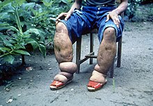
A subset of those affected have continued damage to their lymph vessels. Dysfunctional vessels fail to recirculate lymph fluid, which can pool (called lymphodema) in the nearest extremity – generally the arm, leg, breast, or scrotum.[13] Loss of lymph function (which transports immune cells) results in various repeated infections in the area.[14] Repeated cycles of infection, inflammation, and lymph vessel damage over several years cause the affected extremity to swell to extremely large size.[15] The surrounding skin thickens, becoming dry, discolored, and dotted with wartlike lumps that contain tortuous loops of lymph vessels.[14]
Causes
Life cycle of Wuchereria bancrofti, a parasite that causes lymphatic filariasis
Lymphatic filariasis is caused by infection with three different nematode worms: Wuchereria bancrofti (causes 90% of cases), Brugia malayi, and Brugia timori.[16] The three worms are transmitted by the bite of an infected mosquito – largely of genera Aedes, Anopheles, Culex, or Mansonia. When the mosquito bites, infectious nematode larvae are dropped onto the skin. They crawl into the bite wound, through the subcutaneous tissue, and into nearby lymph vessels. There, they develop into adults over about a year, with adult females up to 10 centimetres (3.9in) long, and males up to half that length.[17] Adult females and males mate, prompting the female to begin releasing a constant stream of larvae called "microfilariae" – more than 10,000 microfilariae each day for the adult's remaining lifespan of around five to eight years.[17] Microfilariae typically circulate in the blood stream at night; during the day they collect in the capillaries of the lungs.[17]
A mosquito that feeds on an infected person can take up microfilariae along with its blood meal. Inside the mosquito, the microfilariae pierce the stomach wall and crawl to the flight muscles, where they mature over 10 to 20 days into their human-infectious form. They then crawl to the mosquito's mouth to be deposited at its next bite, continuing the lifecycle.[17]
The disease itself is a result of a complex interplay between several factors: the worm, the endosymbioticWolbachia bacteria within the worm, the host's immune response, and the numerous opportunistic infections and disorders that arise. The adult worms live in the human lymphatic system and obstruct the flow of lymph throughout the body; this results in chronic lymphedema, most often noted in the lower torso (typically in the legs and genitals).[18] These worms can survive within the human body for up to 8 years, all while reproducing millions of larvae which circulate through the blood.[9]
Diagnosis
The preferred method for diagnosing lymphatic filariasis is by finding the microfilariae via microscopic examination of the blood. The blood sample is typically in the form of a thick smear, stained with Giemsa stain. Technicians analyzing the blood smear must be able to distinguish between W. bancrofti and other parasites potentially present. A blood smear is a simple and fairly accurate diagnostic tool, provided the blood sample is taken during when the microfilariae are in the peripheral circulation. Because the microfilariae only circulate in the blood at night, the blood specimen must be collected at night.[19]
It is often difficult or impossible to detect the causative organism in the peripheral blood, even in advanced cases.[20] In such cases, testing the blood serum for antibodies against the disease may also be used.[4] A polymerase chain reaction test can also be performed to detect a minute fraction, as little as 1 pg, of filarial DNA.[21] Dead, calcified worms can be detected by X-ray examinations. Ultrasonography can also be used to detect the movements and noises caused by the movement of adult worms.[22]
Differential diagnosis
Lymphatic filariasis may be confused with podoconiosis (also known as nonfilarial elephantiasis), a non-infectious disease caused by exposure of bare feet to irritant alkaline clay soils.[23][24] Podoconiosis however typically affects the legs bilaterally, while filariasis is generally unilateral.[23] Also, podoconiosis very rarely affects the groin while filariasis frequently involves the groin. Geographical location may also help to distinguish between these two diseases: podoconiosis is typically found in higher altitude areas with high seasonal rainfall, while filariasis is common in low-lying areas where mosquitos are prevalent.[23]
Protecting against mosquito bites in endemic regions is crucial to the prevention of lymphatic filariasis. Insect repellents and mosquito nets (especially when treated with an insecticide such as deltamethrin or permethrin)[25] have been demonstrated to reduce the transmission of lymphatic filariasis.[26][27] In addition residual spraying and personal protective equipment are known ways to control vectors.[9]
Worldwide eradication of lymphatic filariasis is the definitive goal. This is considered to be achievable since the disease has no known animal reservoir.[26] The World Health Organization (WHO) is coordinating the global effort to eradicate filariasis. The mainstay of this program is mass deworming of entire populations of people who are at risk with antifilarial drugs. The specific treatment depends on the co-endemicity of lymphatic filariasis with other filarial diseases. The WHO's annual MDA guidelines are listed below.
For areas co-endemic with loiasis 400mg of albendazole should be administered
for countries co-endemic with onchocerciasis, 200 mcg/kg of Ivermectin should be administered with 400mg of albendazole
in countries without onchocerciasis 6mg/kg of diethylcarbamazine citrate (DEC) and 400mg of albendazole should be used
in countries without onchocerciasis and the IDA Guidelines are met 200 mcg/kg of ivermectin should be used with 6mg/kg of diethylcarbamazine and 400mg of albendazole.[9][11]
Because the parasite requires a human host to reproduce, consistent treatment of at-risk populations (annually for a duration of four to six years)[2] is expected to break the cycle of transmission and cause the extinction of the causative organisms.[26]
In 2011, Sri Lanka was certified by the WHO as having eradicated lymphatic filariasis. In July 2017, the WHO announced that the disease had been eliminated in Tonga. Elimination of the disease has also occurred in Cambodia, China, Cook Islands, Egypt, Kiribati, Maldives, Marshall Islands, Niue, Palau, South Korea, Thailand, Vanuatu, Vietnam and Wallis and Futuna.[28] In 2020, the WHO announced that the 2030 targets for this program are that lymphatic filariasis will have been eliminated in 80% of endemic countries.[29]
Treatment of lymphatic filariasis depends in part on the geographic location of the area of the world in which the disease was acquired, but almost always involves the combination of 2 or more anthelmintic agents: albendazole, ivermectin, and diethylcarbamazine. In sub-Saharan Africa, the disease is usually treated with albendazole and ivermectin, whereas in the western pacific region of the world, all 3 anthelmintic agents are used. While Diethylcarbamazine in combination with albendazole is often used, it isn't as region specific as the other combinations.[11]
Wolbachia are endosymbiotic bacteria that live inside the gut of the parasites responsible for lymphatic filariasis, and provide nutrients necessary for their survival. Doxycycline kills these bacteria, which in turn prevents the maturation of microfilariae into adults. It also shortens the lifespan of the adult worms, causing them to die within 1 to 2 years instead of their normal 10 to 14-year lifespan.[31] Doxycycline is effective in treating lymphatic filariasis. Limitations of this antibiotic protocol include that it requires 4 to 6 weeks of treatment rather than the single dose of the anthelmintic agents, that doxycycline should not be used in young children and pregnant women, and that it is phototoxic.[32]
Albendazole is classified as an antihelmintics, which specifically works to kill worms.[33] The drug stops the worms from absorbing glucose, evidently leading to starvation and death from fatigue. The effects of albendazole alone have varying results, however, in combination with DEC drugs it has been found more effective.[34] Ivermectin is administered with albendazole, and works by binding to the nerve cells of the parasites, subsequently making them permeable to chloride. This leads to death by paralysis. Ivermectin, however, has been found to only kill the parasites in their early stages of life, and cannot kill an adult, live worm. Therefore, this drug is usually combined with DEC to kill both the microfilariae and the adult worms.[35]
Surgical treatment may be helpful in cases of scrotal elephantiasis and hydrocele. However, surgery is generally ineffective at correcting elephantiasis of the limbs.[36] Acute inflammatory responses due to lymphedema, and hydrocele can be reduced or prevented by practicing good hygiene, skin care, exercise and elevation of infected limbs.[9]
Lymphatic filariasis occurs in tropical and subtropical regions of Africa, Asia, Central America, the Caribbean, and South America, and certain Pacific Island nations. Elephantiasis caused by lymphatic filariasis is one of the most common causes of permanent disability in the world.[7] As of 2018, 51 million people were infected with lymphatic filariasis and at least 863 million people in 50 countries were living in areas that require preventive chemotherapy to stop the spread of infection. By 2022, the prevalence had declined to somewhere around 40 million and the disease remains endemic in 47 countries. These improvements are a direct result of the WHO's Global Programme to Eliminate Lymphatic Filariasis.[5] Since implementation, 740 million people no longer require preventative chemotherapy to treat the disease.[9]
W. bancrofti is responsible for 90% of lymphatic filariasis. Brugia malayi causes most of the remainder of the cases, while Brugia timori is a rare cause.[5]W. bancrofti largely affects areas across the broad equatorial belt (Africa, the Nile Delta, Turkey, India, the East Indies, Southeast Asia, Philippines, Oceanic Islands, and parts of South America). Due to the fact that Lymphatic Filariasis requires multiple mosquito bites over several months to years to spread of infection due to tourism is low.[37] The mosquito vectors of W. bancrofti have a preference for human blood; humans are apparently the only animals naturally infected with W. bancrofti. No reservoir host is known.[38] Lymphatic Filariasis is extremely uncommon in the United States, with only one reported case found in South Carolina in the early 1900s.[7]
In South America, four endemic countries have been working to beset lymphatic filariasis, consisting of Brazil, the Dominican Republic, Guyana, and Haiti.[39] In Latin America, the spread of lymphatic filariasis is through W. brancrofti, the only anthropods within the region, culex quinquefasciatus.[40] The exponential rate of development within the Americas is being combated through the development of an MDA program. MDA program, a 3-step drug administering program, has led to a 67% decrease in the need for the drug program.[39] Brazil targeted the rising endemic by administering DEC drugs through an MDA program to the communities hit hardest by the disease. By providing these drugs annually, as well as offering post-care, through showing family members how to treat the disease, creating connections for jobs, as well as providing a social network to incorporate patients into society, Brazil has made the most effort to provide care.[41] Dominican Republic has administered 5 rounds of DEC drugs annually for five years, spanning from 2002-2007. After the initial drastic action, the Dominican then administered another three rounds of MDA. Guyana also used DEC drugs to focus on preventing the spread of the disease, using a DEC-fortified salt from 2003-2007 and ultimately switching to MDA with DEC from 2014 to the present. Targeting patient education and access to treatment.[42] Haiti then focused on the disease by implementing DEC drug in 2002. It reached full geographical coverage by 2012, subsequently in 2014 about 20 communities had eradicated the need for MDA.
In areas endemic for podoconiosis, prevalence can be 5% or higher.[43] In communities where lymphatic filariasis is endemic, as many as 10% of women can be affected by swollen limbs, and 50% of men can develop mutilating genital symptoms.[44]
History
A man in Japan is helped to carry his enlarged scrotum19-year-old woman with elephantiasis, c. 1878
There is evidence of Lymphatic filariasis cases dating back 4000 years.[45] The ancient Vedic text, the Rig Veda, composed around 1500 BC–1200 BC, makes a possible reference to elephantiasis. The 50th hymn of the 7th book of the Rigveda calls on the gods Mitra, Varuna and Agni for protection against "that which nests inside and swells". The author of the hymn implores the deities to not let the worm wound his foot. The disease is described as causing eruptions to appear on the ankles and the knees.[46] Artifacts from ancient Egypt (2000 BC) and the Nok civilization in West Africa (500 BC) show possible elephantiasis symptoms. The first clear reference to the disease occurs in ancient Greek literature, wherein scholars differentiated the often similar symptoms of lymphatic filariasis from those of leprosy, describing leprosy as elephantiasis graecorum and lymphatic filariasis as elephantiasis arabum.[45]
The first documentation of symptoms occurred in the 16th century, when Jan Huyghen van Linschoten wrote about the disease during the exploration of Goa. Similar symptoms were reported by subsequent explorers in areas of Asia and Africa, though an understanding of the disease did not begin to develop until centuries later.[citation needed]
The causative agents were first identified in the late 19th century.[47] In 1866, Timothy Lewis, building on the work of Jean Nicolas Demarquay[de] and Otto Henry Wucherer, made the connection between microfilariae and elephantiasis, establishing the course of research that would ultimately explain the disease. In 1876, Joseph Bancroft discovered the adult form of the worm.[48] In 1877, the lifecycle involving an arthropod vector was theorized by Patrick Manson, who proceeded to demonstrate the presence of the worms in mosquitoes. Manson incorrectly hypothesized that the disease was transmitted through skin contact with water in which the mosquitoes had laid eggs.[49] In 1900, George Carmichael Low determined the actual transmission method by discovering the presence of the worm in the proboscis of the mosquito vector.[45]
Many people in Malabar, Nayars as well as Brahmans and their wives – in fact about a quarter or a fifth of the total population, including the people of the lowest castes – have very large legs, swollen to a great size; and they die of this, and it is an ugly thing to see. They say that this is due to the water through which they go, because the country is marshy. This is called pericaes in the native language, and all the swelling is the same from the knees downward, and they have no pain, nor do they take any notice of this infirmity.
Researchers at the University of Illinois at Chicago (UIC) have developed a novel vaccine for the prevention of lymphatic filariasis. This vaccine has been shown to elicit strong, protective immune responses in mouse models of lymphatic filariasis infection. The immune response elicited by this vaccine has been demonstrated to be protective against both W. bancrofti and B. malayi infection in the mouse model and may prove useful in the human.[51]
On 20 September 2007, geneticists published the first draft of the complete genome (genetic content) of Brugia malayi, one of the roundworms which causes lymphatic filariasis.[52] This project had been started in 1994 and by 2000, 80% of the genome had been determined. Determining the content of the genes might lead to the development of new drugs and vaccines.[53]
Loa loa filariasis, (Loiasis) is a skin and eye disease caused by the nematode worm Loa loa. Humans contract this disease through the bite of a deer fly or mango fly, the vectors for Loa loa. The adult Loa loa filarial worm can reach from three to seven centimetres long and migrates throughout the subcutaneous tissues of humans, occasionally crossing into subconjunctival tissues of the eye where it can be easily observed. Loa loa does not normally affect vision but can be painful when moving about the eyeball or across the bridge of the nose. Loiasis can cause red itchy swellings below the skin called "Calabar swellings". The disease is treated with the drug diethylcarbamazine (DEC), and when appropriate, surgical methods may be employed to remove adult worms from the conjunctiva. Loiasis belongs to the group of neglected tropical diseases, and there is a call for it to be included in the high priority listing.
Loa loa is a filarial (arthropod-borne) nematode (roundworm) that causes Loa loa filariasis. Loa loa actually means "worm worm", but is commonly known as the "eye worm", as it localizes to the conjunctiva of the eye. Loa loa is commonly found in Africa. It mainly inhabits rain forests in West Africa and has native origins in Ethiopia. The disease caused by Loa loa is called loiasis and is one of the neglected tropical diseases.
Diethylcarbamazine is a medication used in the treatment of filariasis including lymphatic filariasis, tropical pulmonary eosinophilia, and loiasis. It may also be used for prevention of loiasis in those at high risk. While it has been used for onchocerciasis, ivermectin is preferred. It is taken by mouth.
Elephantiasis, often incorrectly called elephantitis, is the enlargement and hardening of limbs or body parts due to tissue swelling. It is characterised by edema, hypertrophy, and fibrosis of skin and subcutaneous tissues, due to obstruction of lymphatic vessels. It may affect the genitalia. The term elephantiasis is often used in reference to parasitic worm infections, but may refer to a variety of diseases that swell parts of the subject's body to exceptionally massive proportions.
Onchocerciasis, also known as river blindness, is a disease caused by infection with the parasitic worm Onchocerca volvulus. Symptoms include severe itching, bumps under the skin, and blindness. It is the second-most common cause of blindness due to infection, after trachoma.
Filariasis, is a filarial infection caused by parasitic nematodes (roundworms) spread by different vectors. They are included in the list of neglected tropical diseases.
Wuchereria bancrofti is a filarial (arthropod-borne) nematode (roundworm) that is the major cause of lymphatic filariasis. It is one of the three parasitic worms, together with Brugia malayi and B. timori, that infect the lymphatic system to cause lymphatic filariasis. These filarial worms are spread by a variety of mosquito vector species. W. bancrofti is the most prevalent of the three and affects over 120 million people, primarily in Central Africa and the Nile delta, South and Central America, the tropical regions of Asia including southern China, and the Pacific islands. If left untreated, the infection can develop into lymphatic filariasis. In rare conditions, it also causes tropical pulmonary eosinophilia. No vaccine is commercially available, but high rates of cure have been achieved with various antifilarial regimens, and lymphatic filariasis is the target of the World Health Organization Global Program to Eliminate Lymphatic Filariasis with the aim to eradicate the disease as a public-health problem by 2020. However, this goal was not met by 2020.
Albendazole is a broad-spectrum antihelmintic and antiprotozoal agent of the benzimidazole type. It is used for the treatment of a variety of intestinal parasite infections, including ascariasis, pinworm infection, hookworm infection, trichuriasis, strongyloidiasis, taeniasis, clonorchiasis, opisthorchiasis, cutaneous larva migrans, giardiasis, and gnathostomiasis, among other diseases.
Brugia malayi is a filarial (arthropod-borne) nematode (roundworm), one of the three causative agents of lymphatic filariasis in humans. Lymphatic filariasis, also known as elephantiasis, is a condition characterized by swelling of the lower limbs. The two other filarial causes of lymphatic filariasis are Wuchereria bancrofti and Brugia timori, which both differ from B. malayi morphologically, symptomatically, and in geographical extent.
Brugia pahangi is a parasitic roundworm belonging to the genus Brugia. It is a filarial nematode known to infect the lymph vessels of domestic cats and wild animals, causing a disease filariasis.
Acanthocheilonemiasis is a rare tropical infectious disease caused by a parasite known as Acanthocheilonema perstans. It can cause skin rashes, abdominal and chest pains, muscle and joint pains, neurological disorders and skin lumps. It is mainly found in Africa. The parasite is transmitted through the bite of small flies. Studies show that there are elevated levels of white blood cells.
Podoconiosis, also known as nonfilarial elephantiasis, is a disease of the lymphatic vessels of the lower extremities that is caused by chronic exposure to irritant soils. It is the second most common cause of tropical lymphedema after lymphatic filariasis, and it is characterized by prominent swelling of the lower extremities, which leads to disfigurement and disability. Methods of prevention include wearing shoes and using floor coverings. Mainstays of treatment include daily foot hygiene, compression bandaging, and when warranted, surgery of overlying nodules.
Mansonella perstans is a filarial (arthropod-borne) nematode (roundworm), transmitted by tiny blood-sucking flies called midges. Mansonella perstans is one of two filarial nematodes that causes serous cavity filariasis in humans. The other filarial nematode is Mansonella ozzardi. M. perstans is widespread in many parts of sub-Saharan Africa, parts of Central and South America, and the Caribbean.
Mansonelliasis is the condition of infection by the nematode Mansonella. The disease exists in Africa and tropical Americas, spread by biting midges or blackflies. It is usually asymptomatic.
Brugia timori is a filarial (arthropod-borne) nematode (roundworm) which causes the disease "Timor filariasis", or "Timorian filariasis". While this disease was first described in 1965, the identity of Brugia timori as the causative agent was not known until 1977. In that same year, Anopheles barbirostris was shown to be its primary vector. There is no known animal reservoir host.
The Filarioidea are a superfamily of highly specialised parasitic nematodes. Species within this superfamily are known as filarial worms or filariae. Infections with parasitic filarial worms cause disease conditions generically known as filariasis. Drugs against these worms are known as filaricides.
Mansonella streptocerca,, is a filarial (arthropod-borne) nematode (roundworm) causing the disease streptocerciasis. It is a common parasite in the skin of humans in the rain forests of Africa, where it is thought to be a parasite of chimpanzees, as well.
Tropical pulmonary eosinophilia, is characterized by cough, bronchospasm, wheezing, abdominal pain, and an enlarged spleen. Occurring most frequently in the Indian subcontinent and Southeast Asia, TPE is a clinical manifestation of lymphatic filariasis, a parasitic infection caused by filarial roundworms that inhabit the lymphatic vessels, lymph nodes, spleen, and bloodstream. Three species of filarial roundworms, all from the Onchocercidae family, cause human lymphatic filariasis: Wuchereria bancrofti, Brugia malayi, and Brugia timori.
Brugia is a genus for a group of small roundworms. They are among roundworms that cause the parasitic disease filariasis. Specifically, of the three species known, Brugia malayi and Brugia timori cause lymphatic filariasis in humans; and Brugia pahangi and Brugia patei infect domestic cats, dogs and other animals. They are transmitted by the bite of mosquitos.
Lymphatic filariasis in India refers to the presence of the disease lymphatic filariasis in India and the social response to the disease. In India, 99% of infections come from a type of mosquito spreading a type of worm through a mosquito bite. The treatment plan provides 400 million people in India with medication to eliminate the parasite. About 50 million people in India were carrying the worm as of the early 2010s, which is 40% of all the cases in the world. In collaboration with other countries around the world, India is participating in a global effort to eradicate lymphatic filariasis. If the worm is eliminated from India then the disease could be permanently eradicated. In October 2019 the Union health minister Harsh Vardhan said that India's current plan is on schedule to eradicate filariasis by 2021.
↑ Zhong M, McCarthy J, Bierwert L, Lizotte-Waniewski M, Chanteau S, Nutman TB, Ottesen EA, Williams SA (1996). "A polymerase chain reaction assay for detection of the parasite Wuchereria bancrofti in human blood samples". Am J Trop Med Hyg. 54 (4): 357–63. doi:10.4269/ajtmh.1996.54.357. PMID8615447.
↑ Amaral F, Dryer G, Figueredo-Silva J, Noroes J, Cavalcanti A, Samico SC, Santos A, Coutinho A (1994). "Live adult worms detected by ultrasonography in human Bancroftian filariasis". Am J Trop Med Hyg. 50 (6): 753–7. doi:10.4269/ajtmh.1994.50.753. PMID8024070.
↑ Davey G, Tekola F, Newport MJ (2007). "Podoconiosis: non-infectious geochemical elephantiasis". Transactions of the Royal Society of Tropical Medicine and Hygiene. 101 (12): 1175–80. doi:10.1016/j.trstmh.2007.08.013. PMID17976670.
↑ Gloviczki P (1995). "The management of lymphatic disorders". In Rutherford RB (ed.). Vascular surgery (4thed.). Philadelphia: WB Saunders. pp.1883–1945.
↑ Grove, David I (1990). A history of human helminthology. Wallingford: CAB International. pp.1–848. ISBN0-85198-689-7.
↑ Grove, David I (2014). Tapeworms, lice and prions: a compendium of unpleasant infections. Oxford: Oxford University Press. pp.1–602. ISBN978-0-19-964102-4.
↑ Williams SA, Lizotte-Waniewski MR, Foster J, Guiliano D, Daub J, Scott AL, Slatko B, Blaxter ML (April 2000). "The filarial genome project: analysis of the nuclear, mitochondrial and endosymbiont genomes of Brugia malayi". International Journal for Parasitology. 30 (4): 411–19. doi:10.1016/s0020-7519(00)00014-x. PMID10731564.
Despommier DD, Griffin DO, Gwadz RW, Hotez PJ, Knirsch CA (2019). "22. Lymphatic Filariae". Parasitic Diseases(PDF) (7ed.). Parasites Without Borders. pp.253–264. Retrieved 18 May 2024.
Hotez PJ (2022). "4. Elephantiasis: Lymphatic filariasis, endemic nonfilarial elephantiasis (podoconiosis), and dracunculiasis (Guinea worm)". Forgotten People, Forgotten Diseases: The Neglected Tropical Diseases and Their Impact on Global Health and Development (3ed.). John Wiley & Sons. ISBN9781683673897.
This page is based on this Wikipedia article Text is available under the CC BY-SA 4.0 license; additional terms may apply. Images, videos and audio are available under their respective licenses.