Mesodinium

Last updated

Mesodinium
Mesodinium species.png
Mesodinium species containing green plastids taken from cryptophyte prey [1]
Scientific classification
Domain:
(unranked):
SAR
(unranked):
Phylum:
Class:
Order:
Family:
Genus:
Mesodinium
Species

Mesodinium is a genus of ciliates that are widely distributed and are abundant in marine and brackish waters. [2] [3]

Currently, six marine species of Mesodinium have been described and grouped by nutritional mode: plastidic (M. chamaeleon, M. coatsi, M. major, and M. rubrum) or heterotrophic (M. pulex and M. pupula). There is some debate as to whether the nutritional mode of plastidic Mesodinium species is phototrophic (permanent plastid) or mixotrophic. [4] [5] Among the plastidic species, wild M. major and M. rubrum populations possess red plastids belonging to genera Teleaulax , Plagioselmis , and Geminigera , [6] [7] [8] while wild M. chamaeleon and M. coatsi populations normally contain green plastids. [9] [10] [11] The availability of suitable cryptophyte prey is important for bloom formation of plastidic Mesodinium species. [1]

The most common species, Mesodinium rubrum , causes red tides in many coastal ecosystems. Although M. rubrum is known as a nontoxic species, [12] blooms of the ciliate can be potentially harmful to aquaculture industries. [13] [14] [1] M. rubrum photosynthesizes by sequestering the nucleus of its cryptophyte prey, in order to maintain stolen plastids and other organelles. [15] In this way, the genus Mesodinium plays an important role in linking cryptophycean prey and diverse predators in the aquatic microbial food web. For example, the dinoflagellates Dinophysis spp., which are a predator of M. rubrum and the source of their cryptophyte-derived plastids, have been frequently observed to precede or to coincide with high densities of M. rubrum. [16] [17] [18] [19] [1]

See also

Related Research Articles

<span class="mw-page-title-main">Dinoflagellate</span> Unicellular algae with two flagella

The dinoflagellates are a monophyletic group of single-celled eukaryotes constituting the phylum Dinoflagellata and are usually considered protists. Dinoflagellates are mostly marine plankton, but they also are common in freshwater habitats. Their populations vary with sea surface temperature, salinity, and depth. Many dinoflagellates are photosynthetic, but a large fraction of these are in fact mixotrophic, combining photosynthesis with ingestion of prey.

<span class="mw-page-title-main">Cryptomonad</span> Subphylum of algae

The cryptomonads are a group of algae, most of which have plastids. They are common in freshwater, and also occur in marine and brackish habitats. Each cell is around 10–50 μm in size and flattened in shape, with an anterior groove or pocket. At the edge of the pocket there are typically two slightly unequal flagella.

<span class="mw-page-title-main">Chromista</span> Eukaryotic biological kingdom

Chromista is a proposed but polyphyletic biological kingdom, refined from the Chromalveolata, consisting of single-celled and multicellular eukaryotic species that share similar features in their photosynthetic organelles (plastids). It includes all eukaryotes whose plastids contain chlorophyll c and are surrounded by four membranes. If the ancestor already possessed chloroplasts derived by endosymbiosis from red algae, all non-photosynthetic Chromista have secondarily lost the ability to photosynthesise. Its members might have arisen independently as separate evolutionary groups from the last eukaryotic common ancestor.

<span class="mw-page-title-main">Kleptoplasty</span> Form of algae symbiosis

Kleptoplasty or kleptoplastidy is a process in symbiotic relationships whereby plastids, notably chloroplasts from algae, are sequestered by the host. The word is derived from Kleptes (κλέπτης) which is Greek for thief. The alga is eaten normally and partially digested, leaving the plastid intact. The plastids are maintained within the host, temporarily continuing photosynthesis and benefiting the host.

<span class="mw-page-title-main">Chromalveolata</span> Group of eukaryotic organisms

Chromalveolata was a eukaryote supergroup present in a major classification of 2005, then regarded as one of the six major groups within the eukaryotes. It was a refinement of the kingdom Chromista, first proposed by Thomas Cavalier-Smith in 1981. Chromalveolata was proposed to represent the organisms descended from a single secondary endosymbiosis involving a red alga and a bikont. The plastids in these organisms are those that contain chlorophyll c.

<i>Paulinella</i> Genus of single-celled organisms

Paulinella is a genus of at least eleven species including both freshwater and marine amoeboids. Like many members of euglyphids it is covered by rows of siliceous scales, and use filose pseudopods to crawl over the substrate of the benthic zone.

<span class="mw-page-title-main">Protozoa</span> Single-celled eukaryotic organisms that feed on organic matter

Protozoa are a polyphyletic group of single-celled eukaryotes, either free-living or parasitic, that feed on organic matter such as other microorganisms or organic debris. Historically, protozoans were regarded as "one-celled animals".

<i>Guillardia</i> Genus of single-celled organisms

Guillardia is a genus of marine biflagellate cryptomonad algae with a plastid obtained through secondary endosymbiosis of a red alga.

<i>Hemiselmis</i> Genus of single-celled organisms

Hemiselmis is a genus of cryptomonads.

Amoebophyra is a genus of dinoflagellates. Amoebophyra is a syndinian parasite that infects free-living dinoflagellates that are attributed to a single species by using several host-specific parasites. It acts as "biological control agents for red tides and in defining species of Amoebophrya." Researchers have found a correlation between a large amount of host specify and the impact host parasites may have on other organisms. Due to the host specificity found in each strain of Amoebophrya's physical makeup, further studies need to be tested to determine whether the Amoebophrya can act as a control against harmful algal blooms.

<i>Dinophysis</i> Genus of single-celled organisms

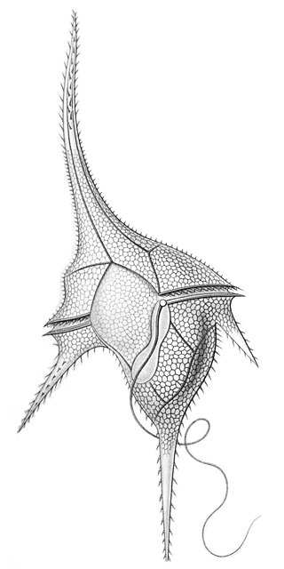
Dinophysis is a genus of dinoflagellates common in tropical, temperate, coastal and oceanic waters. It was first described in 1839 by Christian Gottfried Ehrenberg.

<i>Raphidiophrys contractilis</i> Species of single-celled organism

Raphidiophrys contractilis is a species of freshwater centrohelid.

Mesodinium chamaeleon is a ciliate of the genus Mesodinium. It is known for being able to consume and maintain algae endosymbiotically for days before digesting the algae. It has the ability to eat red and green algae, and afterwards using the chlorophyll granules from the algae to generate energy, turning itself from being a heterotroph into an autotroph. The species was discovered in January 2012 outside the coast of Nivå, Denmark by professor Øjvind Moestrup.

Vahlkampfia avara is a species of excavates. It has a PAS-positive surface layer and forms cysts in culture.

<i>Dinophysis acuminata</i> Species of dinoflagellate

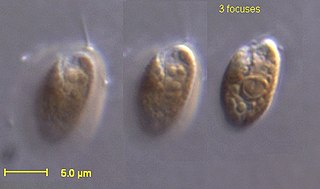
Dinophysis acuminata is a marine plankton species of dinoflagellates that is found in coastal waters of the north Atlantic and Pacific oceans. The genus Dinophysis includes both phototrophic and heterotrophic species. D. acuminata is one of several phototrophic species of Dinophysis classed as toxic, as they produce okadaic acid which can cause diarrhetic shellfish poisoning (DSP). Okadiac acid is taken up by shellfish and has been found in the soft tissue of mussels and the liver of flounder species. When contaminated animals are consumed, they cause severe diarrhoea. D. acuminata blooms are constant threat to and indication of diarrhoeatic shellfish poisoning outbreaks.

<i>Mesodinium rubrum</i> Species of single-celled organism

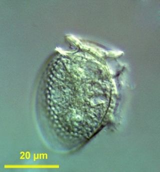
Mesodinium rubrum is a species of ciliates. It constitutes a plankton community and is found throughout the year, most abundantly in spring and fall, in coastal areas. Although discovered in 1908, its scientific importance came into light in the late 1960s when it attracted scientists by the recurrent red colouration it caused by forming massive blooms, that cause red tides in the oceans.

Karyoklepty is a strategy for cellular evolution, whereby a predator cell appropriates the nucleus of a cell from another organism to supplement its own biochemical capabilities.

<span class="mw-page-title-main">Mixotrophic dinoflagellate</span> Plankton

Dinoflagellates are eukaryotic plankton, existing in marine and freshwater environments. Previously, dinoflagellates had been grouped into two categories, phagotrophs and phototrophs. Mixotrophs, however include a combination of phagotrophy and phototrophy. Mixotrophic dinoflagellates are a sub-type of planktonic dinoflagellates and are part of the phylum Dinoflagellata. They are flagellated eukaryotes that combine photoautotrophy when light is available, and heterotrophy via phagocytosis. Dinoflagellates are one of the most diverse and numerous species of phytoplankton, second to diatoms.

<i>Licnophora</i> Genus of single-celled organisms

Licnophora is a genus of ciliates in the family Licnophoridae. They typically have an hourglass-like shape and live as ectocommensals on marine animals.

Arve Elvebakk is a Norwegian mycologist and professor working from the Arctic University of Norway in Tromsø. He has published widely on Arctic biology, and climatology. Additionally, he collaborates with many mycologists across the world, and has published names for lichens in Australia, New Zealand, the South Pacific, and South America, and the Antarctic.

References

  1. 1 2 3 4 Nishitani, G. and Yamaguchi, M. (2018) "Seasonal succession of ciliate Mesodinium spp. with red, green, or mixed plastids and their association with cryptophyte prey". Scientific reports, 8(1): 1–9. doi : 10.1038/s41598-018-35629-4. CC-BY icon.svg Material was copied from this source, which is available under a Creative Commons Attribution 4.0 International License.
  2. Leppanen, J. M. & Bruun, J. E. (1986) The role of pelagic ciliates including the autotrophic Mesodinium rubrum during the spring bloom of 1982 in the open northern Baltic proper. Ophelia 4, 147–157.
  3. Sanders, R. W. (1995) Seasonal distributions of the photosynthesizing ciliates Laboea strobila and Myrionecta rubra (=Mesodinium rubrum) in an estuary of the Gulf of Maine. Aquat. Microb. Ecol. 9, 237–242.
  4. Dajun Qiu; Liangmin Huang; Senjie Lin (10 October 2016). "Cryptophyte farming by symbiotic ciliate host detected in situ". Proceedings of the National Academy of Sciences of the United States of America . 113 (43): 12208–12213. Bibcode:2016PNAS..11312208Q. doi:10.1073/PNAS.1612483113. ISSN   0027-8424. PMC   5087057 . PMID   27791006. Wikidata   Q30826949.
  5. Matthew D Johnson; Erica Lasek-Nesselquist; Holly V Moeller; et al. (1 February 2017). "Mesodinium rubrum: The symbiosis that wasn't". Proceedings of the National Academy of Sciences of the United States of America . 114 (7): E1040–E1042. Bibcode:2017PNAS..114.1040J. doi:10.1073/PNAS.1619247114. ISSN   0027-8424. PMC   5320990 . PMID   28154148. Wikidata   Q42320407.
  6. Goh Nishitani; Satoshi Nagai; Katsuhisa Baba; et al. (May 2010). "High-level congruence of Myrionecta rubra prey and Dinophysis species plastid identities as revealed by genetic analyses of isolates from Japanese coastal waters". Applied and Environmental Microbiology . 76 (9): 2791–8. doi:10.1128/AEM.02566-09. ISSN   0099-2240. PMC   2863437 . PMID   20305031. Wikidata   Q24629387.
  7. L Herfort; TD Peterson; LA McCue; et al. (4 January 2011). "Myrionecta rubra population genetic diversity and its cryptophyte chloroplast specificity in recurrent red tides in the Columbia River estuary". Aquatic Microbial Ecology. 62 (1): 85–97. doi:10.3354/AME01460. ISSN   0948-3055. Wikidata   Q57272516.
  8. Lydie Herfort; Katie Maxey; Ian Voorhees; Holly M Simon; Kolette Grobler; Tawnya D Peterson; Peter Zuber (7 April 2017). "Use of Highly Specific Molecular Markers Reveals Positive Correlation between Abundances of Mesodinium cf. major and Its Preferred Prey, Teleaulax amphioxeia, During Red Water Blooms in the Columbia River Estuary". Journal of Eukaryotic Microbiology . 64 (6): 740–755. doi:10.1111/JEU.12407. ISSN   1066-5234. PMID   28258655. Wikidata   Q46343430.
  9. Lydia Garcia-Cuetos; Øjvind Moestrup; Per J Hansen (18 June 2012). "Studies on the genus Mesodinium II. Ultrastructural and molecular investigations of five marine species help clarifying the taxonomy". Journal of Eukaryotic Microbiology . 59 (4): 374–400. doi:10.1111/J.1550-7408.2012.00630.X. ISSN   1066-5234. PMID   22708786. Wikidata   Q34282405.
  10. Ojvind Moestrup; Lydia Garcia-Cuetos; Per Juel Hansen; Tom Fenchel (1 January 2012). "Studies on the genus Mesodinium I: ultrastructure and description of Mesodinium chamaeleon n. sp., a benthic marine species with green or red chloroplasts". Journal of Eukaryotic Microbiology . 59 (1): 20–39. doi:10.1111/J.1550-7408.2011.00593.X. ISSN   1066-5234. PMID   22221919. Wikidata   Q34030100.
  11. Seung Won Nam; Woongghi Shin; Misun Kang; Wonho Yih; Myung Gil Park (21 August 2014). "Ultrastructure and molecular phylogeny of Mesodinium coatsi sp. nov., a benthic marine ciliate". Journal of Eukaryotic Microbiology . 62 (1): 102–120. doi:10.1111/JEU.12150. ISSN   1066-5234. PMID   25047232. Wikidata   Q30837739.
  12. Lindholm, T. (1985) Mesodinium rubrum–a unique photosynthetic ciliate. Adv. Aquat. Microbiol. 3, 1–48.
  13. Hayes, G. C., Purdie, D. A. & Williams, J. A. (1989) The distribution of ichthyoplankton in Southampton Water in response to low oxygen levels produced by a Mesodinium rubrum bloom. J. Fish Biol. 34, 811–813.
  14. Liu, H. et al. (2012) Potential risk of Mesodinium rubrum bloom in the aquaculture area of Dapeng’ao cove, China: diurnal changes in the ciliate community structure in the surface water. Oceanologia 54, 109–117.
  15. Matthew D Johnson; David Oldach; Charles F. Delwiche; Diane K Stoecker (1 January 2007). "Retention of transcriptionally active cryptophyte nuclei by the ciliate Myrionecta rubra". Nature . 445 (7126): 426–428. doi:10.1038/NATURE05496. ISSN   1476-4687. PMID   17251979. Wikidata   Q34606048.
  16. Mouritsen, L. T. & Richardson, K. (2003) Vertical microscale patchiness in nano- and microplankton distributions in a stratifed estuary. J. Plankton Res. 25, 783–797.
  17. Conny O Sjöqvist; Tore J Lindholm (13 May 2011). "Natural co-occurrence of Dinophysis acuminata (Dinoflagellata) and Mesodinium rubrum (Ciliophora) in thin layers in a coastal inlet". Journal of Eukaryotic Microbiology . 58 (4): 365–372. doi:10.1111/J.1550-7408.2011.00559.X. ISSN   1066-5234. PMID   21569163. Wikidata   Q51598919.
  18. Lips, U. & Lips, I. (2014) Bimodal distribution patterns of motile phytoplankton in relation to physical processes and stratification (Gulf of Finland, Baltic Sea). Deep-Sea Res. II. 101, 107–119.
  19. L. Velo-Suárez; S. González-Gil; Y. Pazos; B. Reguera (March 2014). "The growth season of Dinophysis acuminata in an upwelling system embayment: A conceptual model based on in situ measurements". Deep-Sea Research. Part 2: Topical Studies in Oceanography. 101: 141–151. doi:10.1016/J.DSR2.2013.03.033. ISSN   0967-0645. Wikidata   Q60507600.