Mesodinium rubrum

Last updated

Mesodinium rubrum
Mesodinium rubrum.jpg
Mesodinium rubrum
Scientific classification
Domain:
(unranked):
SAR
(unranked):
Phylum:
Class:
Order:
Family:
Genus:
Species:
M. rubrum
Binomial name
Mesodinium rubrum
Hamburger and Buddenbrock, 1929
Synonyms

Halteria rubraLohmann, 1908
Myrionecta rubraLohmann, 1908
Cyclotrichium meunieriPowers, 1932
Mesodinium pulex Bakker, 1966

Contents

M. rubrum from north-west Black Sea Myrionecta rubra.jpg
M. rubrum from north-west Black Sea

Mesodinium rubrum (or Myrionecta rubra) is a species of ciliates. [1] It constitutes a plankton community and is found throughout the year, most abundantly in spring and fall, in coastal areas. Although discovered in 1908, its scientific importance came into light in the late 1960s when it attracted scientists by the recurrent red colouration it caused by forming massive blooms, [2] that cause red tides in the oceans. [3] [4]

Unlike typical protozoans, M. rubrum can make its own nutrition by photosynthesis. The unusual autotrophic property was discovered in 2006 when genetic sequencing revealed that the photosynthesising organelles, plastids, were derived from the ciliate's principal food, the autotrophic algae called cryptomonads (or cryptophytes), which contain endosymbiont red algae whose internal chloroplasts (evolved via endosymbiosis with cyanobacteria) indirectly enable M. rubrum to photosynthesize using sunlight. [5] The ciliate is thus both autotrophic and heterotrophic at the same time. This also indicates that it is an example of multiple-stage endosymbiosis, an amazing instance of what life are capable of evolving according to the endosymbiotic theory, as well as the concept of stealing of cell organelles called kleptoplastidy. [6] Moreover, M. rubrum represents additional endosymbiosis by transferring its plastids to its predators, the dinoflagellate planktons belonging to the genus Dinophysis. [7] [8]

In 2009, a new species of Gram-negative bacteria called Maritalea myrionectae was discovered from a cell culture of M. rubrum. [9]

Description

M. rubrum is a free-living marine ciliate. It is reddish in colour and form dark-red mass during blooming. Its body is almost spherical, looking like a miniature sunflower with its radiating hair-like cilia on its body surface. It measures up to 100 μm in length and 75 μm in width. The body is superficially divided into two lobes due to formation of a constriction at the centre. The constriction gives rise to a larger anterior lobe and a smaller posterior lobe. The cilia arise from the constriction. Using the cilia it can jump about 10-20 times its body length in one movement. [10] Its nucleus is prominently situated at the centre, and is surrounded by organelles mostly derived from algae. For example, its cytoplasm contains numerous plastids, mitochondria and other nuclei. These organelles are properly separated such that the mitochondria are fully enclosed in a vacuole membrane and two endoplasmic reticulum membranes of the ciliate. [6] This indicates that the ciliate is primarily a heterotroph, but after acquiring algal plastid, it transforms into an autotroph.

The endosymbiont

Genetic analysis showed that in the American coastal areas, the primary food of M. rubrum is the algae most closely related to the free-living Geminigera cryophila . [5] But in Japanese coasts, the major algal species is Teleaulax amphioxeia . [8] When these plastid-containing algae are ingested by the ciliate, they are not digested. The plastids remain functional and provide nutrition to the ciliate by photosynthesis. In order for the plastids to be normally active, they still require enzymes, which are synthesised by the sequestered algal nuclei. The single nucleus can survive and remain genetically active up to 30 days in the cytoplasm of the ciliate. [11] As the retention time of the prey nuclei is short, an average M. rubrum cell may contain eight algal plastids per single prey nucleus and the nuclei need to be replaced by continuous feeding on fresh algae. Thus, the algal organelles are not permanently integrated. [5]

Related Research Articles

<span class="mw-page-title-main">Chloroplast</span> Plant organelle that conducts photosynthesis

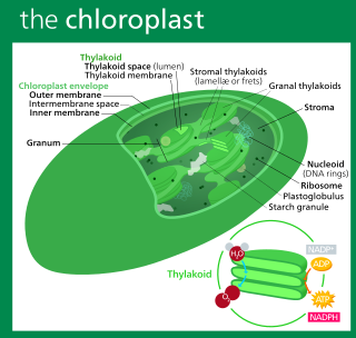
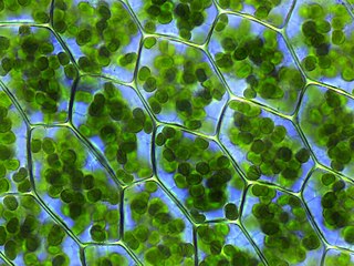
A chloroplast is a type of membrane-bound organelle known as a plastid that conducts photosynthesis mostly in plant and algal cells. The photosynthetic pigment chlorophyll captures the energy from sunlight, converts it, and stores it in the energy-storage molecules ATP and NADPH while freeing oxygen from water in the cells. The ATP and NADPH is then used to make organic molecules from carbon dioxide in a process known as the Calvin cycle. Chloroplasts carry out a number of other functions, including fatty acid synthesis, amino acid synthesis, and the immune response in plants. The number of chloroplasts per cell varies from one, in unicellular algae, up to 100 in plants like Arabidopsis and wheat.

<span class="mw-page-title-main">Symbiogenesis</span> Evolutionary theory holding that eukaryotic organelles evolved through symbiosis with prokaryotes

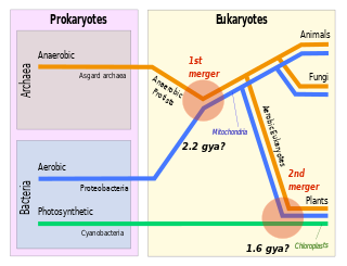
Symbiogenesis is the leading evolutionary theory of the origin of eukaryotic cells from prokaryotic organisms. The theory holds that mitochondria, plastids such as chloroplasts, and possibly other organelles of eukaryotic cells are descended from formerly free-living prokaryotes taken one inside the other in endosymbiosis. Mitochondria appear to be phylogenetically related to Rickettsiales bacteria, while chloroplasts are thought to be related to cyanobacteria.

<span class="mw-page-title-main">Dinoflagellate</span> Unicellular algae with two flagella

The dinoflagellates are a monophyletic group of single-celled eukaryotes constituting the phylum Dinoflagellata and are usually considered protists. Dinoflagellates are mostly marine plankton, but they also are common in freshwater habitats. Their populations vary with sea surface temperature, salinity, and depth. Many dinoflagellates are photosynthetic, but a large fraction of these are in fact mixotrophic, combining photosynthesis with ingestion of prey.

<span class="mw-page-title-main">Alveolate</span> Superphylum of protists

The alveolates are a group of protists, considered a major clade and superphylum within Eukarya. They are currently grouped with the stramenopiles and Rhizaria among the protists with tubulocristate mitochondria into the SAR supergroup.

<span class="mw-page-title-main">Cryptomonad</span> Subphylum of algae

The cryptomonads are a group of algae, most of which have plastids. They are common in freshwater, and also occur in marine and brackish habitats. Each cell is around 10–50 μm in size and flattened in shape, with an anterior groove or pocket. At the edge of the pocket there are typically two slightly unequal flagella.

<span class="mw-page-title-main">Plastid</span> Plant cell organelles that perform photosynthesis and store starch

A plastid, pl.plastids, is a membrane-bound organelle found in the cells of plants, algae, and some other eukaryotic organisms;. They are considered to be intracellular endosymbiotic cyanobacteria.

<span class="mw-page-title-main">Chromista</span> Eukaryotic biological kingdom

Chromista is a proposed but polyphyletic biological kingdom, refined from the Chromalveolata, consisting of single-celled and multicellular eukaryotic species that share similar features in their photosynthetic organelles (plastids). It includes all eukaryotes whose plastids contain chlorophyll c and are surrounded by four membranes. If the ancestor already possessed chloroplasts derived by endosymbiosis from red algae, all non-photosynthetic Chromista have secondarily lost the ability to photosynthesise. Its members might have arisen independently as separate evolutionary groups from the last eukaryotic common ancestor.

<span class="mw-page-title-main">Nucleomorph</span>

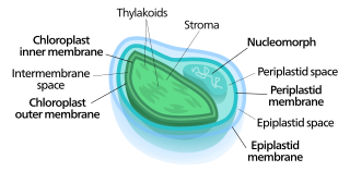
Nucleomorphs are small, vestigial eukaryotic nuclei found between the inner and outer pairs of membranes in certain plastids. They are thought to be vestiges of primitive red and green algal nuclei that were engulfed by a larger eukaryote. Because the nucleomorph lies between two sets of membranes, nucleomorphs support the endosymbiotic theory and are evidence that the plastids containing them are complex plastids. Having two sets of membranes indicate that the plastid, a prokaryote, was engulfed by a eukaryote, an alga, which was then engulfed by another eukaryote, the host cell, making the plastid an example of secondary endosymbiosis.

<span class="mw-page-title-main">Kleptoplasty</span> Form of algae symbiosis

Kleptoplasty or kleptoplastidy is a process in symbiotic relationships whereby plastids, notably chloroplasts from algae, are sequestered by the host. The word is derived from Kleptes (κλέπτης) which is Greek for thief. The alga is eaten normally and partially digested, leaving the plastid intact. The plastids are maintained within the host, temporarily continuing photosynthesis and benefiting the host.

<span class="mw-page-title-main">Cryptophyceae</span> Class of single-celled organisms

The cryptophyceae are a class of algae, most of which have plastids. About 230 species are known, and they are common in freshwater, and also occur in marine and brackish habitats. Each cell is around 10–50 μm in size and flattened in shape, with an anterior groove or pocket. At the edge of the pocket there are typically two slightly unequal flagella.

<span class="mw-page-title-main">Protozoa</span> Single-celled eukaryotic organisms that feed on organic matter

Protozoa are a polyphyletic group of single-celled eukaryotes, either free-living or parasitic, that feed on organic matter such as other microorganisms or organic debris. Historically, protozoans were regarded as "one-celled animals".

<i>Guillardia</i> Genus of single-celled organisms

Guillardia is a genus of marine biflagellate cryptomonad algae with a plastid obtained through secondary endosymbiosis of a red alga.

<i>Dinophysis</i> Genus of single-celled organisms

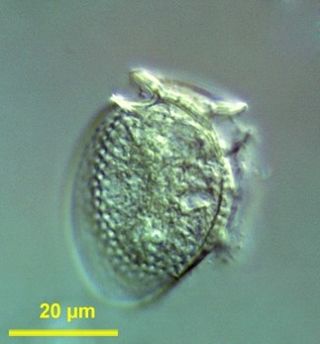
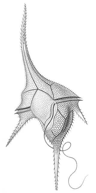
Dinophysis is a genus of dinoflagellates common in tropical, temperate, coastal and oceanic waters. It was first described in 1839 by Christian Gottfried Ehrenberg.

A mixotroph is an organism that can use a mix of different sources of energy and carbon, instead of having a single trophic mode on the continuum from complete autotrophy at one end to heterotrophy at the other. It is estimated that mixotrophs comprise more than half of all microscopic plankton. There are two types of eukaryotic mixotrophs: those with their own chloroplasts, and those with endosymbionts—and those that acquire them through kleptoplasty or through symbiotic associations with prey or enslavement of their organelles.

Mesodinium chamaeleon is a ciliate of the genus Mesodinium. It is known for being able to consume and maintain algae endosymbiotically for days before digesting the algae. It has the ability to eat red and green algae, and afterwards using the chlorophyll granules from the algae to generate energy, turning itself from being a heterotroph into an autotroph. The species was discovered in January 2012 outside the coast of Nivå, Denmark by professor Øjvind Moestrup.

<i>Dinophysis acuminata</i> Species of dinoflagellate

Dinophysis acuminata is a marine plankton species of dinoflagellates that is found in coastal waters of the north Atlantic and Pacific oceans. The genus Dinophysis includes both phototrophic and heterotrophic species. D. acuminata is one of several phototrophic species of Dinophysis classed as toxic, as they produce okadaic acid which can cause diarrhetic shellfish poisoning (DSP). Okadiac acid is taken up by shellfish and has been found in the soft tissue of mussels and the liver of flounder species. When contaminated animals are consumed, they cause severe diarrhoea. D. acuminata blooms are constant threat to and indication of diarrhoeatic shellfish poisoning outbreaks.

Karyoklepty is a strategy for cellular evolution, whereby a predator cell appropriates the nucleus of a cell from another organism to supplement its own biochemical capabilities.

<span class="mw-page-title-main">Floridean starch</span> Type of storage glucan

Floridean starch is a type of a storage glucan found in glaucophytes and in red algae, in which it is usually the primary sink for fixed carbon from photosynthesis. It is found in grains or granules in the cell's cytoplasm and is composed of an α-linked glucose polymer with a degree of branching intermediate between amylopectin and glycogen, though more similar to the former. The polymers that make up floridean starch are sometimes referred to as "semi-amylopectin".

<i>Mesodinium</i> Genus of single-celled organisms

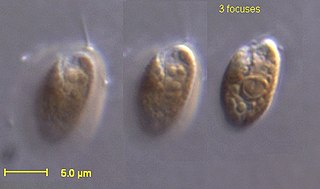
Mesodinium is a genus of ciliates that are widely distributed and are abundant in marine and brackish waters.

<span class="mw-page-title-main">Marine protists</span> Protists that live in saltwater or brackish water

Marine protists are defined by their habitat as protists that live in marine environments, that is, in the saltwater of seas or oceans or the brackish water of coastal estuaries. Life originated as marine single-celled prokaryotes and later evolved into more complex eukaryotes. Eukaryotes are the more developed life forms known as plants, animals, fungi and protists. Protists are the eukaryotes that cannot be classified as plants, fungi or animals. They are mostly single-celled and microscopic. The term protist came into use historically as a term of convenience for eukaryotes that cannot be strictly classified as plants, animals or fungi. They are not a part of modern cladistics because they are paraphyletic.

References

  1. Gustafson, Daniel E.; Stoecker, Diane K.; Johnson, Matthew D.; Van Heukelem, William F.; Sneider, Kerri (2000). "Cryptophyte algae are robbed of their organelles by the marine ciliate Mesodinium rubrum". Nature. 405 (6790): 1049–1052. doi:10.1038/35016570. PMID   10890444. S2CID   205007270.
  2. Ryther, John H. (1967). "Occurrence of red water off Peru". Nature. 214 (5095): 1318–1319. doi:10.1038/2141318a0. S2CID   4151183.
  3. Johnson, William S.; Fylling, Dennis M. Allen (2012). Zooplankton of the Atlantic and Gulf coasts : a guide to their identification and ecology (2 ed.). Baltimore: Johns Hopkins University Press. p. 82. ISBN   978-1-421406183.
  4. Fehling, Johanna; Stoecker, Diane; Baldauf, Sandra L (2007). "Photosynthesis and the eukaryote tree of life". In Falkowski, Paul G.; Knoll, Andrew H. (eds.). Evolution of Primary Producers in the Sea. Amsterdam: Elsevier Academic Press. p. 97. ISBN   978-0-08-055051-0.
  5. 1 2 3 Johnson, Matthew D.; Tengs, Torstein; Oldach, David; Stoecker, Diane K. (2006). "Sequestration, performance, and functional control of cryptophyte plastids in the ciliate Myrionecta rubra (Ciliophora)". Journal of Phycology. 42 (6): 1235–1246. doi:10.1111/j.1529-8817.2006.00275.x. S2CID   85408210.
  6. 1 2 Nowack, E. C. M.; Melkonian, M. (2010). "Endosymbiotic associations within protists". Philosophical Transactions of the Royal Society B: Biological Sciences. 365 (1541): 699–712. doi:10.1098/rstb.2009.0188. PMC   2817226 . PMID   20124339.
  7. Janson, Sven (2004). "Molecular evidence that plastids in the toxin-producing dinoflagellate genus Dinophysis originate from the free-living cryptophyte Teleaulax amphioxeia". Environmental Microbiology. 6 (10): 1102–1106. doi:10.1111/j.1462-2920.2004.00646.x. PMID   15344936.
  8. 1 2 Nishitani, G.; Nagai, S.; Baba, K.; Kiyokawa, S.; Kosaka, Y.; Miyamura, K.; Nishikawa, T.; Sakurada, K.; Shinada, A.; Kamiyama, T. (2010). "High-level congruence of Myrionecta rubra prey and Dinophysis species plastid identities as revealed by genetic analyses of isolates from Japanese coastal waters". Applied and Environmental Microbiology. 76 (9): 2791–2798. doi:10.1128/AEM.02566-09. PMC   2863437 . PMID   20305031.
  9. Hwang, C. Y.; Cho, K. D.; Yih, W.; Cho, B. C. (2009). "Maritalea myrionectae gen. nov., sp. nov., isolated from a culture of the marine ciliate Myrionecta rubra". International Journal of Systematic and Evolutionary Microbiology. 59 (3): 609–614. doi: 10.1099/ijs.0.002881-0 . PMID   19244448.
  10. Taylor, F. J. R.; Blackbourn, D. J.; Blackbourn, Janice (1971). "The red-water ciliate Mesodinium rubrum and its 'incomplete symbionts': A review including new ultrastructural observations". Journal of the Fisheries Research Board of Canada. 28 (3): 391–407. doi:10.1139/f71-052.
  11. Johnson, Matthew D.; Oldach, David; Delwiche, Charles F.; Stoecker, Diane K. (2007). "Retention of transcriptionally active cryptophyte nuclei by the ciliate Myrionecta rubra". Nature. 445 (7126): 426–428. doi:10.1038/nature05496. PMID   17251979. S2CID   4410812.