Related Research Articles

The adrenal glands are endocrine glands that produce a variety of hormones including adrenaline and the steroids aldosterone and cortisol. They are found above the kidneys. Each gland has an outer cortex which produces steroid hormones and an inner medulla. The adrenal cortex itself is divided into three main zones: the zona glomerulosa, the zona fasciculata and the zona reticularis.

The endocrine system is a messenger system comprising feedback loops of the hormones released by internal glands of an organism directly into the circulatory system, regulating distant target organs. In vertebrates, the hypothalamus is the neural control center for all endocrine systems. In humans, the major endocrine glands are the thyroid gland, parathyroid gland, pituitary gland, pineal gland, the testes (male), ovaries (female), and the adrenal glands. The hypothalamus, pancreas, and thymus also function as endocrine glands, among other functions. Other organs, such as the kidneys, also have roles within the endocrine system by secreting certain hormones. The study of the endocrine system and its disorders is known as endocrinology.

In vertebrate anatomy, the pituitary gland is an endocrine gland. In humans, it is about the size of a chickpea and weighs, on average, 0.5 grams (0.018 oz). It is a protrusion off the bottom of the hypothalamus at the base of the brain. The hypophysis rests upon the hypophyseal fossa of the sphenoid bone in the center of the middle cranial fossa and is surrounded by a small bony cavity covered by a dural fold.

The hypothalamus is a part of the brain that contains a number of small nuclei with a variety of functions. One of the most important functions is to link the nervous system to the endocrine system via the pituitary gland. The hypothalamus is located below the thalamus and is part of the limbic system. In the terminology of neuroanatomy, it forms the ventral part of the diencephalon. All vertebrate brains contain a hypothalamus. In humans, it is the size of an almond.

A catecholamine is a monoamine neurotransmitter, an organic compound that has a catechol and a side-chain amine.

The adrenal medulla is part of the adrenal gland. It is located at the center of the gland, being surrounded by the adrenal cortex. It is the innermost part of the adrenal gland, consisting of chromaffin cells that secrete catecholamines, including epinephrine (adrenaline), norepinephrine (noradrenaline), and a small amount of dopamine, in response to stimulation by sympathetic preganglionic neurons.

Somatostatin, also known as growth hormone-inhibiting hormone (GHIH) or by several other names, is a peptide hormone that regulates the endocrine system and affects neurotransmission and cell proliferation via interaction with G protein-coupled somatostatin receptors and inhibition of the release of numerous secondary hormones. Somatostatin inhibits insulin and glucagon secretion.
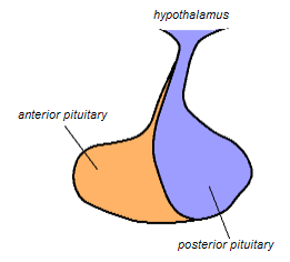
The posterior pituitary is the posterior lobe of the pituitary gland which is part of the endocrine system. The posterior pituitary is not glandular as is the anterior pituitary. Instead, it is largely a collection of axonal projections from the hypothalamus that terminate behind the anterior pituitary, and serve as a site for the secretion of neurohypophysial hormones directly into the blood. The hypothalamic–neurohypophyseal system is composed of the hypothalamus, posterior pituitary, and these axonal projections.
Chromaffin cells, also called pheochromocytes, are neuroendocrine cells found mostly in the medulla of the adrenal glands in mammals. These cells serve a variety of functions such as serving as a response to stress, monitoring carbon dioxide and oxygen concentrations in the body, maintenance of respiration and the regulation of blood pressure. They are in close proximity to pre-synaptic sympathetic ganglia of the sympathetic nervous system, with which they communicate, and structurally they are similar to post-synaptic sympathetic neurons. In order to activate chromaffin cells, the splanchnic nerve of the sympathetic nervous system releases acetylcholine, which then binds to nicotinic acetylcholine receptors on the adrenal medulla. This causes the release of catecholamines. The chromaffin cells release catecholamines: ~80% of adrenaline (epinephrine) and ~20% of noradrenaline (norepinephrine) into systemic circulation for systemic effects on multiple organs, and can also send paracrine signals. Hence they are called neuroendocrine cells.

The supraoptic nucleus (SON) is a nucleus of magnocellular neurosecretory cells in the hypothalamus of the mammalian brain. The nucleus is situated at the base of the brain, adjacent to the optic chiasm. In humans, the SON contains about 3,000 neurons.

The paraventricular nucleus is a nucleus in the hypothalamus. Anatomically, it is adjacent to the third ventricle and many of its neurons project to the posterior pituitary. These projecting neurons secrete oxytocin and a smaller amount of vasopressin, otherwise the nucleus also secretes corticotropin-releasing hormone (CRH) and thyrotropin-releasing hormone (TRH). CRH and TRH are secreted into the hypophyseal portal system and act on different targets neurons in the anterior pituitary. PVN is thought to mediate many diverse functions through these different hormones, including osmoregulation, appetite, and the response of the body to stress.
Magnocellular neurosecretory cells are large neuroendocrine cells within the supraoptic nucleus and paraventricular nucleus of the hypothalamus. They are also found in smaller numbers in accessory cell groups between these two nuclei, the largest one being the circular nucleus. There are two types of magnocellular neurosecretory cells, oxytocin-producing cells and vasopressin-producing cells, but a small number can produce both hormones. These cells are neuroendocrine neurons, are electrically excitable, and generate action potentials in response to afferent stimulation. Vasopressin is produced from the vasopressin-producing cells via the AVP gene, a molecular output of circadian pathways.

The arcuate nucleus of the hypothalamus is an aggregation of neurons in the mediobasal hypothalamus, adjacent to the third ventricle and the median eminence. The arcuate nucleus includes several important and diverse populations of neurons that help mediate different neuroendocrine and physiological functions, including neuroendocrine neurons, centrally projecting neurons, and astrocytes. The populations of neurons found in the arcuate nucleus are based on the hormones they secrete or interact with and are responsible for hypothalamic function, such as regulating hormones released from the pituitary gland or secreting their own hormones. Neurons in this region are also responsible for integrating information and providing inputs to other nuclei in the hypothalamus or inputs to areas outside this region of the brain. These neurons, generated from the ventral part of the periventricular epithelium during embryonic development, locate dorsally in the hypothalamus, becoming part of the ventromedial hypothalamic region. The function of the arcuate nucleus relies on its diversity of neurons, but its central role is involved in homeostasis. The arcuate nucleus provides many physiological roles involved in feeding, metabolism, fertility, and cardiovascular regulation.
Neuroendocrine cells are cells that receive neuronal input and, as a consequence of this input, release messenger molecules (hormones) into the blood. In this way they bring about an integration between the nervous system and the endocrine system, a process known as neuroendocrine integration. An example of a neuroendocrine cell is a cell of the adrenal medulla, which releases adrenaline to the blood. The adrenal medullary cells are controlled by the sympathetic division of the autonomic nervous system. These cells are modified postganglionic neurons. Autonomic nerve fibers lead directly to them from the central nervous system. The adrenal medullary hormones are kept in vesicles much in the same way neurotransmitters are kept in neuronal vesicles. Hormonal effects can last up to ten times longer than those of neurotransmitters. Sympathetic nerve fiber impulses stimulate the release of adrenal medullary hormones. In this way the sympathetic division of the autonomic nervous system and the medullary secretions function together.
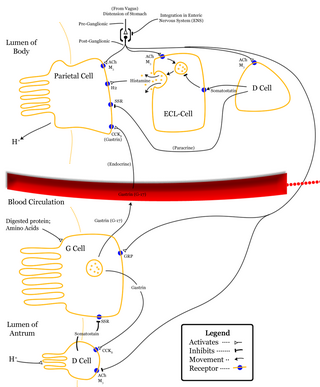
Enterochromaffin-like cells or ECL cells are a type of neuroendocrine cell found in the gastric glands of the gastric mucosa beneath the epithelium, in particular in the vicinity of parietal cells, that aid in the production of gastric acid via the release of histamine. They are also considered a type of enteroendocrine cell.

Enterochromaffin (EC) cells are a type of enteroendocrine cell, and neuroendocrine cell. They reside alongside the epithelium lining the lumen of the digestive tract and play a crucial role in gastrointestinal regulation, particularly intestinal motility and secretion. They were discovered by Nikolai Kulchitsky.
Neuroendocrinology is the branch of biology which studies the interaction between the nervous system and the endocrine system; i.e. how the brain regulates the hormonal activity in the body. The nervous and endocrine systems often act together in a process called neuroendocrine integration, to regulate the physiological processes of the human body. Neuroendocrinology arose from the recognition that the brain, especially the hypothalamus, controls secretion of pituitary gland hormones, and has subsequently expanded to investigate numerous interconnections of the endocrine and nervous systems.

A paraganglion is a group of non-neuronal cells derived of the neural crest. They are named for being generally in close proximity to sympathetic ganglia. They are essentially of two types: (1) chromaffin or sympathetic paraganglia made of chromaffin cells and (2) nonchromaffin or parasympathetic ganglia made of glomus cells. They are neuroendocrine cells, the former with primary endocrine functions and the latter with primary chemoreceptor functions.

Neurophysin II is a carrier protein with a size of 19,687.3 Da and is made up of a dimer of two virtually identical chains of amino acids. Neurophysin II is a cleavage product of the AVP gene. It is a neurohypophysial hormone that is transported in vesicles with vasopressin, the other cleavage product, along axons, from magnocellular neurons of the hypothalamus to the posterior lobe of the pituitary. Although it is stored in neurosecretory granules with vasopressin and released with vasopressin into the bloodstream, its biological action is unclear. Neurophysin II is also known as a stimulator of prolactin secretion.
Parvocellular neurosecretory cells are small neurons that produce hypothalamic releasing and inhibiting hormones. The cell bodies of these neurons are located in various nuclei of the hypothalamus or in closely related areas of the basal brain, mainly in the medial zone of the hypothalamus. All or most of the axons of the parvocellular neurosecretory cells project to the median eminence, at the base of the brain, where their nerve terminals release the hypothalamic hormones. These hormones are then immediately absorbed into the blood vessels of the hypothalamo-pituitary portal system, which carry them to the anterior pituitary gland, where they regulate the secretion of hormones into the systemic circulation.
References
- ↑ Purves WK, Sadava D, Orians GH, Heller HC (2001). Life: The Science of Biology (6th ed.). Massachusetts: Sinauer Associates. p. 718. ISBN 978-0-7167-3873-2.
- ↑ Nelson. 2005 An Introduction To Behavioral Endocrinology, Third Edition
- ↑ Purves et al. p. 714.
- ↑ Unsicker K, Huber K, Schütz G, Kalcheim C (Jun–Jul 2005). "The chromaffin cell and its development". Neurochemical Research. 30 (6–7): 921–5. doi:10.1007/s11064-005-6966-5. PMID 16187226.
- ↑ Andrew A (June 1974). "Further evidence that enterochromaffin cells are not derived from the neural crest". Journal of Embryology and Experimental Morphology. 31 (3): 589–98. PMID 4448939.
- ↑ Meites J, Sonntag WE (April 1981). "Hypothalamic hypophysiotropic hormones and neurotransmitter regulation: current views". Annual Review of Pharmacology and Toxicology. 21: 295–322. doi:10.1146/annurev.pa.21.040181.001455. PMID 6112966.
- ↑ Nillni EA (April 2010). "Regulation of the hypothalamic thyrotropin releasing hormone (TRH) neuron by neuronal and peripheral inputs". Frontiers in Neuroendocrinology. 31 (2): 134–56. doi:10.1016/j.yfrne.2010.01.001. PMC 2849853 . PMID 20074584.
- ↑ Brown AG (2001). Nerve cells and nervous systems : an introduction to neuroscience. London: Springer. p. 200. ISBN 978-3-540-76090-0.
- ↑ Burbach JP, Luckman SM, Murphy D, Gainer H (July 2001). "Gene regulation in the magnocellular hypothalamo-neurohypophysial system". Physiological Reviews. 81 (3): 1197–267. doi:10.1152/physrev.2001.81.3.1197. PMID 11427695.
- ↑ Chung KF, Sicard F, Vukicevic V, Hermann A, Storch A, Huttner WB, Bornstein SR, Ehrhart-Bornstein M (October 2009). "Isolation of neural crest derived chromaffin progenitors from adult adrenal medulla". Stem Cells. 27 (10): 2602–13. doi: 10.1002/stem.180 . PMID 19609938.
- ↑ Gasman S, Chasserot-Golaz S, Bader MF, Vitale N (October 2003). "Regulation of exocytosis in adrenal chromaffin cells: focus on ARF and Rho GTPases". Cellular Signalling. 15 (10): 893–9. doi:10.1016/S0898-6568(03)00052-4. PMID 12873702.
- ↑ Bornstein SR, Ehrhart-Bornstein M (December 1992). "Ultrastructural evidence for a paracrine regulation of the rat adrenal cortex mediated by the local release of catecholamines from chromaffin cells". Endocrinology. 131 (6): 3126–8. doi:10.1210/endo.131.6.1446648. PMID 1446648.
- ↑ Prinz C, Zanner R, Gerhard M, Mahr S, Neumayer N, Höhne-Zell B, Gratzl M (November 1999). "The mechanism of histamine secretion from gastric enterochromaffin-like cells". The American Journal of Physiology. 277 (5 Pt 1): C845-55. PMID 10564076.
- ↑ Rhee SH, Pothoulakis C, Mayer EA (May 2009). "Principles and clinical implications of the brain-gut-enteric microbiota axis". Nature Reviews. Gastroenterology & Hepatology. 6 (5): 306–14. doi:10.1038/nrgastro.2009.35. PMC 3817714 . PMID 19404271.
- ↑ Haas HL, Sergeeva OA, Selbach O (July 2008). "Histamine in the nervous system". Physiological Reviews. 88 (3): 1183–241. doi:10.1152/physrev.00043.2007. PMID 18626069.
- ↑ Rodriguez-Diaz R, Dando R, Jacques-Silva MC, Fachado A, Molina J, Abdulreda MH, Ricordi C, Roper SD, Berggren PO, Caicedo A (June 2011). "Alpha cells secrete acetylcholine as a non-neuronal paracrine signal priming beta cell function in humans". Nature Medicine. 17 (7): 888–92. doi:10.1038/nm.2371. PMC 3132226 . PMID 21685896.
- ↑ Sandor A, Kidd M, Lawton GP, Miu K, Tang LH, Modlin IM (April 1996). "Neurohormonal modulation of rat enterochromaffin-like cell histamine secretion". Gastroenterology. 110 (4): 1084–92. doi: 10.1053/gast.1996.v110.pm8612997 . PMID 8612997.