Nucleic acid analogue

Last updated
RNA with its nucleobases to the left and DNA to the right. Difference DNA RNA-EN.svg
RNA with its nucleobases to the left and DNA to the right.

Nucleic acid analogues are compounds which are analogous (structurally similar) to naturally occurring RNA and DNA, used in medicine and in molecular biology research. Nucleic acids are chains of nucleotides, which are composed of three parts: a phosphate backbone, a pentose sugar, either ribose or deoxyribose, and one of four nucleobases. An analogue may have any of these altered. [1] Typically the analogue nucleobases confer, among other things, different base pairing and base stacking properties. Examples include universal bases, which can pair with all four canonical bases, and phosphate-sugar backbone analogues such as PNA, which affect the properties of the chain (PNA can even form a triple helix). [2] Nucleic acid analogues are also called xeno nucleic acids and represent one of the main pillars of xenobiology, the design of new-to-nature forms of life based on alternative biochemistries.

Contents

Artificial nucleic acids include peptide nucleic acids (PNA), Morpholino and locked nucleic acids (LNA), as well as glycol nucleic acids (GNA), threose nucleic acids (TNA) and hexitol nucleic acids (HNA). Each of these is distinguished from naturally occurring DNA or RNA by changes to the backbone of the molecule. However, the polyelectrolyte theory of the gene proposes that a genetic molecule require a charged backbone to function.

In May 2014, researchers announced that they had successfully introduced two new artificial nucleotides into bacterial DNA, and by including individual artificial nucleotides in the culture media, were able to passage the bacteria 24 times; they did not create mRNA or proteins able to use the artificial nucleotides. The artificial nucleotides featured 2 fused aromatic rings.

Medicine

Several nucleoside analogues are used as antiviral or anticancer agents. The viral polymerase incorporates these compounds with non-canonical bases. These compounds are activated in the cells by being converted into nucleotides, they are administered as nucleosides since charged nucleotides cannot easily cross cell membranes.[ citation needed ]

Molecular biology

Common changes in nucleotide analogues Nucleoside analogues.svg
Common changes in nucleotide analogues

Nucleic acid analogues are used in molecular biology for several purposes:

Backbone analogues

Hydrolysis resistant RNA-analogues

Chemical structure of Morpholino Morpholino-monomer.svg
Chemical structure of Morpholino

Ribose's 2' hydroxy group reacts with the phosphate linked 3' hydroxy group, making RNA too unstable to be used or synthesized reliably. To overcome this, a ribose analogue can be used. The most common RNA analogues are 2'-O-methyl-substituted RNA, locked nucleic acid (LNA) or bridged nucleic acid (BNA), morpholino, [5] [6] and peptide nucleic acid (PNA). Although these oligonucleotides have a different backbone sugar—or, in the case of PNA, an amino acid residue in place of the ribose phosphate—they still bind to RNA or DNA according to Watson and Crick pairing while being immune to nuclease activity. They cannot be synthesized enzymatically and can only be obtained synthetically using the phosphoramidite strategy or, for PNA, other methods of peptide synthesis.[ citation needed ]

Other notable analogues used as tools

Dideoxynucleotides are used in sequencing. These nucleoside triphosphates possess a non-canonical sugar, dideoxyribose, which lacks the 3' hydroxyl group normally present in DNA and therefore cannot bond with the next base. The lack of the 3' hydroxyl group terminates the chain reaction as the DNA polymerases mistake it for a regular deoxyribonucleotide. Another chain-terminating analogue that lacks a 3' hydroxyl and mimics adenosine is called cordycepin. Cordycepin is an anticancer drug that targets RNA replication. Another analogue in sequencing is a nucleobase analogue, 7-deaza-GTP and is used to sequence CG rich regions, instead 7-deaza-ATP is called tubercidin, an antibiotic.[ citation needed ]

Precursors to the RNA world

RNA may be too complex to be the first nucleic acid, so before the RNA world several simpler nucleic acids that differ in the backbone, such as GNA, PNA, and TNA have been offered as candidates for the first nucleic acids.[ citation needed ]

Base analogues

Nucleobase structure and nomenclature

Naturally occurring bases can be divided into two classes according to their structure:

Artificial nucleotides (Unnatural Base Pairs (UBPs) named d5SICS UBP and dNaM UBP) have been inserted into bacterial DNA but these genes did not template mRNA or induce protein synthesis. The artificial nucleotides featured two fused aromatic rings which formed a (d5SICS–dNaM) complex mimicking the natural (dG–dC) base pair. [7] [8] [9]

Mutagens

One of the most common base analogs is 5-bromouracil (5BU), the abnormal base found in the mutagenic nucleotide analog BrdU. When a nucleotide containing 5-bromouracil is incorporated into the DNA, it is most likely to pair with adenine; however, it can spontaneously shift into another isomer which pairs with a different nucleobase, guanine. If this happens during DNA replication, a guanine will be inserted as the opposite base analog, and in the next DNA replication, that guanine will pair with a cytosine. This results in a change in one base pair of DNA, specifically a transition mutation.[ citation needed ]

Additionally, nitrous acid (HNO2) is a potent mutagen that acts on replicating and non-replicating DNA. It can cause deamination of the amino groups of adenine, guanine and cytosine. Adenine is deaminated to hypoxanthine, which base pairs to cytosine instead of thymine. Cytosine is deaminated to uracil, which base pairs with adenine instead of guanine. Deamination of guanine is not mutagenic. Nitrous acid-induced mutations also are induced to mutate back to wild-type.[ citation needed ]

Fluorophores

Structure of aminoallyl-uridine Aminoallyl uracil.svg
Structure of aminoallyl-uridine

Commonly fluorophores (such as rhodamine or fluorescein) are linked to the ring linked to the sugar (in para) via a flexible arm, presumably extruding from the major groove of the helix. Due to low processivity of the nucleotides linked to bulky adducts such as florophores by [Taq polymerase]s, the sequence is typically copied using a nucleotide with an arm and later coupled with a reactive fluorophore (indirect labelling):

Fluorophores find a variety of uses in medicine and biochemistry.

Fluorescent base analogues

The most commonly used and commercially available fluorescent base analogue, 2-aminopurine (2-AP), has a high-fluorescence quantum yield free in solution (0.68) that is considerably reduced (appr. 100 times but highly dependent on base sequence) when incorporated into nucleic acids. [10] The emission sensitivity of 2-AP to immediate surroundings is shared by other promising and useful fluorescent base analogues like 3-MI, 6-MI, 6-MAP, [11] pyrrolo-dC (also commercially available), [12] modified and improved derivatives of pyrrolo-dC, [13] furan-modified bases [14] and many other ones (see recent reviews). [15] [16] [17] [18] [19] This sensitivity to the microenvironment has been utilized in studies of e.g. structure and dynamics within both DNA and RNA, dynamics and kinetics of DNA-protein interaction and electron transfer within DNA.[ citation needed ]

A newly developed and very interesting group of fluorescent base analogues that has a fluorescence quantum yield that is nearly insensitive to their immediate surroundings is the tricyclic cytosine family. 1,3-Diaza-2-oxophenothiazine, tC, has a fluorescence quantum yield of approximately 0.2 both in single- and in double-strands irrespective of surrounding bases. [20] [21] Also the oxo-homologue of tC called tCO (both commercially available), 1,3-diaza-2-oxophenoxazine, has a quantum yield of 0.2 in double-stranded systems. [22] However, it is somewhat sensitive to surrounding bases in single-strands (quantum yields of 0.14–0.41). The high and stable quantum yields of these base analogues make them very bright, and, in combination with their good base analogue properties (leaves DNA structure and stability next to unperturbed), they are especially useful in fluorescence anisotropy and FRET measurements, areas where other fluorescent base analogues are less accurate. Also, in the same family of cytosine analogues, a FRET-acceptor base analogue, tCnitro, has been developed. [23] Together with tCO as a FRET-donor this constitutes the first nucleic acid base analogue FRET-pair ever developed. The tC-family has, for example, been used in studies related to polymerase DNA-binding and DNA-polymerization mechanisms.

Natural non-canonical bases

In a cell, there are several non-canonical bases present: CpG islands in DNA (often methylated), all eukaryotic mRNA (capped with a methyl-7-guanosine), and several bases of rRNAs (methylated). Often, tRNAs are heavily modified postranscriptionally in order to improve their conformation or base pairing, in particular in or near the anticodon: inosine can base pair with C, U, and even with A, whereas thiouridine (with A) is more specific than uracil (with a purine). [24] Other common tRNA base modifications are pseudouridine (which gives its name to the TΨC loop), dihydrouridine (which does not stack as it is not aromatic), queuosine, wyosine, and so forth. Nevertheless, these are all modifications to normal bases and are not placed by a polymerase. [24]

Base-pairing

Canonical bases may have either a carbonyl or an amine group on the carbons surrounding the nitrogen atom furthest away from the glycosidic bond, which allows them to base pair (Watson-Crick base pairing) via hydrogen bonds (amine with ketone, purine with pyrimidine). Adenine and 2-aminoadenine have one/two amine group(s), whereas thymine has two carbonyl groups, and cytosine and guanine are mixed amine and carbonyl (inverted in respect to each other).[ citation needed ]

Natural basepairs
Base pair GC.svg Base pair AT.svg
A GC basepair: purine carbonyl/amine forms three intermolecular hydrogen bonds with pyrimidine amine/carbonylAn AT basepair: purine amine/- forms two intermolecular hydrogen bonds with pyrimidine carbonyl/carbonyl

The precise reason why there are only four nucleotides is debated, but there are several unused possibilities. Furthermore, adenine is not the most stable choice for base pairing: in Cyanophage S-2L, diaminopurine (DAP) is used instead of adenine. [25] Diaminopurine basepairs perfectly with thymine as it is identical to adenine but has an amine group at position 2 forming 3 intramolecular hydrogen bonds, eliminating the major difference between the two types of basepairs (weak A-T vs strong C-G). This improved stability affects protein-binding interactions that rely on those differences. Other combination include:

Unused basepair arrangements
DiampurineT DNA base pair.svg Base pair X-DAP.svg IG-iC DNA base pair.svg
A DAP-T base: purine amine/amine forms three intermolecular hydrogen bonds with pyrimidine ketone/ketoneAn X-DAP base: purine ketone/ketone forms three intermolecular hydrogen bonds with pyrimidine amine/amineA iG-iC base: purine amine/ketone forms three intermolecular hydrogen bonds with pyrimidine ketone/amine

However, correct DNA structure can form even when the bases are not paired via hydrogen bonding; that is, the bases pair thanks to hydrophobicity, as studies have shown with DNA isosteres (analogues with same number of atoms) such as the thymine analogue 2,4-difluorotoluene (F) or the adenine analogue 4-methylbenzimidazole (Z). [27] An alternative hydrophobic pair could be isoquinoline and pyrrolo[2,3-b]pyridine [28]

Other noteworthy basepairs:

Novel basepairs with special properties
F-Z DNA base pair.svg S-Da DNA base pair.svg XA-T DNA base pair.svg
A F-Z base: methylbenzimidazole does not form intermolecular hydrogen bonds with toluene F/FAn S-Pa base: purine thienyl/amine forms three intermolecular hydrogen bonds with pyrrole -/carbaldehydeAn xA-T base: same bonding as A-T


Metal base-pairs

In metal base-pairing, the Watson-Crick hydrogen bonds are replaced by the interaction between a metal ion with nucleosides acting as ligands. The possible geometries of the metal that would allow for duplex formation with two bidentate nucleosides around a central metal atom are tetrahedral, dodecahedral, and square planar. Metal-complexing with DNA can occur by the formation of non-canonical base pairs from natural nucleobases with participation by metal ions and also by the exchanging the hydrogen atoms that are part of the Watson-Crick base pairing by metal ions. [32] Introduction of metal ions into a DNA duplex has shown to have potential magnetic [33] or conducting properties, [34] as well as increased stability. [35]

Metal complexing has been shown to occur between natural nucleobases. A well-documented example is the formation of T-Hg-T, which involves two deprotonated thymine nucleobases that are brought together by Hg2+ and forms a connected metal-base pair. [36] This motif does not accommodate stacked Hg2+ in a duplex due to an intrastrand hairpin formation process that is favored over duplex formation. [37] Two thymines across from each other do not form a Watson-Crick base pair in a duplex; this is an example where a Watson-Crick basepair mismatch is stabilized by the formation of the metal-base pair. Another example of a metal complexing to natural nucleobases is the formation of A-Zn-T and G-Zn-C at high pH; Co+2 and Ni+2 also form these complexes. These are Watson-Crick base pairs where the divalent cation in coordinated to the nucleobases. The exact binding is debated. [38]

A large variety of artificial nucleobases have been developed for use as metal base pairs. These modified nucleobases exhibit tunable electronic properties, sizes, and binding affinities that can be optimized for a specific metal. For example, a nucleoside modified with a pyridine-2,6-dicarboxylate has shown to bind tightly to Cu2+, whereas other divalent ions are only loosely bound. The tridentate character contributes to this selectivity. The fourth coordination site on the copper is saturated by an oppositely arranged pyridine nucleobase. [39] The asymmetric metal base pairing system is orthogonal to the Watson-Crick base pairs. Another example of an artificial nucleobase is that with hydroxypyridone nucleobases, which are able to bind Cu2+ inside the DNA duplex. Five consecutive copper-hydroxypyridone base pairs were incorporated into a double strand, which were flanked by only one natural nucleobase on both ends. EPR data showed that the distance between copper centers was estimated to be 3.7 ± 0.1 Å, while a natural B-type DNA duplex is only slightly larger (3.4 Å). [40] The appeal for stacking metal ions inside a DNA duplex is the hope to obtain nanoscopic self-assembling metal wires, though this has not been realized yet.

Unnatural base pair (UBP)

An unnatural base pair (UBP) is a designed subunit (or nucleobase) of DNA that is created in a laboratory and does not occur in nature. In 2012, a group of American scientists led by Floyd Romesberg, a chemical biologist at the Scripps Research Institute in San Diego, California, published that his team had designed two unnatural base pairs named d5SICS and dNaM. [41] More technically, these artificial nucleotides bearing hydrophobic nucleobases feature two fused aromatic rings that form a d5SICS–dNaM complex or base pair in DNA. [9] [42] In 2014, the same team reported that they had synthesized a plasmid containing natural T-A and C-G base pairs along with the best-performing UBP Romesberg's laboratory had designed and inserted it into cells of the common bacterium E. coli, which successfully replicated the unnatural base pairs through multiple generations. [43] This is the first known example of a living organism passing along an expanded genetic code to subsequent generations. [9] [44] This was in part achieved by the addition of a supportive algal gene that expresses a nucleotide triphosphate transporter which efficiently imports the triphosphates of both d5SICSTP and dNaMTP into E. coli bacteria. [9] Then, the natural bacterial replication pathways use them to accurately replicate the plasmid containing d5SICS–dNaM.[ citation needed ]

The successful incorporation of a third base pair is a significant breakthrough toward the goal of greatly expanding the number of amino acids which can be encoded by DNA, from the existing 20 amino acids to a theoretically possible 172, thereby expanding the potential for living organisms to produce novel proteins. [43] Earlier, the artificial strings of DNA did not encode for anything, but scientists speculated they could be designed to manufacture new proteins which could have industrial or pharmaceutical uses. [45] Transcription of DNA containing unnatural base pairs and translation of corresponding mRNA were actually achieved recently. In November 2017, the same team at the Scripps Research Institute that first introduced two extra nucleobases into bacterial DNA reported having constructed a semi-synthetic E. coli bacteria able to make proteins using such DNA. Its DNA contained six different nucleobases: four canonical and two artificially added, dNaM and dTPT3 (these two form a pair). The bacteria had two corresponding RNA bases included in two new codons, additional tRNAs recognizing these new codons (these tRNAs also contained two new RNA bases within their anticodons) and additional amino acids, enabling the bacteria to synthesize "unnatural" proteins. [46] [47]

Another demonstration of UBPs were achieved by Ichiro Hirao's group at RIKEN institute in Japan. In 2002, they developed an unnatural base pair between 2-amino-8-(2-thienyl)purine (s) and pyridine-2-one (y) that functions in vitro in transcription and translation, for the site-specific incorporation of non-standard amino acids into proteins. [48] In 2006, they created 7-(2-thienyl)imidazo[4,5-b]pyridine (Ds) and pyrrole-2-carbaldehyde (Pa) as a third base pair for replication and transcription. [49] Afterward, Ds and 4-[3-(6-aminohexanamido)-1-propynyl]-2-nitropyrrole (Px) was discovered as a high fidelity pair in PCR amplification. [50] [51] In 2013, they applied the Ds-Px pair to DNA aptamer generation by in vitro selection (SELEX) and demonstrated the genetic alphabet expansion significantly augment DNA aptamer affinities to target proteins. [52]

Orthogonal system

The possibility has been proposed and studied, both theoretically and experimentally, of implementing an orthogonal system inside cells independent of the cellular genetic material in order to make a completely safe system, [53] with the possible increase in encoding potentials. [54] Several groups have focused on different aspects:

See also

Related Research Articles

<span class="mw-page-title-main">Base pair</span> Unit consisting of two nucleobases bound to each other by hydrogen bonds

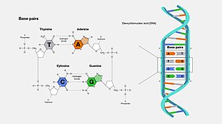
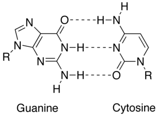
A base pair (bp) is a fundamental unit of double-stranded nucleic acids consisting of two nucleobases bound to each other by hydrogen bonds. They form the building blocks of the DNA double helix and contribute to the folded structure of both DNA and RNA. Dictated by specific hydrogen bonding patterns, "Watson–Crick" base pairs allow the DNA helix to maintain a regular helical structure that is subtly dependent on its nucleotide sequence. The complementary nature of this based-paired structure provides a redundant copy of the genetic information encoded within each strand of DNA. The regular structure and data redundancy provided by the DNA double helix make DNA well suited to the storage of genetic information, while base-pairing between DNA and incoming nucleotides provides the mechanism through which DNA polymerase replicates DNA and RNA polymerase transcribes DNA into RNA. Many DNA-binding proteins can recognize specific base-pairing patterns that identify particular regulatory regions of genes.

<span class="mw-page-title-main">DNA</span> Molecule that carries genetic information

Deoxyribonucleic acid is a polymer composed of two polynucleotide chains that coil around each other to form a double helix. The polymer carries genetic instructions for the development, functioning, growth and reproduction of all known organisms and many viruses. DNA and ribonucleic acid (RNA) are nucleic acids. Alongside proteins, lipids and complex carbohydrates (polysaccharides), nucleic acids are one of the four major types of macromolecules that are essential for all known forms of life.

<span class="mw-page-title-main">Nucleic acid</span> Class of large biomolecules essential to all known life

Nucleic acids are large biomolecules that are crucial in all cells and viruses. They are composed of nucleotides, which are the monomer components: a 5-carbon sugar, a phosphate group and a nitrogenous base. The two main classes of nucleic acids are deoxyribonucleic acid (DNA) and ribonucleic acid (RNA). If the sugar is ribose, the polymer is RNA; if the sugar is deoxyribose, a variant of ribose, the polymer is DNA.

<span class="mw-page-title-main">Nucleotide</span> Biological molecules constituting nucleic acids

Nucleotides are organic molecules composed of a nitrogenous base, a pentose sugar and a phosphate. They serve as monomeric units of the nucleic acid polymers – deoxyribonucleic acid (DNA) and ribonucleic acid (RNA), both of which are essential biomolecules within all life-forms on Earth. Nucleotides are obtained in the diet and are also synthesized from common nutrients by the liver.

<span class="mw-page-title-main">Nucleobase</span> Nitrogen-containing biological compounds that form nucleosides

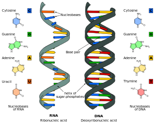
Nucleobases are nitrogen-containing biological compounds that form nucleosides, which, in turn, are components of nucleotides, with all of these monomers constituting the basic building blocks of nucleic acids. The ability of nucleobases to form base pairs and to stack one upon another leads directly to long-chain helical structures such as ribonucleic acid (RNA) and deoxyribonucleic acid (DNA). Five nucleobases—adenine (A), cytosine (C), guanine (G), thymine (T), and uracil (U)—are called primary or canonical. They function as the fundamental units of the genetic code, with the bases A, G, C, and T being found in DNA while A, G, C, and U are found in RNA. Thymine and uracil are distinguished by merely the presence or absence of a methyl group on the fifth carbon (C5) of these heterocyclic six-membered rings. In addition, some viruses have aminoadenine (Z) instead of adenine. It differs in having an extra amine group, creating a more stable bond to thymine.

<span class="mw-page-title-main">Nucleic acid sequence</span> Succession of nucleotides in a nucleic acid

A nucleic acid sequence is a succession of bases within the nucleotides forming alleles within a DNA or RNA (GACU) molecule. This succession is denoted by a series of a set of five different letters that indicate the order of the nucleotides. By convention, sequences are usually presented from the 5' end to the 3' end. For DNA, with its double helix, there are two possible directions for the notated sequence; of these two, the sense strand is used. Because nucleic acids are normally linear (unbranched) polymers, specifying the sequence is equivalent to defining the covalent structure of the entire molecule. For this reason, the nucleic acid sequence is also termed the primary structure.

<span class="mw-page-title-main">DNA synthesis</span>

DNA synthesis is the natural or artificial creation of deoxyribonucleic acid (DNA) molecules. DNA is a macromolecule made up of nucleotide units, which are linked by covalent bonds and hydrogen bonds, in a repeating structure. DNA synthesis occurs when these nucleotide units are joined to form DNA; this can occur artificially or naturally. Nucleotide units are made up of a nitrogenous base, pentose sugar (deoxyribose) and phosphate group. Each unit is joined when a covalent bond forms between its phosphate group and the pentose sugar of the next nucleotide, forming a sugar-phosphate backbone. DNA is a complementary, double stranded structure as specific base pairing occurs naturally when hydrogen bonds form between the nucleotide bases.

<span class="mw-page-title-main">Ribonucleotide</span> Nucleotide containing ribose as its pentose component

In biochemistry, a ribonucleotide is a nucleotide containing ribose as its pentose component. It is considered a molecular precursor of nucleic acids. Nucleotides are the basic building blocks of DNA and RNA. Ribonucleotides themselves are basic monomeric building blocks for RNA. Deoxyribonucleotides, formed by reducing ribonucleotides with the enzyme ribonucleotide reductase (RNR), are essential building blocks for DNA. There are several differences between DNA deoxyribonucleotides and RNA ribonucleotides. Successive nucleotides are linked together via phosphodiester bonds.

A nucleoside triphosphate is a nucleoside containing a nitrogenous base bound to a 5-carbon sugar, with three phosphate groups bound to the sugar. They are the molecular precursors of both DNA and RNA, which are chains of nucleotides made through the processes of DNA replication and transcription. Nucleoside triphosphates also serve as a source of energy for cellular reactions and are involved in signalling pathways.

Xenobiology (XB) is a subfield of synthetic biology, the study of synthesizing and manipulating biological devices and systems. The name "xenobiology" derives from the Greek word xenos, which means "stranger, alien". Xenobiology is a form of biology that is not (yet) familiar to science and is not found in nature. In practice, it describes novel biological systems and biochemistries that differ from the canonical DNA–RNA-20 amino acid system. For example, instead of DNA or RNA, XB explores nucleic acid analogues, termed xeno nucleic acid (XNA) as information carriers. It also focuses on an expanded genetic code and the incorporation of non-proteinogenic amino acids, or “xeno amino acids” into proteins.

<span class="mw-page-title-main">Isoguanine</span> Chemical compound

Isoguanine or 2-hydroxyadenine is a purine base that is an isomer of guanine. It is a product of oxidative damage to DNA and has been shown to cause mutation. It is also used in combination with isocytosine in studies of unnatural nucleic acid analogues of the normal base pairs in DNA.

Artificially Expanded Genetic Information System (AEGIS) is a synthetic DNA analog experiment that uses some unnatural base pairs from the laboratories of the Foundation for Applied Molecular Evolution in Gainesville, Florida. AEGIS is a NASA-funded project to try to understand how extraterrestrial life may have developed.

<span class="mw-page-title-main">Nucleic acid structure</span> Biomolecular structure of nucleic acids such as DNA and RNA

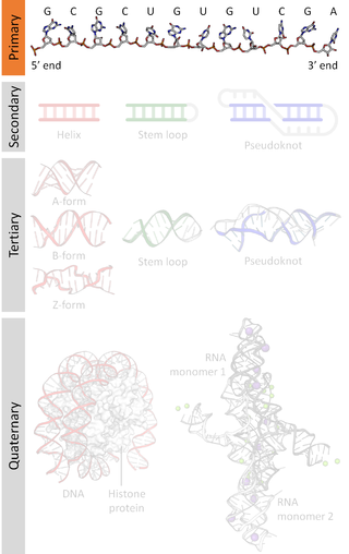
Nucleic acid structure refers to the structure of nucleic acids such as DNA and RNA. Chemically speaking, DNA and RNA are very similar. Nucleic acid structure is often divided into four different levels: primary, secondary, tertiary, and quaternary.

<span class="mw-page-title-main">Nucleic acid secondary structure</span>

Nucleic acid secondary structure is the basepairing interactions within a single nucleic acid polymer or between two polymers. It can be represented as a list of bases which are paired in a nucleic acid molecule. The secondary structures of biological DNAs and RNAs tend to be different: biological DNA mostly exists as fully base paired double helices, while biological RNA is single stranded and often forms complex and intricate base-pairing interactions due to its increased ability to form hydrogen bonds stemming from the extra hydroxyl group in the ribose sugar.

<span class="mw-page-title-main">Complementarity (molecular biology)</span> Lock-and-key pairing between two structures

In molecular biology, complementarity describes a relationship between two structures each following the lock-and-key principle. In nature complementarity is the base principle of DNA replication and transcription as it is a property shared between two DNA or RNA sequences, such that when they are aligned antiparallel to each other, the nucleotide bases at each position in the sequences will be complementary, much like looking in the mirror and seeing the reverse of things. This complementary base pairing allows cells to copy information from one generation to another and even find and repair damage to the information stored in the sequences.

xDNA Benzo-homologated DNA analogue

xDNA is a size-expanded nucleotide system synthesized from the fusion of a benzene ring and one of the four natural bases: adenine, guanine, cytosine, and thymine. This size expansion produces an 8 letter alphabet which has a larger information storage capacity than natural DNA's 4 letter alphabet. As with normal base-pairing, A pairs with xT, C pairs with xG, G pairs with xC, and T pairs with xA. The double helix is thus 2.4Å wider than a natural double helix. While similar in structure to B-DNA, xDNA has unique absorption, fluorescence, and stacking properties.

<span class="mw-page-title-main">Xeno nucleic acid</span> Synthetic nucleic acid analogues

Xeno nucleic acids (XNA) are synthetic nucleic acid analogues that have a different backbone than the ribose and deoxyribose found in the nucleic acids of naturally occurring RNA and DNA.

<span class="mw-page-title-main">Hachimoji DNA</span> Synthetic DNA

Hachimoji DNA is a synthetic nucleic acid analog that uses four synthetic nucleotides in addition to the four present in the natural nucleic acids, DNA and RNA. This leads to four allowed base pairs: two unnatural base pairs formed by the synthetic nucleobases in addition to the two normal pairs. Hachimoji bases have been demonstrated in both DNA and RNA analogs, using deoxyribose and ribose respectively as the backbone sugar.

This glossary of cellular and molecular biology is a list of definitions of terms and concepts commonly used in the study of cell biology, molecular biology, and related disciplines, including genetics, biochemistry, and microbiology. It is split across two articles:

This glossary of cellular and molecular biology is a list of definitions of terms and concepts commonly used in the study of cell biology, molecular biology, and related disciplines, including genetics, biochemistry, and microbiology. It is split across two articles:

References

  1. Singer E (July 19, 2015). "Chemists Invent New Letters for Nature's Genetic Alphabet". Wired . Retrieved July 20, 2015.
  2. Petersson B, Nielsen BB, Rasmussen H, Larsen IK, Gajhede M, Nielsen PE, Kastrup JS (February 2005). "Crystal structure of a partly self-complementary peptide nucleic acid (PNA) oligomer showing a duplex-triplex network". Journal of the American Chemical Society. 127 (5): 1424–30. doi:10.1021/ja0458726. PMID   15686374.
  3. 1 2 Taylor AI, Pinheiro VB, Smola MJ, Morgunov AS, Peak-Chew S, Cozens C, Weeks KM, Herdewijn P, Holliger P (February 2015). "Catalysts from synthetic genetic polymers". Nature. 518 (7539): 427–30. Bibcode:2015Natur.518..427T. doi:10.1038/nature13982. PMC   4336857 . PMID   25470036.
  4. Wang Q, Chen L, Long Y, Tian H, Wu J (2013). "Molecular beacons of xeno-nucleic acid for detecting nucleic acid". Theranostics. 3 (6): 395–408. doi:10.7150/thno.5935. PMC   3677410 . PMID   23781286.
  5. Summerton J, Weller D (June 1997). "Morpholino antisense oligomers: design, preparation, and properties". Antisense & Nucleic Acid Drug Development. 7 (3): 187–95. doi:10.1089/oli.1.1997.7.187. PMID   9212909. S2CID   19372403.
  6. Summerton J (December 1999). "Morpholino antisense oligomers: the case for an RNase H-independent structural type". Biochimica et Biophysica Acta (BBA) - Gene Structure and Expression. 1489 (1): 141–58. doi:10.1016/s0167-4781(99)00150-5. PMID   10807004.
  7. Pollack A (May 7, 2014). "Researchers Report Breakthrough in Creating Artificial Genetic Code". New York Times . Retrieved May 7, 2014.
  8. Callaway E (May 7, 2014). "First life with 'alien' DNA". Nature . doi:10.1038/nature.2014.15179. S2CID   86967999.
  9. 1 2 3 4 Malyshev DA, Dhami K, Lavergne T, Chen T, Dai N, Foster JM, Corrêa IR, Romesberg FE (May 2014). "A semi-synthetic organism with an expanded genetic alphabet". Nature. 509 (7500): 385–88. Bibcode:2014Natur.509..385M. doi:10.1038/nature13314. PMC   4058825 . PMID   24805238.
  10. Ward DC, Reich E, Stryer L (March 1969). "Fluorescence studies of nucleotides and polynucleotides. I. Formycin, 2-aminopurine riboside, 2,6-diaminopurine riboside, and their derivatives". The Journal of Biological Chemistry. 244 (5): 1228–37. doi: 10.1016/S0021-9258(18)91833-8 . PMID   5767305.
  11. Hawkins ME (2001). "Fluorescent pteridine nucleoside analogs: a window on DNA interactions". Cell Biochemistry and Biophysics. 34 (2): 257–81. doi:10.1385/cbb:34:2:257. PMID   11898867. S2CID   12134698.
  12. Berry DA, Jung KY, Wise DS, Sercel AD, Pearson WH, Mackie H, Randolph JB, Somers RL (2004). "Pyrrolo-dC and pyrrolo-C: fluorescent analogs of cytidine and 2 '-deoxycytidine for the study of oligonucleotides". Tetrahedron Lett. 45 (11): 2457–61. doi:10.1016/j.tetlet.2004.01.108.
  13. Wojciechowski F, Hudson RH (September 2008). "Fluorescence and hybridization properties of peptide nucleic acid containing a substituted phenylpyrrolocytosine designed to engage Guanine with an additional H-bond". Journal of the American Chemical Society. 130 (38): 12574–75. doi:10.1021/ja804233g. PMID   18761442.
  14. Greco NJ, Tor Y (August 2005). "Simple fluorescent pyrimidine analogues detect the presence of DNA abasic sites". Journal of the American Chemical Society. 127 (31): 10784–85. doi:10.1021/ja052000a. PMID   16076156.
  15. Rist MJ, Marino JP (2002). "Fluorescent nucleotide base analogs as probes of nucleic acid structure, dynamics and interactions". Curr. Org. Chem. 6 (9): 775–93. doi:10.2174/1385272023373914.
  16. Wilson JN, Kool ET (December 2006). "Fluorescent DNA base replacements: Reporters and sensors for biological systems". Organic & Biomolecular Chemistry. 4 (23): 4265–74. doi:10.1039/b612284c. PMID   17102869.
  17. Wilhelmsson and Tor (2016). Fluorescent Analogs of Biomolecular Building Blocks: Design and Applications. New Jersey: Wiley. ISBN   978-1-118-17586-6.
  18. Wilhelmsson LM (May 2010). "Fluorescent nucleic acid base analogues". Quarterly Reviews of Biophysics. 43 (2): 159–83. doi:10.1017/s0033583510000090. PMID   20478079. S2CID   10783202.
  19. Sinkeldam RW, Greco NJ, Tor Y (May 2010). "Fluorescent analogs of biomolecular building blocks: design, properties, and applications". Chemical Reviews. 110 (5): 2579–619. doi:10.1021/cr900301e. PMC   2868948 . PMID   20205430.
  20. Wilhelmsson LM, Holmén A, Lincoln P, Nielsen PE, Nordén B (2001). "A highly fluorescent DNA base analogue that forms Watson-Crick base pairs with guanine". J. Am. Chem. Soc. 123 (10): 2434–35. doi:10.1021/ja0025797. PMID   11456897.
  21. Sandin P, Wilhelmsson LM, Lincoln P, Powers VE, Brown T, Albinsson B (2005). "Fluorescent properties of DNA base analogue tC upon incorporation into DNA – negligible influence of neighbouring bases on fluorescence quantum yield". Nucleic Acids Research. 33 (16): 5019–25. doi:10.1093/nar/gki790. PMC   1201328 . PMID   16147985.
  22. Sandin P, Börjesson K, Li H, Mårtensson J, Brown T, Wilhelmsson LM, Albinsson B (January 2008). "Characterization and use of an unprecedentedly bright and structurally non-perturbing fluorescent DNA base analogue". Nucleic Acids Research. 36 (1): 157–67. doi:10.1093/nar/gkm1006. PMC   2248743 . PMID   18003656.
  23. Börjesson K, Preus S, El-Sagheer AH, Brown T, Albinsson B, Wilhelmsson LM (April 2009). "Nucleic acid base analog FRET-pair facilitating detailed structural measurements in nucleic acid containing systems". Journal of the American Chemical Society. 131 (12): 4288–93. doi:10.1021/ja806944w. PMID   19317504.
  24. 1 2 Rodriguez-Hernandez A, Spears JL, Gaston KW, Limbach PA, Gamper H, Hou YM, Kaiser R, Agris PF, Perona JJ (October 2013). "Structural and mechanistic basis for enhanced translational efficiency by 2-thiouridine at the tRNA anticodon wobble position". Journal of Molecular Biology. 425 (20): 3888–906. doi:10.1016/j.jmb.2013.05.018. PMC   4521407 . PMID   23727144.
  25. Kirnos MD, Khudyakov IY, Alexandrushkina NI, Vanyushin BF (November 1977). "2-aminoadenine is an adenine substituting for a base in S-2L cyanophage DNA". Nature. 270 (5635): 369–70. Bibcode:1977Natur.270..369K. doi:10.1038/270369a0. PMID   413053. S2CID   4177449.
  26. Johnson SC, Sherrill CB, Marshall DJ, Moser MJ, Prudent JR (2004). "A third base pair for the polymerase chain reaction: inserting isoC and isoG". Nucleic Acids Research. 32 (6): 1937–41. doi:10.1093/nar/gkh522. PMC   390373 . PMID   15051811.
  27. Taniguchi Y, Kool ET (July 2007). "Nonpolar isosteres of damaged DNA bases: effective mimicry of mutagenic properties of 8-oxopurines". Journal of the American Chemical Society. 129 (28): 8836–44. doi:10.1021/ja071970q. PMID   17592846.
  28. Hwang GT, Romesberg FE (November 2008). "Unnatural substrate repertoire of A, B, and X family DNA polymerases". Journal of the American Chemical Society. 130 (44): 14872–82. doi:10.1021/ja803833h. PMC   2675700 . PMID   18847263.
  29. Kimoto M, Mitsui T, Harada Y, Sato A, Yokoyama S, Hirao I (2007). "Fluorescent probing for RNA molecules by an unnatural base-pair system". Nucleic Acids Research. 35 (16): 5360–69. doi:10.1093/nar/gkm508. PMC   2018647 . PMID   17693436.
  30. Atwell, Shane; Meggers, Eric; Spraggon, Glen; Schultz, Peter G. (Dec 2001). "Structure of a Copper-Mediated Base Pair in DNA". Journal of the American Chemical Society. 123 (49): 12364–12367. doi:10.1021/ja011822e. ISSN   0002-7863. PMID   11734038.
  31. Liu H, Gao J, Lynch SR, Saito YD, Maynard L, Kool ET (October 2003). "A four-base paired genetic helix with expanded size". Science. 302 (5646): 868–71. Bibcode:2003Sci...302..868L. doi:10.1126/science.1088334. PMID   14593180. S2CID   37244007.
  32. Wettig SD, Lee JS (2003). "Thermodynamic investigation of M-DNA: a novel metal ion–DNA complex". Journal of Inorganic Biochemistry. 94 (1–2): 94–99. doi:10.1016/S0162-0134(02)00624-4. PMID   12620678.
  33. Zhang HY, Calzolari A, Di Felice R (August 2005). "On the magnetic alignment of metal ions in a DNA-mimic double helix". The Journal of Physical Chemistry B. 109 (32): 15345–48. doi:10.1021/jp052202t. PMID   16852946.
  34. Aich P, Skinner RJ, Wettig SD, Steer RP, Lee JS (August 2002). "Long range molecular wire behaviour in a metal complex of DNA". Journal of Biomolecular Structure & Dynamics. 20 (1): 93–98. doi:10.1080/07391102.2002.10506826. PMID   12144356. S2CID   41568646.
  35. Clever GH, Polborn K, Carell T (2005). "Ein hochgradig DNA-Duplex-stabilisierendes Metall-Salen-Basenpaar". Angew. Chem. Int. Ed. 117 (44): 7370–74. Bibcode:2005AngCh.117.7370C. doi:10.1002/ange.200501589.
  36. Buncel E, Boone C, Joly H, Kumar R, Norris AR (1985). "Metal ion-biomolecule interactions. XII. 1H and 13C NMR evidence for the preferred reaction of thymidine over guanosine in exchange and competition reactions with mercury (II) and methylmercury (II)". Inorg. Biochem. 25: 61–73. doi:10.1016/0162-0134(85)83009-9.
  37. Ono A, Togashi H (August 2004). "Highly selective oligonucleotide-based sensor for mercury(II) in aqueous solutions". Angewandte Chemie. 43 (33): 4300–02. doi:10.1002/anie.200454172. PMID   15368377.
  38. Meggers E, Holland PL, Tolman WB, Romesberg FE, Schultz PG (2000). "A Novel Copper-Mediated DNA Base Pair". J. Am. Chem. Soc. 122 (43): 10714–15. doi:10.1021/ja0025806.
  39. Lee JS, Latimer LJ, Reid RS (1993). "A cooperative conformational change in duplex DNA induced by Zn2+ and other divalent metal ions". Biochemistry and Cell Biology. 71 (3–4): 162–68. doi:10.1139/o93-026. PMID   8398074.
  40. Tanaka K, Tengeiji A, Kato T, Toyama N, Shionoya M (February 2003). "A discrete self-assembled metal array in artificial DNA". Science. 299 (5610): 1212–13. Bibcode:2003Sci...299.1212T. doi:10.1126/science.1080587. PMID   12595687. S2CID   22413126.
  41. Malyshev DA, Dhami K, Quach HT, Lavergne T, Ordoukhanian P, Torkamani A, Romesberg FE (July 2012). "Efficient and sequence-independent replication of DNA containing a third base pair establishes a functional six-letter genetic alphabet". Proceedings of the National Academy of Sciences of the United States of America. 109 (30): 12005–10. Bibcode:2012PNAS..10912005M. doi: 10.1073/pnas.1205176109 . PMC   3409741 . PMID   22773812.
  42. Callaway E (May 7, 2014). "Scientists Create First Living Organism With 'Artificial' DNA". Nature News. Huffington Post. Retrieved 8 May 2014.
  43. 1 2 Fikes BJ (May 8, 2014). "Life engineered with expanded genetic code". San Diego Union Tribune. Retrieved 8 May 2014.
  44. Sample I (May 7, 2014). "First life forms to pass on artificial DNA engineered by US scientists". The Guardian. Retrieved 8 May 2014.
  45. Pollack A (May 7, 2014). "Scientists Add Letters to DNA's Alphabet, Raising Hope and Fear". New York Times. Retrieved 8 May 2014.
  46. Zhang Y, Ptacin JL, Fischer EC, Aerni HR, Caffaro CE, San Jose K, Feldman AW, Turner CR, Romesberg FE (2017). "A semi-synthetic organism that stores and retrieves increased genetic information". Nature. 551 (7682): 644–47. Bibcode:2017Natur.551..644Z. doi:10.1038/nature24659. PMC   5796663 . PMID   29189780.
  47. 'Unnatural' microbe can make proteins. BBC News. 29 November 2017.
  48. Hirao I, Ohtsuki T, Fujiwara T, Mitsui T, Yokogawa T, Okuni T, Nakayama H, Takio K, Yabuki T, Kigawa T, Kodama K, Yokogawa T, Nishikawa K, Yokoyama S (February 2002). "An unnatural base pair for incorporating amino acid analogs into proteins". Nature Biotechnology. 20 (2): 177–82. doi:10.1038/nbt0202-177. PMID   11821864. S2CID   22055476.
  49. Hirao I, Kimoto M, Mitsui T, Fujiwara T, Kawai R, Sato A, Harada Y, Yokoyama S (September 2006). "An unnatural hydrophobic base pair system: site-specific incorporation of nucleotide analogs into DNA and RNA". Nature Methods. 3 (9): 729–35. doi:10.1038/nmeth915. PMID   16929319. S2CID   6494156.
  50. Kimoto M, Kawai R, Mitsui T, Yokoyama S, Hirao I (February 2009). "An unnatural base pair system for efficient PCR amplification and functionalization of DNA molecules". Nucleic Acids Research. 37 (2): e14. doi:10.1093/nar/gkn956. PMC   2632903 . PMID   19073696.
  51. Yamashige R, Kimoto M, Takezawa Y, Sato A, Mitsui T, Yokoyama S, Hirao I (March 2012). "Highly specific unnatural base pair systems as a third base pair for PCR amplification". Nucleic Acids Research. 40 (6): 2793–806. doi:10.1093/nar/gkr1068. PMC   3315302 . PMID   22121213.
  52. Kimoto M, Yamashige R, Matsunaga K, Yokoyama S, Hirao I (May 2013). "Generation of high-affinity DNA aptamers using an expanded genetic alphabet". Nature Biotechnology. 31 (5): 453–57. doi:10.1038/nbt.2556. PMID   23563318. S2CID   23329867.
  53. Schmidt M. "Xenobiology: a new form of life as the ultimate biosafety tool" Bioessays Vol 32(4):322–31
  54. Herdewijn P, Marlière P (June 2009). "Toward safe genetically modified organisms through the chemical diversification of nucleic acids". Chemistry & Biodiversity. 6 (6): 791–808. doi:10.1002/cbdv.200900083. PMID   19554563. S2CID   8572188.
  55. Shinkai A, Patel PH, Loeb LA (June 2001). "The conserved active site motif A of Escherichia coli DNA polymerase I is highly mutable". The Journal of Biological Chemistry. 276 (22): 18836–42. doi: 10.1074/jbc.M011472200 . PMID   11278911.
  56. Rackham O, Chin JW (August 2005). "A network of orthogonal ribosome x mRNA pairs". Nature Chemical Biology. 1 (3): 159–66. doi:10.1038/nchembio719. PMID   16408021. S2CID   37181098.