Multicopy single-stranded DNA

Last updated
msDNA from Stigmatella aurantiaca compared with msDNA from the closely related Myxococcus xanthus. The hypervariable domain in the DNA sequence is shaded gray. The highly conserved AGC RNA sequence including the branch G is shaded pink. An RNA cleavage site between precursor and product forms of msDNA is indicated by a red triangle. Redrawn from Dhundale et al. Myxobacterial msDNA.svg
msDNA from Stigmatella aurantiaca compared with msDNA from the closely related Myxococcus xanthus. The hypervariable domain in the DNA sequence is shaded gray. The highly conserved AGC RNA sequence including the branch G is shaded pink. An RNA cleavage site between precursor and product forms of msDNA is indicated by a red triangle. Redrawn from Dhundale et al.

Multicopy single-stranded DNA (msDNA) is a type of extrachromosomal satellite DNA that consists of a single-stranded DNA molecule covalently linked via a 2'-5'phosphodiester bond to an internal guanosine of an RNA molecule. The resultant DNA/RNA chimera possesses two stem-loops joined by a branch similar to the branches found in RNA splicing intermediates. The coding region for msDNA, called a "retron", also encodes a type of reverse transcriptase, which is essential for msDNA synthesis. [2]

Contents

Discovery

Before the discovery of msDNA in myxobacteria, [3] [4] a group of swarming, soil-dwelling bacteria, it was thought that the enzymes known as reverse transcriptases (RT) existed only in eukaryotes and viruses. The discovery led to an increase in research of the area. As a result, msDNA has been found to be widely distributed among bacteria, including various strains of Escherichia coli and pathogenic bacteria. [5] Further research discovered similarities between HIV-encoded reverse transcriptase and an open reading frame (ORF) found in the msDNA coding region. Tests confirmed the presence of reverse transcriptase activity in crude lysates of retron-containing strains. [6] Although an RNase H domain was tentatively identified in the retron ORF, it was later found that the RNase H activity required for msDNA synthesis is actually supplied by the host. [7]

Retrons

The discovery of msDNA has led to broader questions regarding where reverse transcriptase originated, as genes encoding for reverse transcriptase (not necessarily associated with msDNA) have been found in prokaryotes, eukaryotes, viruses and even archaea. After a DNA fragment coding for the production of msDNA in E. coli was discovered, [8] it was conjectured that bacteriophages might have been responsible for the introduction of the RT gene into E. coli. [9] These discoveries suggest that reverse transcriptase played a role in the evolution of viruses from bacteria, with one hypothesis stating that, with the help of reverse transcriptase, viruses may have arisen as a breakaway msDNA gene that acquired a protein coat. Since nearly all RT genes function in retrovirus replication and/or the movement of transposable elements, it is reasonable to imagine that retrons might be mobile genetic elements, but there has been little supporting evidence for such a hypothesis, save for the observed fact that msDNA is widely yet sporadically dispersed among bacterial species in a manner suggestive of both horizontal and vertical transfer. [5] [10] [11] Since it is not known whether retron sequences per se represent mobile elements, retrons are functionally defined by their ability to produce msDNA while deliberately avoiding speculation about other possible activities.

Function

The function of msDNA remains unknown even though many copies are present within cells. Knockout mutations that do not express msDNA are viable, so the production of msDNA is not essential to life under laboratory conditions. Over-expression of msDNA is mutagenic, apparently as a result of titrating out repair proteins by the mismatched base pairs that are typical of their structure. [10] It has been suggested that msDNA may have some role in pathogenicity or the adaptation to stressful conditions. [12] Sequence comparison of msDNAs from Myxococcus xanthus , Stigmatella aurantiaca , [1] and many other bacteria [5] [12] reveal conserved and hypervariable domains reminiscent of conserved and hypervariable sequences found in allorecognition molecules. [13] The major msDNAs of M. xanthus and S. aurantiaca, for instance, share 94% sequence homology except within a 19 base-pair domain that shares sequence homology of only 42%. [1] The presence of such domains is significant because myxobacteria exhibit complex cooperative social behaviors including swarming and formation of fruiting bodies, while E. coli and other pathogenic bacteria form biofilms that exhibit enhanced antibiotic and detergent resistance. The sustainability of social assemblies that require significant individual investment of energy is generally dependent on the evolution of allorecognition mechanisms that enable groups to distinguish self versus non-self. [14]

Biosynthesis

Proposed mechanism for the synthesis of msDNA. (A) Folding of the primer-template RNA into a secondary structure allows the 2'-OH group of a specific branching G residue to serve as a primer to initiate cDNA synthesis by the retron reverse transcriptase. (B) Synthesis of cDNA is accompanied by RNase H digestion of the template strand. (C) In the completed msDNA molecule, part of the RNA template remains joined to the 5' end of the cDNA. Msdna synthesis.png
Proposed mechanism for the synthesis of msDNA. (A) Folding of the primer-template RNA into a secondary structure allows the 2'-OH group of a specific branching G residue to serve as a primer to initiate cDNA synthesis by the retron reverse transcriptase. (B) Synthesis of cDNA is accompanied by RNase H digestion of the template strand. (C) In the completed msDNA molecule, part of the RNA template remains joined to the 5' end of the cDNA.

Biosynthesis of msDNA is purported to follow a unique pathway found nowhere else in DNA/RNA biochemistry. Because of the similarity of the 2'-5' branch junction to the branch junctions found in RNA splicing intermediates, it might at first have been expected that branch formation would be via spliceosome- or ribozyme-mediated ligation. Surprisingly, however, experiments in cell-free systems using purified retron reverse transcriptase indicate that cDNA synthesis is directly primed from the 2'-OH group of the specific internal G residue of the primer RNA. [15] The RT recognizes specific stem-loop structures in the precursor RNA, rendering synthesis of msDNA by the RT highly specific to its own retron. [16] The priming of msDNA synthesis offers a fascinating challenge to our understanding of DNA synthesis. DNA polymerases (which include RT) share highly conserved structural features, which means that their active catalytic sites vary little from species to species, or even between DNA polymerases using DNA as a template, versus DNA polymerases using RNA as a template. The catalytic region of eukaryotic reverse transcriptase comprises three domains termed the "fingers", "palm", and "thumb" which hold the double-stranded primer-template in a right-hand grip with the 3'-OH of the primer buried in the active site of the polymerase, [17] a cluster of highly conserved acidic and polar residues situated on the palm between what would be the index and middle fingers. In eukaryotic RTs, the RNase H domain lies on the wrist below the base of the thumb, but retron RTs lack RNase H activity. The nucleic acid binding cleft, extending from the polymerase active site to the RNase H active site, is about 60 Å in length in eukaryotic RTs, corresponding to nearly two helical turns. [18] When eukaryotic RT extends a conventional primer, the growing DNA/RNA double helix spirals along the cleft, and as the double helix passes the RNase H domain, the template RNA is digested to release the nascent strand of cDNA. In the case of msDNA primer extension, however, a long strand of RNA remains attached to the 3'-OH of the priming G. Although it is possible to model an RT-primer template complex which would make the 2'-OH accessible for the priming reaction, [16] further extension of the DNA strand presents a problem: as DNA synthesis progresses, the bulky RNA strand extending from the 3'-OH needs somehow to spiral down the binding cleft without being blocked by steric hindrance. To overcome this issue, the msDNA reverse transcriptase clearly would require special features not shared by other RTs. [10]

Related Research Articles

<span class="mw-page-title-main">Complementary DNA</span> Single-stranded DNA synthesized from RNA

In genetics, complementary DNA (cDNA) is DNA synthesized from a single-stranded RNA template in a reaction catalyzed by the enzyme reverse transcriptase. cDNA is often used to express a specific protein in a cell that does not normally express that protein, or to sequence or quantify mRNA molecules using DNA based methods. cDNA that codes for a specific protein can be transferred to a recipient cell for expression, often bacterial or yeast expression systems. cDNA is also generated to analyze transcriptomic profiles in bulk tissue, single cells, or single nuclei in assays such as microarrays, qPCR, and RNA-seq.

<span class="mw-page-title-main">DNA replication</span> Biological process

In molecular biology, DNA replication is the biological process of producing two identical replicas of DNA from one original DNA molecule. DNA replication occurs in all living organisms acting as the most essential part for biological inheritance. This is essential for cell division during growth and repair of damaged tissues, while it also ensures that each of the new cells receives its own copy of the DNA. The cell possesses the distinctive property of division, which makes replication of DNA essential.

<span class="mw-page-title-main">Polymerase chain reaction</span> Laboratory technique to multiply a DNA sample for study

The polymerase chain reaction (PCR) is a method widely used to rapidly make millions to billions of copies of a specific DNA sample, allowing scientists to take a very small sample of DNA and amplify it to a large enough amount to study in detail. PCR was invented in 1983 by the American biochemist Kary Mullis at Cetus Corporation; Mullis and biochemist Michael Smith, who had developed other essential ways of manipulating DNA, were jointly awarded the Nobel Prize in Chemistry in 1993.

<span class="mw-page-title-main">Primer (molecular biology)</span> Short strand of RNA or DNA that serves as a starting point for DNA synthesis

A primer is a short single-stranded nucleic acid used by all living organisms in the initiation of DNA synthesis. DNA polymerase enzymes are only capable of adding nucleotides to the 3’-end of an existing nucleic acid, requiring a primer be bound to the template before DNA polymerase can begin a complementary strand. DNA polymerase adds nucleotides after binding to the RNA primer and synthesizes the whole strand. Later, the RNA strands must be removed accurately and replace them with DNA nucleotides forming a gap region known as a nick that is filled in using an enzyme called ligase. The removal process of the RNA primer requires several enzymes, such as Fen1, Lig1, and others that work in coordination with DNA polymerase, to ensure the removal of the RNA nucleotides and the addition of DNA nucleotides. Living organisms use solely RNA primers, while laboratory techniques in biochemistry and molecular biology that require in vitro DNA synthesis usually use DNA primers, since they are more temperature stable. Primers can be designed in laboratory for specific reactions such as polymerase chain reaction (PCR). When designing PCR primers, there are specific measures that must be taken into consideration, like the melting temperature of the primers and the annealing temperature of the reaction itself. Moreover, the DNA binding sequence of the primer in vitro has to be specifically chosen, which is done using a method called basic local alignment search tool (BLAST) that scans the DNA and finds specific and unique regions for the primer to bind. 

<span class="mw-page-title-main">Retrovirus</span> Family of viruses

A retrovirus is a type of virus that inserts a DNA copy of its RNA genome into the DNA of a host cell that it invades, thus changing the genome of that cell. Once inside the host cell's cytoplasm, the virus uses its own reverse transcriptase enzyme to produce DNA from its RNA genome, the reverse of the usual pattern, thus retro (backwards). The new DNA is then incorporated into the host cell genome by an integrase enzyme, at which point the retroviral DNA is referred to as a provirus. The host cell then treats the viral DNA as part of its own genome, transcribing and translating the viral genes along with the cell's own genes, producing the proteins required to assemble new copies of the virus. Many retroviruses cause serious diseases in humans, other mammals, and birds.

<span class="mw-page-title-main">Reverse transcriptase</span> Enzyme which generates DNA

A reverse transcriptase (RT) is an enzyme used to generate complementary DNA (cDNA) from an RNA template, a process termed reverse transcription. Reverse transcriptases are used by viruses such as HIV and hepatitis B to replicate their genomes, by retrotransposon mobile genetic elements to proliferate within the host genome, and by eukaryotic cells to extend the telomeres at the ends of their linear chromosomes. Contrary to a widely held belief, the process does not violate the flows of genetic information as described by the classical central dogma, as transfers of information from RNA to DNA are explicitly held possible.

<span class="mw-page-title-main">RNA polymerase</span> Enzyme that synthesizes RNA from DNA

In molecular biology, RNA polymerase, or more specifically DNA-directed/dependent RNA polymerase (DdRP), is an enzyme that synthesizes RNA from a DNA template.

<span class="mw-page-title-main">Reverse transcription polymerase chain reaction</span> Laboratory technique to multiply an RNA sample for study

Reverse transcription polymerase chain reaction (RT-PCR) is a laboratory technique combining reverse transcription of RNA into DNA and amplification of specific DNA targets using polymerase chain reaction (PCR). It is primarily used to measure the amount of a specific RNA. This is achieved by monitoring the amplification reaction using fluorescence, a technique called real-time PCR or quantitative PCR (qPCR). Combined RT-PCR and qPCR are routinely used for analysis of gene expression and quantification of viral RNA in research and clinical settings.

<span class="mw-page-title-main">DNA polymerase</span> Form of DNA replication

A DNA polymerase is a member of a family of enzymes that catalyze the synthesis of DNA molecules from nucleoside triphosphates, the molecular precursors of DNA. These enzymes are essential for DNA replication and usually work in groups to create two identical DNA duplexes from a single original DNA duplex. During this process, DNA polymerase "reads" the existing DNA strands to create two new strands that match the existing ones. These enzymes catalyze the chemical reaction

DNA primase is an enzyme involved in the replication of DNA and is a type of RNA polymerase. Primase catalyzes the synthesis of a short RNA segment called a primer complementary to a ssDNA template. After this elongation, the RNA piece is removed by a 5' to 3' exonuclease and refilled with DNA.

<span class="mw-page-title-main">Ribonuclease H</span> Enzyme family

Ribonuclease H is a family of non-sequence-specific endonuclease enzymes that catalyze the cleavage of RNA in an RNA/DNA substrate via a hydrolytic mechanism. Members of the RNase H family can be found in nearly all organisms, from bacteria to archaea to eukaryotes.

<span class="mw-page-title-main">DNA polymerase I</span> Family of enzymes

DNA polymerase I is an enzyme that participates in the process of prokaryotic DNA replication. Discovered by Arthur Kornberg in 1956, it was the first known DNA polymerase. It was initially characterized in E. coli and is ubiquitous in prokaryotes. In E. coli and many other bacteria, the gene that encodes Pol I is known as polA. The E. coli Pol I enzyme is composed of 928 amino acids, and is an example of a processive enzyme — it can sequentially catalyze multiple polymerisation steps without releasing the single-stranded template. The physiological function of Pol I is mainly to support repair of damaged DNA, but it also contributes to connecting Okazaki fragments by deleting RNA primers and replacing the ribonucleotides with DNA.

A cDNA library is a combination of cloned cDNA fragments inserted into a collection of host cells, which constitute some portion of the transcriptome of the organism and are stored as a "library". cDNA is produced from fully transcribed mRNA found in the nucleus and therefore contains only the expressed genes of an organism. Similarly, tissue-specific cDNA libraries can be produced. In eukaryotic cells the mature mRNA is already spliced, hence the cDNA produced lacks introns and can be readily expressed in a bacterial cell. While information in cDNA libraries is a powerful and useful tool since gene products are easily identified, the libraries lack information about enhancers, introns, and other regulatory elements found in a genomic DNA library.

<span class="mw-page-title-main">Replisome</span> Molecular complex

The replisome is a complex molecular machine that carries out replication of DNA. The replisome first unwinds double stranded DNA into two single strands. For each of the resulting single strands, a new complementary sequence of DNA is synthesized. The Total result is formation of two new double stranded DNA sequences that are exact copies of the original double stranded DNA sequence.

Nucleic acid sequence-based amplification, commonly referred to as NASBA, is a method in molecular biology which is used to produce multiple copies of single stranded RNA. NASBA is a two-step process that takes RNA and anneals specially designed primers, then utilizes an enzyme cocktail to amplify it.

<span class="mw-page-title-main">Retron</span>

A retron is a distinct DNA sequence found in the genome of many bacteria species that codes for reverse transcriptase and a unique single-stranded DNA/RNA hybrid called multicopy single-stranded DNA (msDNA). Retron msr RNA is the non-coding RNA produced by retron elements and is the immediate precursor to the synthesis of msDNA. The retron msr RNA folds into a characteristic secondary structure that contains a conserved guanosine residue at the end of a stem loop. Synthesis of DNA by the retron-encoded reverse transcriptase (RT) results in a DNA/RNA chimera which is composed of small single-stranded DNA linked to small single-stranded RNA. The RNA strand is joined to the 5′ end of the DNA chain via a 2′–5′ phosphodiester linkage that occurs from the 2′ position of the conserved internal guanosine residue.

The versatility of polymerase chain reaction (PCR) has led to a large number of variants of PCR.

<span class="mw-page-title-main">Hepatitis B virus DNA polymerase</span>

Hepatitis B virus DNA polymerase is a hepatitis B viral protein. It is a DNA polymerase that can use either DNA or RNA templates and a ribonuclease H that cuts RNA in the duplex. Both functions are supplied by the reverse transcriptase (RT) domain.

<span class="mw-page-title-main">In vitro recombination</span>

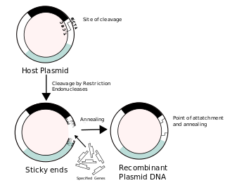
Recombinant DNA (rDNA), or molecular cloning, is the process by which a single gene, or segment of DNA, is isolated and amplified. Recombinant DNA is also known as in vitro recombination. A cloning vector is a DNA molecule that carries foreign DNA into a host cell, where it replicates, producing many copies of itself along with the foreign DNA. There are many types of cloning vectors such plasmids and phages. In order to carry out recombination between vector and the foreign DNA, it is necessary the vector and DNA to be cloned by digestion, ligase the foreign DNA into the vector with the enzyme DNA ligase. And DNA is inserted by introducing the DNA into bacteria cells by transformation.

<span class="mw-page-title-main">Retroviral ribonuclease H</span>

The retroviral ribonuclease H is a catalytic domain of the retroviral reverse transcriptase (RT) enzyme. The RT enzyme is used to generate complementary DNA (cDNA) from the retroviral RNA genome. This process is called reverse transcription. To complete this complex process, the retroviral RT enzymes need to adopt a multifunctional nature. They therefore possess 3 of the following biochemical activities: RNA-dependent DNA polymerase, ribonuclease H, and DNA-dependent DNA polymerase activities. Like all RNase H enzymes, the retroviral RNase H domain cleaves DNA/RNA duplexes and will not degrade DNA or unhybridized RNA.

References

  1. 1 2 3 Dhundale A, Lampson B, Furuichi T, Inouye M, Inouye S (December 1987). "Structure of msDNA from Myxococcus xanthus: evidence for a long, self-annealing RNA precursor for the covalently linked, branched RNA". Cell. 51 (6): 1105–12. doi:10.1016/0092-8674(87)90596-4. PMID   2446773. S2CID   21762469.
  2. Inouye S, Herzer PJ, Inouye M (February 1990). "Two independent retrons with highly diverse reverse transcriptases in Myxococcus xanthus". Proceedings of the National Academy of Sciences of the United States of America. 87 (3): 942–5. Bibcode:1990PNAS...87..942I. doi: 10.1073/pnas.87.3.942 . PMC   53385 . PMID   1689062.
  3. Yee T, Furuichi T, Inouye S, Inouye M (August 1984). "Multicopy single-stranded DNA isolated from a gram-negative bacterium, Myxococcus xanthus". Cell. 38 (1): 203–9. doi:10.1016/0092-8674(84)90541-5. PMID   6088065. S2CID   41165293.
  4. Furuichi T, Inouye S, Inouye M (January 1987). "Biosynthesis and structure of stable branched RNA covalently linked to the 5' end of multicopy single-stranded DNA of Stigmatella aurantiaca". Cell. 48 (1): 55–62. doi:10.1016/0092-8674(87)90355-2. PMID   2431795. S2CID   32376617.
  5. 1 2 3 Das R, Shimamoto T, Hosen SM, Arifuzzaman M (2011). "Comparative Study of different msDNA (multicopy single-stranded DNA) structures and phylogenetic comparison of reverse transcriptases (RTs): evidence for vertical inheritance" (PDF). Bioinformation. 7 (4): 176–9. doi:10.6026/97320630007176. PMC   3218519 . PMID   22102774.
  6. Lampson BC, Sun J, Hsu MY, Vallejo-Ramirez J, Inouye S, Inouye M (February 1989). "Reverse transcriptase in a clinical strain of Escherichia coli: production of branched RNA-linked msDNA" (PDF). Science. 243 (4894 Pt 1): 1033–8. Bibcode:1989Sci...243.1033L. doi:10.1126/science.2466332. PMID   2466332. Archived from the original (PDF) on 2014-12-22. Retrieved 2012-02-08.
  7. Lima TM, Lim D (May 1995). "Isolation and characterization of host mutants defective in msDNA synthesis: role of ribonuclease H in msDNA synthesis". Plasmid. 33 (3): 235–8. doi: 10.1006/plas.1995.1026 . PMID   7568472.
  8. Hsu MY, Inouye M, Inouye S (December 1990). "Retron for the 67-base multicopy single-stranded DNA from Escherichia coli: a potential transposable element encoding both reverse transcriptase and Dam methylase functions". Proceedings of the National Academy of Sciences of the United States of America. 87 (23): 9454–8. Bibcode:1990PNAS...87.9454H. doi: 10.1073/pnas.87.23.9454 . PMC   55184 . PMID   1701261.
  9. Inouye S.; Inouye M. (1993). "Bacterial Reverse Transcriptase". In Goff, Stephen and Anna M. Skalka (ed.). Reverse transcriptase. Cold Spring Harbor monograph series. Vol. 23. Plainview, N.Y: Cold Spring Harbor Laboratory Press. ISBN   978-0-87969-382-4.
  10. 1 2 3 4 Lampson BC, Inouye M, Inouye S (2005). "Retrons, msDNA, and the bacterial genome" (PDF). Cytogenetic and Genome Research. 110 (1–4): 491–9. doi:10.1159/000084982. PMID   16093702. S2CID   24854188. Archived from the original (PDF) on 2016-03-05. Retrieved 2012-02-08.
  11. Simon DM, Zimmerly S (December 2008). "A diversity of uncharacterized reverse transcriptases in bacteria". Nucleic Acids Research. 36 (22): 7219–29. doi:10.1093/nar/gkn867. PMC   2602772 . PMID   19004871.
  12. 1 2 Das R, Shimamoto T, Arifuzzaman M (2011). "A Novel msDNA (Multicopy Single-Stranded DNA) Strain Present in Yersinia frederiksenii ATCC 33641 Contig01029 Enteropathogenic Bacteria with the Genomic Analysis of It's Retron". Journal of Pathogens. 2011 (693769): 693769. doi:10.4061/2011/693769. PMC   3335539 . PMID   22567337.
  13. Sherman LA, Chattopadhyay S (1993). "The molecular basis of allorecognition". Annual Review of Immunology. 11: 385–402. doi:10.1146/annurev.iy.11.040193.002125. PMID   8476567.
  14. Buss, Leo (2006). The Evolution of Individuality . Princeton University Press. ISBN   978-0-691-08469-5.
  15. Shimamoto T, Kawanishi H, Tsuchiya T, Inouye S, Inouye M (June 1998). "In vitro synthesis of multicopy single-stranded DNA, using separate primer and template RNAs, by Escherichia coli reverse transcriptase". Journal of Bacteriology. 180 (11): 2999–3002. doi:10.1128/JB.180.11.2999-3002.1998. PMC   107272 . PMID   9603895.
  16. 1 2 Inouye S, Hsu MY, Xu A, Inouye M (October 1999). "Highly specific recognition of primer RNA structures for 2'-OH priming reaction by bacterial reverse transcriptases". The Journal of Biological Chemistry. 274 (44): 31236–44. doi: 10.1074/jbc.274.44.31236 . PMID   10531319.
  17. Jacobo-Molina A, Ding J, Nanni RG, Clark AD, Lu X, Tantillo C, Williams RL, Kamer G, Ferris AL, Clark P (July 1993). "Crystal structure of human immunodeficiency virus type 1 reverse transcriptase complexed with double-stranded DNA at 3.0 A resolution shows bent DNA". Proceedings of the National Academy of Sciences of the United States of America. 90 (13): 6320–4. Bibcode:1993PNAS...90.6320J. doi: 10.1073/pnas.90.13.6320 . PMC   46920 . PMID   7687065.
  18. Sarafianos SG, Das K, Tantillo C, Clark AD, Ding J, Whitcomb JM, Boyer PL, Hughes SH, Arnold E (March 2001). "Crystal structure of HIV-1 reverse transcriptase in complex with a polypurine tract RNA:DNA". The EMBO Journal. 20 (6): 1449–61. doi:10.1093/emboj/20.6.1449. PMC   145536 . PMID   11250910.

Further reading