Related Research Articles
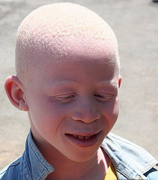
Albinism is a congenital condition characterized in humans by the partial or complete absence of pigment in the skin, hair and eyes. Albinism is associated with a number of vision defects, such as photophobia, nystagmus, and amblyopia. Lack of skin pigmentation makes for more susceptibility to sunburn and skin cancers. In rare cases such as Chédiak–Higashi syndrome, albinism may be associated with deficiencies in the transportation of melanin granules. This also affects essential granules present in immune cells, leading to increased susceptibility to infection.

Human skin color ranges from the darkest brown to the lightest hues. Differences in skin color among individuals is caused by variation in pigmentation, which is the result of genetics, exposure to the sun, disorders, or some combination thereof. Differences across populations evolved through natural selection or sexual selection, because of social norms and differences in environment, as well as regulations of the biochemical effects of ultraviolet radiation penetrating the skin.

Melanin consist of oligomers or polymers arranged in a disordered manner which among other functions provide the pigments of many organisms. Melanin pigments are produced in a specialized group of cells known as melanocytes. They have been described as "among the last remaining biological frontiers with the unknown".

Melanocytes are melanin-producing neural crest-derived cells located in the bottom layer of the skin's epidermis, the middle layer of the eye, the inner ear, vaginal epithelium, meninges, bones, and heart. Melanin is a dark pigment primarily responsible for skin color. Once synthesized, melanin is contained in special organelles called melanosomes which can be transported to nearby keratinocytes to induce pigmentation. Thus darker skin tones have more melanosomes present than lighter skin tones. Functionally, melanin serves as protection against UV radiation. Melanocytes also have a role in the immune system.

Freckles are clusters of concentrated melaninized cells which are most easily visible on people with a fair complexion. Freckles do not have an increased number of the melanin-producing cells, or melanocytes, but instead have melanocytes that overproduce melanin granules (melanosomes) changing the coloration of the outer skin cells (keratinocytes). As such, freckles are different from lentigines and moles, which are caused by accumulation of melanocytes in a small area. Freckles can appear on all types of skin tones. Of the six Fitzpatrick skin types, they are most common on skin tones 1 and 2, which usually belong to North Europeans. However, it can also be found on people all over the world. In England a historical term for freckles is summer-voys, sometimes spelt summervoise, which may be related to the German "summersprosse".

Human hair color is the pigmentation of human hair follicles due to two types of melanin: eumelanin and pheomelanin. Generally, if more melanin is present, the color of the hair is darker; if less melanin is present, the hair is lighter. The tone of the hair is dependent on the ratio of black or brown eumelanin to yellow or red pheomelanin. Levels of melanin can vary over time causing a person's hair color to change, and it is possible to have hair follicles of more than one color on the same person. Some hair colors are associated with some ethnic groups due to observed higher frequency of particular hair color within their geographical region, e.g. straight dark hair amongst East Asians, Southeast Asians, Polynesians and Native Americans, a large variety of dark, fair, curly, straight, wavy and bushy hair amongst Europeans, West Asians and North Africans, curly, dark, and uniquely helical hair with Sub Saharan Africans, whilst gray, white or "silver" hair is often associated with age.

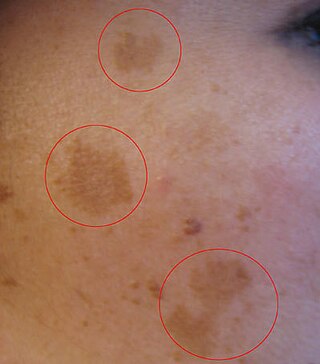
Hyperpigmentation is the darkening of an area of skin or nails caused by increased melanin.

Hypopigmentation is characterized specifically as an area of skin becoming lighter than the baseline skin color, but not completely devoid of pigment. This is not to be confused with depigmentation, which is characterized as the absence of all pigment. It is caused by melanocyte or melanin depletion, or a decrease in the amino acid tyrosine, which is used by melanocytes to make melanin. Some common genetic causes include mutations in the tyrosinase gene or OCA2 gene. As melanin pigments tend to be in the skin, eye, and hair, these are the commonly affected areas in those with hypopigmentation.
Melasma is a tan or dark skin discoloration. Melasma is thought to be caused by sun exposure, genetic predisposition, hormone changes, and skin irritation. Although it can affect anyone, it is particularly common in women, especially pregnant women and those who are taking oral or patch contraceptives or hormone replacement therapy medications.

Tyrosinase is an oxidase that is the rate-limiting enzyme for controlling the production of melanin. The enzyme is mainly involved in two distinct reactions of melanin synthesis otherwise known as the Raper Mason pathway. Firstly, the hydroxylation of a monophenol and secondly, the conversion of an o-diphenol to the corresponding o-quinone. o-Quinone undergoes several reactions to eventually form melanin. Tyrosinase is a copper-containing enzyme present in plant and animal tissues that catalyzes the production of melanin and other pigments from tyrosine by oxidation. It is found inside melanosomes which are synthesized in the skin melanocytes. In humans, the tyrosinase enzyme is encoded by the TYR gene.
Skin whitening, also known as skin lightening and skin bleaching, is the practice of using chemical substances in an attempt to lighten the skin or provide an even skin color by reducing the melanin concentration in the skin. Several chemicals have been shown to be effective in skin whitening, while some have proven to be toxic or have questionable safety profiles. This includes mercury compounds which may cause neurological problems and kidney problems.

Griscelli syndrome is a rare autosomal recessive disorder characterized by albinism (hypopigmentation) with immunodeficiency, that usually causes death by early childhood. Researchers have developed three different classifications of the form of disorder, characterised by different signs and symptoms. Type 1 Griscelli Syndrome is assosciated with severe brain function issues along with distinctive discolouring of the hair and skin. Type 2 Griscelli Syndrome have immune system abnormalities in addition to hypopigmentation of skin and hair. Finally, Type 3 is seen as those only affected by hypopigmentation of the skin and hair. This type is not associated with immune deficiencies or neurological abnormalities.
Piebaldism refers to the absence of mature melanin-forming cells (melanocytes) in certain areas of the skin and hair. It is a rare autosomal dominant disorder of melanocyte development. Common characteristics include a congenital white forelock, scattered normal pigmented and hypopigmented macules and a triangular shaped depigmented patch on the forehead. There is nevertheless great variation in the degree and pattern of presentation, even within affected families. In some cases, piebaldism occurs together with severe developmental problems, as in Waardenburg syndrome and Hirschsprung's disease.

Smoker's melanosis is seen with the naked eye as a brown to black pigmentation of the oral tissue i.e. the gums, cheeks or palate as well as in larynx. It is most often seen in the lower labial gingiva of tobacco users. Most easily it is found in Caucasians, due to their lack of a genetically caused melanin pigmentation.

Light skin is a human skin color that has a base level of eumelanin pigmentation that has adapted to environments of low UV radiation. Light skin is most commonly found amongst the native populations of Europe, Central Asia, and Northeast Asia as measured through skin reflectance. People with light skin pigmentation are often referred to as "white" although these usages can be ambiguous in some countries where they are used to refer specifically to certain ethnic groups or populations.

Ocular albinism type 1(OA1) is the most common type of ocular albinism, with a prevalence rate of 1:50,000. It is an inheritable classical Mendelian type X-linked recessive disorder wherein the retinal pigment epithelium lacks pigment while hair and skin appear normal. Since it is usually an X-linked disorder, it occurs mostly in males, while females are carriers unless they are homozygous. About 60 missense and nonsense mutations, insertions, and deletions have been identified in Oa1. Mutations in OA1 have been linked to defective glycosylation and thus improper intracellular transportation.
Drug induced pigmentation may take on many different appearances, one of the most common being a change in the color, or pigmentation, of the skin.
Postinflammatory hypopigmentation is a cutaneous condition characterized by decreased pigment in the skin following inflammation of the skin.
Oral pigmentation is asymptomatic and does not usually cause any alteration to the texture or thickness of the affected area. The colour can be uniform or speckled and can appear solitary or as multiple lesions. Depending on the site, depth, and quantity of pigment, the appearance can vary considerably.
Postinflammatory hyperpigmentation (PIH) is a skin condition characterized by the darkening of the skin (hyperpigmentation) following an inflammatory injury, such as acne, dermatitis, infectious disease, or trauma. Less frequently, it may occur as a complication of a medical procedure performed on the skin. It is a common cause of skin discoloration and can affect individuals of all skin types.
References
- 1 2 James, William D.; Elston, Dirk; Treat, James R.; Rosenbach, Misha A.; Neuhaus, Isaac (2020). "36. Disturbances of pigmentation". Andrews' Diseases of the Skin: Clinical Dermatology (13th ed.). Edinburgh: Elsevier. pp. 862–880. ISBN 978-0-323-54753-6.
- ↑ "MedlinePlus: Skin Pigmentation Disorders".
- ↑ "Introduction: Pigment Disorders: Merck Manual Home Edition".
- 1 2 3 4 5 6 7 Thawabteh, Amin Mahmood; Jibreen, Alaa; Karaman, Donia; Thawabteh, Alà; Karaman, Rafik (2023-06-18). "Skin Pigmentation Types, Causes and Treatment—A Review". Molecules. MDPI AG. 28 (12): 4839. doi: 10.3390/molecules28124839 . ISSN 1420-3049. PMC 10304091 .