Keratosis pilaris

Last updated
Keratosis pilaris
Other namesFollicular keratosis, lichen pilaris
Keratosis pilaris (moderate).jpg
Condition on a calf
Specialty Dermatology

Keratosis pilaris (KP; also follicular keratosis, lichen pilaris, or colloquially chicken skin [1] ) is a common, autosomal-dominant, genetic condition of the skin's hair follicles characterized by the appearance of possibly itchy, small, gooseflesh-like bumps, with varying degrees of reddening or inflammation. [2] It most often appears on the outer sides of the upper arms (the forearms can also be affected), thighs, face, back, and buttocks; [2] KP can also occur on the hands, and tops of legs, sides, or any body part except glabrous (hairless) skin (like the palms or soles of feet). [3] Often the lesions can appear on the face, which may be mistaken for acne [4] or folliculitis.

Contents

The several types of KP have been associated with pregnancy, type 1 diabetes mellitus, obesity, dry skin, allergic diseases (e.g., atopic dermatitis), and rarely cancer. [1] Many rarer types of the disorder are part of inherited genetic syndromes. [1]

The cause of KP is not completely understood. As of 2018, KP is thought to be due to abnormalities in the process of depositing the protein keratin in hair follicles, abnormalities in the hair shaft, or both. [1] KP is usually diagnosed by a medical professional based on the appearance of the skin, but dermoscopy can be used, as well, if the diagnosis is unclear. [1] Variants of the ABCA12 gene have been associated with KP. [5]

KP is the most common disorder of the hair follicle in children. [1] How common it is in adults is unclear since keratosis pilaris is an underreported condition and the actual prevalence of the condition may be higher than estimated. [6] No single approach has been found to completely cure KP, but treatments can improve the cosmetic appearance of the condition. Treatment includes the application of topical preparations of moisturizers and medications such as glycolic acid, lactic acid, salicylic acid, urea, or retinoids to the skin. [1] Fractional carbon dioxide lasers and Nd:YAG laser therapies are also effective. [1]

Signs and symptoms

KP results in small, rough bumps on the surface of the skin. They are skin-colored bumps the size of a grain of sand, many of which are surrounded by a slight pink color in light-skinned people and dark spots in dark-skinned people. [4] Most people with KP do not have symptoms, but the bumps in the skin can occasionally be itchy. [7] Irritation due to scratching KP bumps can result in redness and inflammation.

Though people with KP experience the condition year-round, the problem can become exacerbated, with the bumps likely to look and feel more pronounced in color and texture, during the colder months, when moisture levels in the air are lower. [8] The symptoms may also worsen during pregnancy or after childbirth. [9] Increased sun exposure might mitigate the symptoms of KP. [ citation needed ]

Pathophysiology

KP occurs when the human body produces excess amounts of the skin protein keratin, resulting in the formation of small, raised bumps in the skin, often with surrounding redness. [7] The excess keratin, which is the same color as the person's natural skin tone, surrounds and entraps the hair follicles in the pore. This causes the formation of hard plugs (a process known as hyperkeratinization). [4] Many KP bumps contain an ingrown hair that has coiled. [2] This is a result of the keratinized skin's "capping off" the hair follicle, preventing the hair from exiting. The hair grows encapsulated inside the follicle. KP is more common in patients affected by atopic diseases such as allergic rhinitis and atopic dermatitis.

KP subtypes are occasionally part of genetically inherited syndromes associated with intellectual disability, neuro-cardio-facial-cutaneous syndromes, RASopathies, ectodermal dysplasias, and certain myopathies. [1]

Diagnosis

Physicians can often diagnose KP simply by examining the skin without specialized tests, [4] but a dermatologist can use dermoscopy to confirm the diagnosis and assess if a person with KP is responding to treatment. [2] Physicians often consider family history and the presence of symptoms when making the diagnosis. [10] Those with this condition are generally encouraged to contact a physician if the bumps are bothersome and do not improve with over-the-counter lotions. [4]

Differential diagnosis

Several medications that can cause a skin eruption similar to KP include cyclosporine, BRAF inhibitors, and tyrosine kinase inhibitors. [1]

Classification

The several different types of KP include KP rubra (red, inflamed bumps, which can be on arms, head, legs), KP alba (rough, bumpy skin with no irritation), KP rubra faceii (reddish rash on the cheeks), KP atrophicans, keratosis follicularis spinulosa decalvans, atrophoderma vermiculatum, KP atrophicans faciei, erythromelanosis follicularis faciei et colli, and papular profuse precocious KP. [1] [7]

KP is commonly described in association with other dry-skin conditions, such as ichthyosis vulgaris, dry skin, and atopic dermatitis, including those of asthma and allergies. [1] KP does not bear any known, long-term health implications, nor is it associated with increased mortality or morbidity. [11] It is not related to goose bumps, which result from muscle contractions, except that both occur in the area where the hair shaft exits the skin.

Treatment

KP is medically harmless, [2] but many individuals may seek treatment, as the condition can cause emotional distress. [9] Topical creams and lotions are currently the most commonly used treatment for KP, specifically those consisting of moisturizing or keratolytic treatments, including urea, lactic acid, glycolic acid, salicylic acid, vitamin D, fish oil, or topical retinoids such as tretinoin. [12] Improvement of the skin often takes months, and the bumps are likely to return. Limiting time in the shower and using gentle exfoliation to unclog pores can help. [9] [13] Many products are available that apply abrasive materials, with alpha or beta hydroxy acids to assist with exfoliation.

Some cases of KP have been successfully treated with laser therapy, which involves passing intense bursts of light into targeted areas of the skin. [14] Depending on the body's response to the treatment, multiple sessions over the course of a few months may be necessary.

Epidemiology

Worldwide, KP affects an estimated 30 to 50% of the adult population, and around 50 to 80% of all adolescents. It is more common in women than in men, and it is often present in otherwise healthy individuals. [12] The skin condition is prevalent in people of all ethnicities, and no particular ethnicity is at higher risk for developing KP. Although KP may manifest in people of any age, it usually appears within the first decade of life and is more common in young children. [11] In most cases, the condition gradually improves before age 30, but it can persist longer. [9]

See also

Related Research Articles

<span class="mw-page-title-main">Dermatitis</span> Inflammatory disease of the skin

Dermatitis is inflammation of the skin, typically characterized by itchiness, redness and a rash. In cases of short duration, there may be small blisters, while in long-term cases the skin may become thickened. The area of skin involved can vary from small to covering the entire body. Dermatitis is often called eczema, and the difference between those terms is not standardized.

<span class="mw-page-title-main">Sebaceous gland</span> Gland to lubricate the hair and skin

A sebaceous gland or oil gland is a microscopic exocrine gland in the skin that opens into a hair follicle to secrete an oily or waxy matter, called sebum, which lubricates the hair and skin of mammals. In humans, sebaceous glands occur in the greatest number on the face and scalp, but also on all parts of the skin except the palms of the hands and soles of the feet. In the eyelids, meibomian glands, also called tarsal glands, are a type of sebaceous gland that secrete a special type of sebum into tears. Surrounding the female nipple, areolar glands are specialized sebaceous glands for lubricating the nipple. Fordyce spots are benign, visible, sebaceous glands found usually on the lips, gums and inner cheeks, and genitals.

<span class="mw-page-title-main">Ichthyosis vulgaris</span> Skin disorder

Ichthyosis vulgaris is a skin disorder causing dry, scaly skin. It is the most common form, and one of the mildest forms, of ichthyosis, affecting around 1 in 250 people. For this reason it is known as common ichthyosis. It is usually an autosomal dominant inherited disease, although a rare non-heritable version called acquired ichthyosis exists.

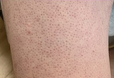
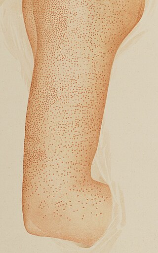
Hyperkeratinization is a disorder of the cells lining the inside of a hair follicle. It is the normal function of these cells to detach or slough off from the skin lining at normal intervals. The dead cells are then forced out of the follicle. However, in hyperkeratinization, this process is interrupted and a number of these dead skin cells do not leave the follicle because of an excess of keratin, a natural protein found in the skin. This excess of keratin, which is influenced by genetics, results in an increased adherence/bonding of dead skin cells together. This cohesion of cells will block or "cap" the hair follicle or clog the sebaceous/oil duct. Pathogens may also play a role in causing, perpetuating, or simply taking advantage of this phenomenon, such as virulent sub-strains of Cutibacterium acnes and irregular migration of Staphylococcus epidermidis from the outer surface of the skin into the follicle, where commensal strains of C. acnes exclusively habitate. It itches mildly at times, and strongly at others. Very often it cannot be felt at all.

<span class="mw-page-title-main">Pseudofolliculitis barbae</span> Medical condition

Pseudofolliculitis barbae (PFB) is a type of irritant folliculitis that commonly affects people who have curly or coarse facial hair. It occurs when hair curls back into the skin after shaving, causing inflammation, redness, and bumps. This can lead to ingrown hairs, scarring, and skin discoloration. PFB can be treated with various methods, including changing shaving habits, using topical creams or ointments, and undergoing laser hair removal. Prevention measures include proper shaving techniques, using sharp razors, and avoiding too close a shave.

<span class="mw-page-title-main">Hyperkeratosis</span> Medical condition

Hyperkeratosis is thickening of the stratum corneum, often associated with the presence of an abnormal quantity of keratin, and is usually accompanied by an increase in the granular layer. As the corneum layer normally varies greatly in thickness in different sites, some experience is needed to assess minor degrees of hyperkeratosis.

<span class="mw-page-title-main">Pityriasis rubra pilaris</span> Medical condition

Pityriasis rubra pilaris refers to a group of chronic disorders characterized by reddish orange, scaling plaques and keratotic follicular papules. Symptoms may include reddish-orange patches on the skin, severe flaking, uncomfortable itching, thickening of the skin on the feet and hands, and thickened bumps around hair follicles. For some, early symptoms may also include generalized swelling of the legs, feet and other parts of the body. PRP has a varied clinical progression and a varied rate of improvement. There is currently no known cause or cure for PRP.

<span class="mw-page-title-main">Comedo</span> Medical condition

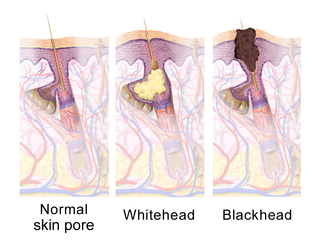
A comedo is a clogged hair follicle (pore) in the skin. Keratin combines with oil to block the follicle. A comedo can be open (blackhead) or closed by skin (whitehead) and occur with or without acne. The word "comedo" comes from the Latin comedere, meaning "to eat up", and was historically used to describe parasitic worms; in modern medical terminology, it is used to suggest the worm-like appearance of the expressed material.

<span class="mw-page-title-main">Erythroderma</span> Inflammatory skin disease with redness and scaling

Erythroderma is an inflammatory skin disease with redness and scaling that affects nearly the entire cutaneous surface. This term applies when 90% or more of the skin is affected.

<span class="mw-page-title-main">Perioral dermatitis</span> Medical condition

Perioral dermatitis, also known as periorificial dermatitis, is a common type of skin rash. Symptoms include multiple small (1–2 mm) bumps and blisters sometimes with background redness and scale, localized to the skin around the mouth and nostrils. Less commonly the eyes and genitalia may be involved. It can be persistent or recurring and resembles particularly rosacea and to some extent acne and allergic dermatitis. The term "dermatitis" is a misnomer because this is not an eczematous process.

<span class="mw-page-title-main">Monilethrix</span> Medical condition

Monilethrix is a rare autosomal dominant hair disease that results in short, fragile, broken hair that appears beaded. It comes from the Latin word for necklace (monile) and the Greek word for hair (thrix). Hair becomes brittle, and breaks off at the thinner parts between the beads. It appears as a thinning or baldness of hair and was first described in 1897 by Walter Smith

<span class="mw-page-title-main">Discoid lupus erythematosus</span> Autoimmune skin condition

Discoid lupus erythematosus is the most common type of chronic cutaneous lupus (CCLE), an autoimmune skin condition on the lupus erythematosus spectrum of illnesses. It presents with red, painful, inflamed and coin-shaped patches of skin with a scaly and crusty appearance, most often on the scalp, cheeks, and ears. Hair loss may occur if the lesions are on the scalp. The lesions can then develop severe scarring, and the centre areas may appear lighter in color with a rim darker than the normal skin. These lesions can last for years without treatment.

Madarosis is a condition that results in the loss of eyelashes, and sometimes eyebrows. The term "madarosis" is derived from the ancient Greek "madaros", meaning "bald". It originally was a disease of only losing eyelashes but it currently is the loss of both eyelashes and eyebrows. Eyebrows and eyelashes are both important in the prevention of bacteria and other foreign objects from entering the eye. A majority of patients with madarosis have leprosy, and it was reported that 76% of patients with varying types of leprosy had madarosis.

Pruritic folliculitis of pregnancy is a skin condition that occurs in one in 3000 people, about 0.2% of cases, who are in their second to third trimester of pregnancy where the hair follicle becomes inflamed or infected, resulting in a pus filled bump. Some dermatologic conditions aside from pruritic folliculitis during pregnancy include "pruritic urticarial papules and plaques of pregnancy, atopic eruption of pregnancy, pemphigoid gestationis, intrahepatic cholestasis of pregnancy, and pustular psoriasis of pregnancy". This pruritic folliculitis of pregnancy differs from typical pruritic folliculitis; in pregnancy, it is characterized by sterile hair follicles becoming inflamed mainly involving the trunk, contrasting how typical pruritic folliculitis is mainly localized on "the upper back, shoulders, and chest." This condition was first observed after some pregnant individuals showed signs of folliculitis that were different than seen before. The inflammation was thought to be caused by hormonal imbalance, infection from bacteria, fungi, viruses or even an ingrown hair. However, there is no known definitive cause as of yet. These bumps usually begin on the belly and then spread to upper regions of the body as well as the thighs.

Keratosis pilaris atropicans is a group of idiopathic genodermatoses that consists of three unique clinical entities: atrophoderma vermiculatum, keratosis follicularis spinulosa decalvans, and keratosis pilaris atrophicans faciei.

Erythromelanosis follicularis faciei et colli is an erythematous pigmentary disease involving the follicles, characterized by a reddish-brown, sharply demarcated, symmetrical discoloration involving the preauricular and maxillary regions.

Traumatic anserine folliculosis is a curious gooseflesh-like follicular hyperkeratosis that may result from persistent pressure and lateral friction of one skin surface against another. Traumatic anserine folliculosis is caused by trauma. Topical keratolytics are the treatment of choice.

<span class="mw-page-title-main">Fungal folliculitis</span> Inflammation of hair follicles due to fungal infection

Majocchi's granuloma is a skin condition characterized by deep, pustular plaques, and is a form of tinea corporis. It is a localized form of fungal folliculitis. Lesions often have a pink and scaly central component with pustules or folliculocentric papules at the periphery. The name comes from Domenico Majocchi, who discovered the disorder in 1883. Majocchi was a professor of dermatology at the University of Parma and later the University of Bologna. The most common dermatophyte is called Trichophyton rubrum.

Childhood granulomatous periorificial dermatitis (CGPD), is a rare benign granulomatous skin disease of unknown cause. The disorder was first described in 1970 by Gianotti in a case series of five children. CGPD is more common in boys than girls.

References

  1. 1 2 3 4 5 6 7 8 9 10 11 12 Wang, JF; Orlow, SJ (July 2018). "Keratosis Pilaris and its Subtypes: Associations, New Molecular and Pharmacologic Etiologies, and Therapeutic Options". American Journal of Clinical Dermatology. 19 (5): 733–757. doi:10.1007/s40257-018-0368-3. PMID   30043128. S2CID   51715063.
  2. 1 2 3 4 5 Panchaprateep R, Tanus A, Tosti A (March 2015). "Clinical, dermoscopic, and histopathologic features of body hair disorders". Journal of the American Academy of Dermatology. 72 (5): 890–900. doi:10.1016/j.jaad.2015.01.024. PMID   25748313.
  3. Alai, Nili. "Keratosis Pilaris (KP)". MedicineNet. Retrieved 2008-10-06.
  4. 1 2 3 4 5 Berman, Kevin. "Keratosis pilaris". MedlinePlus. Retrieved 2008-06-19.
  5. Liu F, Yang Y, Zheng Y, Liang YH, Zeng K (September 2018). "Mutation and expression of ABCA12 in keratosis pilaris and nevus comedonicus". Molecular Medicine Reports. 18 (3): 3153–3158. doi: 10.3892/mmr.2018.9342 . PMC   6102636 . PMID   30066947.
  6. Pennycook KB, McCready TA (June 27, 2022). Keratosis Pilaris in StatPearls. PMID   31536314.
  7. 1 2 3 Hwang S, Schwartz RA (September 2008). "Keratosis pilaris: a common follicular hyperkeratosis". Cutis. 82 (3): 177–80. PMID   18856156.
  8. "Keratosis pilaris ("chicken skin")". NHS Choices. 2017-10-24.
  9. 1 2 3 4 "Keratosis pilaris: Self-management". Mayo Clinic.
  10. Mayo Clinic Staff. "Tests and Diagnosis". Mayo Clinic. Retrieved 2013-10-31
  11. 1 2 Alai, Nili; Arash Michael Saemi; Raul Del Rosario. "Keratosis Pilaris". eMedicine. Medscape (WebMD). and "Pathophysiology". Medscape (WebMD). Retrieved February 5, 2018.
  12. 1 2 Yosipovitch G, DeVore A, Dawn A (June 2007). "Obesity and the skin: skin physiology and skin manifestations of obesity". Journal of the American Academy of Dermatology. 56 (6): 901–16. doi:10.1016/j.jaad.2006.12.004. PMID   17504714.
  13. Mayo Clinic Staff (5 January 2016). "Self-management - Keratosis Pilaris". mayoclinic.org. Mayo Foundation. Retrieved 11 April 2016.
  14. Schoch, Jennifer J.; Tollefson, Megha M.; Witman, Patricia; Davis, Dawn M. R. (July 2016). "Successful Treatment of Keratosis Pilaris Rubra with Pulsed Dye Laser". Pediatric Dermatology. 33 (4): 443–446. doi:10.1111/pde.12892. ISSN   1525-1470. PMID   27282957. S2CID   32102128.