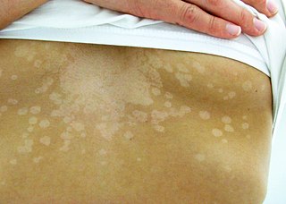
Tinea versicolor is a condition characterized by a skin eruption on the trunk and proximal extremities. The majority of tinea versicolor is caused by the fungus Malassezia globosa, although Malassezia furfur is responsible for a small number of cases. These yeasts are normally found on the human skin and become troublesome only under certain circumstances, such as a warm and humid environment, although the exact conditions that cause initiation of the disease process are poorly understood.

Tinea corporis is a fungal infection of the body, similar to other forms of tinea. Specifically, it is a type of dermatophytosis that appears on the arms and legs, especially on glabrous skin; however, it may occur on any superficial part of the body.

Tinea barbae is a fungal infection of the hair. Tinea barbae is due to a dermatophytic infection around the bearded area of men. Generally, the infection occurs as a follicular inflammation, or as a cutaneous granulomatous lesion, i.e. a chronic inflammatory reaction. It is one of the causes of folliculitis. It is most common among agricultural workers, as the transmission is more common from animal-to-human than human-to-human. The most common causes are Trichophyton mentagrophytes and T. verrucosum.

Erythema marginatum is a type of erythema involving pink rings on the torso and inner surfaces of the limbs which come and go for as long as several months. It is found primarily on extensor surfaces.
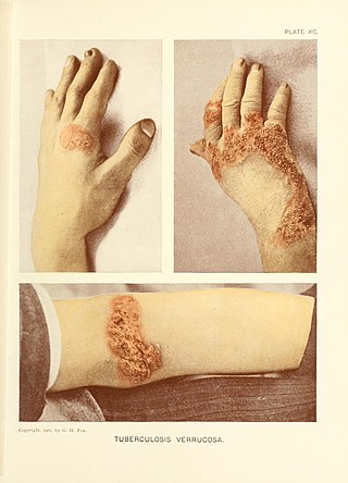
Tuberculosis verrucosa cutis is a rash of small, red papules and nodules in the skin that may appear two to four weeks after inoculation by Mycobacterium tuberculosis in a previously infected and immunocompetent individual.

Grover's disease (GD) is a polymorphic, pruritic, papulovesicular dermatosis characterized histologically by acantholysis with or without dyskeratosis. Once confirmed, most cases of Grover's disease last six to twelve months, which is why it was originally called "transient". However it may last much longer. Nevertheless, it is not to be confused with relapsing linear acantholytic dermatosis.

Spongiosis is mainly intercellular edema in the epidermis, and is characteristic of eczematous dermatitis, manifested clinically by intraepidermal vesicles, "juicy" papules, and/or lichenification. It is a severe case of eczema that affects the epidermis, dermis and/or subcutaneous skin tissues. The three types of spongiotic dermatitis are acute, subacute and chronic. A dermatologist can diagnose acute spongiotic dermatitis by examining the skin during an office visit but a biopsy is needed for an accurate diagnosis of the type.

Lupus miliaris disseminatus faciei, also known as acne agminata, is a disease with a similar appearance to acne vulgaris. The cause of LMDF is unknown.

Dermatitis repens is a rare, sterile, pustular eruption of the fingers and toes that slowly extends proximally.
Tumid lupus erythematosus is a rare, but distinctive entity in which patients present with edematous erythematous plaques, usually on the trunk.
Subacute cutaneous lupus erythematosus is a clinically distinct subset of cases of lupus erythematosus that is most often present in white women aged 15 to 40, consisting of skin lesions that are scaly and evolve as poly-cyclic annular lesions or plaques similar to those of plaque psoriasis.
Telangiectasia macularis eruptiva perstans is persistent, pigmented, asymptomatic eruption of macules usually less than 0.5 cm in diameter with a slightly reddish-brown tinge.
Small plaque parapsoriasis characteristically occurs with skin lesions that are round, oval, discrete patches or thin plaques, mainly on the trunk.
Bullous drug reaction most commonly refers to a drug reaction in the erythema multiforme group. These are uncommon reactions to medications, with an incidence of 0.4 to 1.2 per million person-years for toxic epidermal necrolysis and 1.2 to 6.0 per million person-years for Stevens–Johnson syndrome. The primary skin lesions are large erythemas, most often irregularly distributed and of a characteristic purplish-livid color, at times with flaccid blisters.
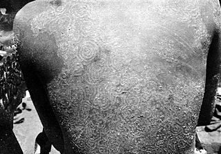
Tinea imbricata is a superficial fungal infection of the skin limited to southwest Polynesia, Melanesia, Southeast Asia, India, and Central America. The skin lesions are often itchy, and mainly in the torso and limbs. The name is derived from the Latin for "tiled" (imbricata) since the lesions are often lamellar. It is often treated with griseofulvin or terbinafine.
A diabetic bulla is a cutaneous condition characterized by a noninflammatory, spontaneous, painless blister, often in acral locations, seen in diabetic patients.
Primary cutaneous adenoid cystic carcinoma is a cutaneous condition characterized by a tumor that usually presents on the chest, scalp, or vulva of middle- to older-aged persons.
Recurrent toxin-mediated perineal erythema is an unusual condition that presents 2–3 days after a throat infection as a fine diffuse macular erythema of the perineal region.

Sarcoidosis, an inflammatory disease, involves the skin in about 25% of patients. The most common lesions are erythema nodosum, plaques, maculopapular eruptions, subcutaneous nodules, and lupus pernio. Treatment is not required, since the lesions usually resolve spontaneously in two to four weeks. Although it may be disfiguring, cutaneous sarcoidosis rarely causes major problems.

