Seborrhoeic dermatitis

Last updated
Seborrhoeic dermatitis
Other namesSebopsoriasis, seborrhoeic eczema, pityriasis capitis [1]
Seborrhoeic dermatitis highres.jpg
Seborrhoeic dermatitis of the face
Specialty Dermatology
Symptoms Flaking, dry or greasy, red, itchy, and inflamed skin [2] [3]
DurationSeveral weeks to lifelong [4]
CausesMultiple factors [4]
Risk factors Stress, dry skin, winter, poor immune function, Parkinson disease [4]
Diagnostic method Based on symptoms [4]
Differential diagnosis Psoriasis, atopic dermatitis, tinea capitis, rosacea, systemic lupus erythematosus [4]
Treatment Humidifier
Medication Antifungal cream, anti-inflammatory agents, coal tar, phototherapy [3]
Frequency~5% (adults), [4] ~10% (babies) [5]
Cradle cap, which is seborrhoeic dermatitis of the infant scalp Cradle cap new photo help in diagnosis.jpg
Cradle cap, which is seborrhoeic dermatitis of the infant scalp

Seborrhoeic dermatitis is a long-term skin disorder. [4] Symptoms include flaky, scaly, greasy, and occasionally itchy and inflamed skin. [2] [3] Areas of the skin rich in oil-producing glands are often affected including the scalp, face, and chest. [4] It can result in social or self-esteem problems. [4] In babies, when the scalp is primarily involved, it is called cradle cap. [2] Seborrhoeic dermatitis of the scalp may be described in lay terms as dandruff due to the dry, flaky character of the skin. [6] However, as dandruff may refer to any dryness or scaling of the scalp, not all dandruff is seborrhoeic dermatitis. [6] Seborrhoeic dermatitis is sometimes inaccurately referred to as seborrhoea. [4]

Contents

The cause is unclear but believed to involve a number of genetic and environmental factors. [2] [4] Risk factors for seborrhoeic dermatitis include poor immune function, Parkinson's disease, and alcoholic pancreatitis. [4] [6] The condition may worsen with stress or during the winter. [4] Malassezia yeast is believed to play a role. [6] It is not a result of poor hygiene. [7] Diagnosis is typically clinical and based on the symptoms present. [4] [8] The condition is not contagious. [9]

The typical treatment is topical antifungal cream and anti-inflammatory agents. [3] Specifically, ketoconazole or ciclopirox are effective. [10] Seborrhoeic dermatitis of the scalp is often treated with shampoo preparations of ketoconazole or zinc pyrithione. [11]

The condition is common in infants within the first three months of age or in adults aged 30 to 70 years. [2] [4] [5] It tends to affect more males. [12] Seborrhoeic dermatitis is more common in African Americans, among individuals who are immune compromised, such as with HIV, and individuals with Parkinson's disease. [11] [12]

Signs and symptoms

Seborrhoeic dermatitis on upper face/head Sequeira Plate 18.jpg
Seborrhoeic dermatitis on upper face/head
Seborrhoeic dermatitis on the shoulder Sequeira Plate 19.jpg
Seborrhoeic dermatitis on the shoulder
Seborrhoeic dermatitis on eyelids Dermatitis of eyelids.jpg
Seborrhoeic dermatitis on eyelids

Seborrhoeic dermatitis typically appears as dry, white, flaky skin. The flakes can be fine, loose, and diffuse or thick and adherent. [11] [8] Additionally, flakes can appear yellow and oily or greasy. [8] [12] In addition to flaky skin, seborrhoeic dermatitis can have areas of red, inflamed, and itchy skin that coincide with the area of skin flaking, but not all individuals have this symptom. [8] Seborrhoeic dermatitis of the scalp can appear similarly to dandruff. [11] When the scalp is affected, there can be associated temporary hair loss. [11] Such hair loss varies in appearance from diffuse thinning to patchy areas of hair loss. [11] On close inspection, the locations where hair has thinned may have broken stubs of hair and pustules around the hair follicles. [11] Individuals with more pigmented skin tones may experience increased or decreased skin pigmentation in affected areas. [12]

Various locations can be affected by seborrhoeic dermatitis. Commonly affected areas include the face, ears, scalp, and across the body. It is less common in intertriginous areas, which are areas where the skin folds and comes into contact with itself, such as the groin or underarm. [11]

Seborrhoeic dermatitis' symptoms are typically mild and appear gradually but are often persistent, lasting weeks to years. [8] [11] [13] Individuals with seborrhoeic dermatitis are subject to recurrent bouts and it may be a lifelong condition. [8] Seborrhoeic dermatitis can also occur quickly and severely in patients with Human Immunodeficiency Virus (HIV). In fact, this is sometimes the first indication of HIV. [12]

Causes

The cause of seborrhoeic dermatitis has not been fully clarified. [1] [14]

In addition to the presence of Malassezia, genetic, environmental, hormonal, and immune-system factors are necessary for and/or modulate the expression of seborrhoeic dermatitis. [15] [16] The condition may be aggravated by illness, psychological stress, fatigue, sleep deprivation, change of season, and reduced general health. [17]

Fungi

The condition is thought to be due to a local inflammatory response to overgrowth by Malassezia fungi species in sebum-producing skin areas including the scalp, face, chest, back, underarms, and groin. [3] [14] This is based on observations of high counts of Malassezia species in skin affected by seborrhoeic dermatitis and on the effectiveness of antifungals in treating the condition. [14] Species of Malassezia implicated in Seborrhoeic dermatitis include M. furfur (formerly Pityrosporum ovale ), M. globosa , M. restricta , M. sympodialis , and M. slooffiae . [3] Malassezia appears to be the significant factor in seborrhoeic dermatitis but it is thought that other factors are necessary for the presence of Malassezia to result in the seborrhoeic dermatitis. [14] For example, summer growth of Malassezia in the skin alone does not result in seborrhoeic dermatitis. [14] Besides antifungals, the effectiveness of anti-inflammatory drugs, which reduce inflammation, and antiandrogens, which reduce sebum production, provide further insights into the pathophysiology of seborrhoeic dermatitis. [3] [18] [19]

Bacteria

Several bacteria, including Propionibacterium species and Staphylococcus aureus, have been shown to have some level of interaction with seborrhoeic dermatitis, though their exact impact is not known. [20] [12]

Nutrition

Seborrhoeic dermatitis-like eruptions are also associated with pyridoxine (vitamin B6) and riboflavin (vitamin B2) deficiency. [21] [8] In children and babies, issues with Δ6-desaturase enzymes [17] have been correlated with increased risk.

Immune dysfunction

Those with immunodeficiency (especially infection with HIV) and with neurological disorders that may impact immune system function such as Parkinson's disease (for which the condition is an autonomic sign) and stroke are particularly prone to it. [22]

Climate

Climate can affect seborrheic dermatitis, but there is a lack of consensus about which climates tend to exacerbate seborrheic dermatitis the most. Some studies show low humidity and low temperature are responsible for high frequency of seborrheic dermatitis. [23] Others suggest hot environments may also worsen seborrhoeic dermatitis. [12] Yet another described that high humidity and low UV exposure are culpable. [24] Dry skin and an impaired skin barrier contribute to the condition. [12] [20] It is likely that climate and weather variations affect the water and lipid content of skin. [20]

Mechanism

Seborrhoeic dermatitis is a complex condition with many interacting factors that are not yet fully explained. [14] In general, the major factors that influence the development and severity include Malassezia yeast presents on and in the skin, skin production of oily sebum, and a subsequent inflammatory response against Malassezia and their byproducts. [12] Additional factors involved in the condition are a compromised skin barrier, the makeup and amount of sebum produced, the character of the immune response and inflammation, and the presence of other microbes species inhabiting the skin. [14] [12] A suggested series of events leading to seborrhoeic dermatitis are initial damaged skin barrier and abnormal sebum production which leads to a change in the microbiome of the skin that in turn elicits an immune response. [14] An alternative explanation is an increase in sebum production feeding an increase in the Malassezia population that instigates inflammation; the inflammation then causes cellular changes that damage the skin barrier. This barrier disruption then encourages additional Malassezia growth and inflammation and again worsened skin barrier function. [12]

Diagnosis

Typically, seborrhoeic dermatitis is a clinical diagnosis based on a physician's expertise in identifying and differentiating skin conditions based on the history of the individual and the appearance of the skin. [8] However, seborrhoeic dermatitis may also be diagnosed with additional testing. The least invasive test is a visual inspection in the clinic using a Wood's Lamp. [11] A KOH test can also be used, where skin scraping of the affected skin may also be taken and prepared with potassium hydroxide (KOH) and visualized under a microscope to look for Malassezia or other microbiological cells. Additionally, a fungal culture of the affected skin may be taken to attempt to grow and identify the causative organism. [11]

Differential diagnosis

Seborrhoeic dermatitis can look similar to other skin conditions that share its characteristic dry, flaky, scaly, and inflamed appearance but have different causes and treatments. Physicians use the history of the individual with the skin condition as well as other tests to identify which disorder is present. Other conditions that may be confused with seborrhoeic dermatitis based on appearance are listed below. [8] [11]

Management

Medications

A variety of different types of medications are able to reduce symptoms of seborrhoeic dermatitis. [3] These include certain antifungals, anti-inflammatory agents like corticosteroids and nonsteroidal anti-inflammatory drugs, antiandrogens, and antihistamines, among others. [3] [1] Treatments must take into consideration potential side effects, especially with long-term use given the chronic nature of seborrhoeic dermatitis. Initial therapy is usually a topical preparation with an agreeable side effect profile. [12]

Antifungals

Regular use of an over-the-counter or prescription antifungal shampoo or cream is a common treatment. The topical antifungal medications ketoconazole and ciclopirox have the best evidence. [10] Ketoconazole should be used twice per week. [8] Shampoo or soap containing zinc pyrithione or selenium sulfide is also used. [8] These options should be used on a daily basis but may also be used in conjunction with a ketoconazole shampoo regimen on alternate days. [8] It is unclear if other antifungals are equally effective as this has not been sufficiently studied. [10] Antifungals that have been studied and found to be effective in the treatment of seborrhoeic dermatitis include ketoconazole, fluconazole, miconazole, bifonazole, sertaconazole, clotrimazole, flutrimazole, ciclopirox, terbinafine, butenafine, selenium disulfide, and lithium salts such as lithium gluconate and lithium succinate. [10] [3] Topical climbazole appears to have little effectiveness in the treatment of seborrhoeic dermatitis. [10] Systemic therapy with oral antifungals including itraconazole, fluconazole, ketoconazole is effective, but adverse side effects have been documented for fluconazole and ketoconazole, with the latter not recommended for use, while itraconazole, with its good safety profile, is the most commonly prescribed. [3] Terbinafine is said to be effective, but with adverse side effects, while other sources state it is not effective and should not be used. [3] [11]

Anti-inflammatory treatments

Topical corticosteroids have been shown to be effective in short-term treatment of seborrhoeic dermatitis. They cannot be used long term or for maintenance because of their skin-thinning side effect. Accordingly, these are used for only a few weeks at a time. [11] There is also evidence for the effectiveness of topical calcineurin inhibitors like tacrolimus and pimecrolimus as well as lithium salt therapy. [25] Calcineurin inhibitors were also effective in reducing growth of Malassezia, offering two routes by which they may treat seborrhoeic dermatitis. [24] Medications such as the calcineurin inhibitors should not be used in individuals with seborrhoeic dermatitis who are immune compromised because they cause further immune suppression. [11]

Oral immunosuppressive treatment, such as with prednisone, has been used in short courses for seborrhoeic dermatitis, as a last resort due to its potential side effects. [26]

Antiandrogens

Seborrhoea, which is sometimes associated with seborrhoeic dermatitis, [27] [28] [29] is recognized as an androgen-sensitive condition – that is, it is caused or aggravated by androgen sex hormones such as testosterone and dihydrotestosterone – and is a common symptom of hyperandrogenism (e.g., that seen in polycystic ovary syndrome). [30] [31] In addition, seborrhoeic dermatitis, as well as acne, are commonly associated with puberty due to the steep increase of androgen levels at that time. [32] Males have increased activity of sebacious glands and are more likely to have seborrhoeic dermatitis. [20] Skin with increased sebacious glands is also more likely to be affected. [20]

In accordance with the involvement of androgens in seborrhoeic dermatitis, antiandrogens, such as cyproterone acetate, [33] spironolactone, [34] flutamide, [35] [36] and nilutamide, [37] [38] are highly effective in alleviating the condition. [30] [39] As such, they are used in the treatment of seborrhoeic dermatitis, [30] [39] particularly severe cases. [40] While beneficial in seborrhoeic dermatitis, effectiveness may vary with different antiandrogens; for instance, spironolactone (which is regarded as a relatively weak antiandrogen) has been found to produce a 50% improvement after three months of treatment, whereas flutamide has been found to result in an 80% improvement within three months. [30] [36] Cyproterone acetate, similarly more potent and effective than spironolactone, results in considerable improvement or disappearance of acne and seborrhoea in 90% of patients within three months. [41]

Systemic antiandrogen therapy is generally used to treat seborrhoeic dermatitis only in women, not in men, as these medications can result in feminization (e.g., gynecomastia), sexual dysfunction, and infertility in males. [42] [43] In addition, antiandrogens theoretically have the potential to feminize male fetuses in pregnant women and, for this reason, are usually combined with effective birth control in sexually active women who can or may become pregnant. [41]

Antihistamines

Antihistamines are used primarily to reduce itching, if present. However, research studies suggest that some antihistamines have anti-inflammatory properties. [44]

Keratolytics

Keratolytics help the skin via exfoliation built-up skin flakes and thereby remove scale. They are applied topically to the affected area. Keratolytics include urea, salicylic acid, coal tar, lactic acid, pyrithione zinc and propylene glycol. [24] Coal tar shampoo formulations can be effective. [8] [24] Although no significant increased risk of cancer in human treatment with coal tar shampoos has been found, caution is advised since coal tar is carcinogenic in animals, and heavy human occupational exposures do increase cancer risks. [45]

Other treatments

  • Isotretinoin, a sebosuppressive agent, may be used to reduce sebaceous gland activity as a last resort in refractory disease. [29] However, isotretinoin has potentially serious side effects, and few patients with seborrhoeic dermatitis are appropriate candidates for therapy. [26]
  • Topical 0.75% and 1% Metronidazole [10] [11]
  • Topical 4% nicotinamide [3]
  • Topical sulfacetamide [11]
  • Tea tree oil [12]
  • Cannabidiol shampoo [24]
  • Frequent washing to avoid the build-up of scale, especially on the scalp, but while avoiding overly drying the skin [12] [11] [20]
  • Avoiding damaging skin with harsh grooming or chemical irritants [20]

Phototherapy

Another option is natural and artificial UV radiation since it can inhibit the growth of Malassezia yeast. [46] Some recommend photodynamic therapy using UV-A and UV-B laser or red and blue LED light to inhibit the growth of Malassezia fungus and reduce seborrhoeic inflammation. [46] [47] [48]

Outcome

Seborrhoeic dermatitis is generally a chronic and recurring condition. Individuals may have the condition for several weeks to months, but it may also last years or their lifetime. There may be periods of relapse and worsening. [11] [8]

Epidemiology

Seborrhoeic dermatitis affects 1 to 5% of the general population. [1] [49] [50] It is slightly more common in men, but affected women tend to have more severe symptoms. [50] The condition usually recurs throughout a person's lifetime. [51] Seborrhoeic dermatitis can occur in any age group [51] but often occurs during the first three months of life then again at puberty and peaks in incidence at around 40 years of age. [52] [20] It can reportedly affect as many as 31% of older people. [50] Infants may also have this condition, though it is typically milder, and is referred to as cradle cap. [12] Seborrhoeic dermatitis is more common in African-Americans. [12]

Severity is worse in dry climates [51] as well as hot weather as dry skin can exacerbate the condition. [12] COVID-19 related mask usage may also cause or exacerbate facial seborrhoeic dermatitis. [12]

Individuals who are immune compromised have increased risk of seborrhoeic dermatitis. [12] Conditions that are associated with increased rates of seborrhoeic dermatitis include individuals with HIV, hepatitis C, alcoholic pancreatitis, Parkinson's disease, and alcohol abuse. [12] Seborrhoeic dermatitis is common in people with alcoholism, between 7 and 11 percent, which is twice the normal expected occurrence. [53]

Related Research Articles

<span class="mw-page-title-main">Dermatitis</span> Inflammatory disease of the skin

Dermatitis is inflammation of the skin, typically characterized by itchiness, redness and a rash. In cases of short duration, there may be small blisters, while in long-term cases the skin may become thickened. The area of skin involved can vary from small to covering the entire body. Dermatitis is often called eczema, and the difference between those terms is not standardized.

<span class="mw-page-title-main">Acne</span> Skin condition characterized by pimples

Acne, also known as acne vulgaris, is a long-term skin condition that occurs when dead skin cells and oil from the skin clog hair follicles. Typical features of the condition include blackheads or whiteheads, pimples, oily skin, and possible scarring. It primarily affects skin with a relatively high number of oil glands, including the face, upper part of the chest, and back. The resulting appearance can lead to lack of confidence, anxiety, reduced self-esteem, and, in extreme cases, depression or thoughts of suicide.

<span class="mw-page-title-main">Dandruff</span> Skin condition of the scalp

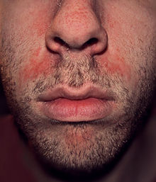
Dandruff is a skin condition that mainly affects the scalp. Symptoms include flaking and sometimes mild itchiness. It can result in social or self-esteem problems. A more severe form of the condition, which includes inflammation of the skin, is known as seborrhoeic dermatitis.

<span class="mw-page-title-main">Tinea versicolor</span> Skin disease

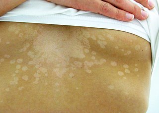
Tinea versicolor is a condition characterized by a skin eruption on the trunk and proximal extremities. The majority of tinea versicolor is caused by the fungus Malassezia globosa, although Malassezia furfur is responsible for a small number of cases. These yeasts are normally found on the human skin and become troublesome only under certain conditions, such as a warm and humid environment, although the exact conditions that cause initiation of the disease process are poorly understood.

<span class="mw-page-title-main">Sebaceous gland</span> Gland to lubricate the hair and skin

A sebaceous gland or oil gland is a microscopic exocrine gland in the skin that opens into a hair follicle to secrete an oily or waxy matter, called sebum, which lubricates the hair and skin of mammals. In humans, sebaceous glands occur in the greatest number on the face and scalp, but also on all parts of the skin except the palms of the hands and soles of the feet. In the eyelids, meibomian glands, also called tarsal glands, are a type of sebaceous gland that secrete a special type of sebum into tears. Surrounding the female nipple, areolar glands are specialized sebaceous glands for lubricating the nipple. Fordyce spots are benign, visible, sebaceous glands found usually on the lips, gums and inner cheeks, and genitals.

<span class="mw-page-title-main">Zinc pyrithione</span> Chemical compound

Zinc pyrithione is a coordination complex of zinc. It has fungistatic and bacteriostatic properties and is used in the treatment of seborrhoeic dermatitis and dandruff.

<span class="mw-page-title-main">Ketoconazole</span> Antifungal chemical compound

Ketoconazole, sold under the brand name Nizoral among others, is an antiandrogen, antifungal, and antiglucocorticoid medication used to treat a number of fungal infections. Applied to the skin it is used for fungal skin infections such as tinea, cutaneous candidiasis, pityriasis versicolor, dandruff, and seborrheic dermatitis. Taken by mouth it is a less preferred option and only recommended for severe infections when other agents cannot be used. Other uses include treatment of excessive male-patterned hair growth in women and Cushing's syndrome.

<span class="mw-page-title-main">Tinea capitis</span> Cutaneous fungal infection of the scalp

Tinea capitis is a cutaneous fungal infection (dermatophytosis) of the scalp. The disease is primarily caused by dermatophytes in the genera Trichophyton and Microsporum that invade the hair shaft. The clinical presentation is typically single or multiple patches of hair loss, sometimes with a 'black dot' pattern, that may be accompanied by inflammation, scaling, pustules, and itching. Uncommon in adults, tinea capitis is predominantly seen in pre-pubertal children, more often boys than girls.

<span class="mw-page-title-main">Ciclopirox</span> Antifungal medication

Ciclopirox is a synthetic antifungal agent for topical dermatologic treatment of superficial mycoses. It is most useful against tinea versicolor. It is often used clinically as ciclopirox olamine, the olamine salt of ciclopirox.

The management of hair loss, includes prevention and treatment of alopecia, baldness, and hair thinning, and regrowth of hair.

<span class="mw-page-title-main">Cradle cap</span> Human medical condition

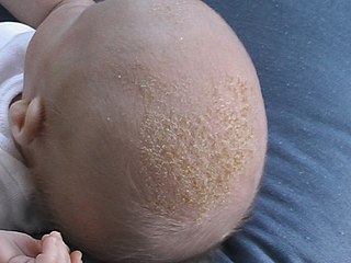
Cradle cap causes crusty or oily scaly patches on a baby's scalp. The condition is not painful or itchy, but it can cause thick white or yellow scales that are not easy to remove. Cradle cap most commonly begins sometime in the first three months but can occur in later years. Similar symptoms in older children are more likely to be dandruff than cradle cap. The rash is often prominent around the ear, the eyebrows or the eyelids. It may appear in other locations as well, where it is called infantile seborrhoeic dermatitis. Cradle cap is just a special—and more benign—case of this condition. The exact cause of cradle cap is not known. Cradle cap is not spread from person to person (contagious). It is also not caused by poor hygiene. It is not an allergy, and it is not dangerous. Cradle cap often lasts a few months. In some children, the condition can last until age 2 or 3.

Selenium disulfide, also known as selenium sulfide, is a chemical compound and medication used to treat seborrheic dermatitis, dandruff, and pityriasis versicolor. It is applied to the affected area as a lotion or shampoo. Symptoms frequently return if treatment is stopped.

<i>Malassezia furfur</i> Species of fungus

Malassezia furfur is a species of yeast that is naturally found on the skin surfaces of humans and some other mammals. It is associated with a variety of dermatological conditions caused by fungal infections, notably seborrhoeic dermatitis and tinea versicolor. As an opportunistic pathogen, it has further been associated with dandruff, malassezia folliculitis, pityriasis versicolor (alba), and malassezia intertrigo, as well as catheter-related fungemia and pneumonia in patients receiving hematopoietic transplants.

<span class="mw-page-title-main">Neonatal acne</span> Medical condition

Neonatal acne, also known as acne neonatorum, is a type of acne that develops in newborns, typically before 6 weeks of life. It presents with open and closed comedones on the cheeks, chin and forehead.

<span class="mw-page-title-main">Pityriasis amiantacea</span> Medical condition

Pityriasis amiantacea is an eczematous condition of the scalp in which thick tenaciously adherent scale infiltrates and surrounds the base of a group of scalp hairs. It does not result in scarring or alopecia.

<span class="mw-page-title-main">Climbazole</span> Chemical compound

Climbazole is a topical antifungal agent commonly used in the treatment of human fungal skin infections such as dandruff, seborrhoeic dermatitis and eczema. Climbazole has shown a high in vitro and in vivo efficacy against Malassezia spp. that appear to play an important role in the pathogenesis of dandruff. Its chemical structure and properties are similar to other azole fungicides such as ketoconazole, clotrimazole and miconazole.

Anti-seborrheics are drugs effective in seborrheic dermatitis. Selenium sulfide, zinc pyrithione, corticosteroids, imidazole antifungals, and salicylic acid are common anti-seborrheics.

Malassezia sympodialis is a species in the genus Malassezia. It is characterized by a pronounced lipophily, unilateral, percurrent or sympodial budding and an irregular, corrugated cell wall ultrastructure. It is one of the most common species found on the skin of healthy and diseased individuals. It is considered to be part of the skin's normal human microbiota and begins to colonize the skin of humans shortly after birth. Malassezia sympodialis, often has a symbiotic or commensal relationship with its host, but it can act as a pathogen causing a number of different skin diseases, such as atopic dermatitis.

<span class="mw-page-title-main">Pyrithione</span> Chemical compound

Pyrithione is the common name of an organosulfur compound with molecular formula C
5
H
5
NOS
, chosen as an abbreviation of pyridinethione, and found in the Persian shallot. It exists as a pair of tautomers, the major form being the thione 1-hydroxy-2(1H)-pyridinethione and the minor form being the thiol 2-mercaptopyridine N-oxide; it crystallises in the thione form. It is usually prepared from either 2-bromopyridine, 2-chloropyridine, or 2-chloropyridine N-oxide, and is commercially available as both the neutral compound and its sodium salt. It is used to prepare zinc pyrithione, which is used primarily to treat dandruff and seborrhoeic dermatitis in medicated shampoos, though is also an anti-fouling agent in paints.

Topical antifungaldrugs are used to treat fungal infections on the skin, scalp, nails, vagina or inside the mouth. These medications come as creams, gels, lotions, ointments, powders, shampoos, tinctures and sprays. Most antifungal drugs induce fungal cell death by destroying the cell wall of the fungus. These drugs inhibit the production of ergosterol, which is a fundamental component of the fungal cell membrane and wall.

References

  1. 1 2 3 4 Dessinioti C, Katsambas A (Jul–Aug 2013). "Seborrheic dermatitis: etiology, risk factors, and treatments: facts and controversies". Clinics in Dermatology. 31 (4): 343–351. doi:10.1016/j.clindermatol.2013.01.001. PMID   23806151.
  2. 1 2 3 4 5 "Seborrheic Dermatitis - Dermatologic Disorders". Merck Manuals Professional Edition. Archived from the original on 26 January 2020. Retrieved 22 November 2019.
  3. 1 2 3 4 5 6 7 8 9 10 11 12 13 Borda LJ, Perper M, Keri JE (March 2019). "Treatment of seborrheic dermatitis: a comprehensive review". The Journal of Dermatological Treatment. 30 (2): 158–169. doi:10.1080/09546634.2018.1473554. PMID   29737895. S2CID   13686180.
  4. 1 2 3 4 5 6 7 8 9 10 11 12 13 14 15 Ijaz N, Fitzgerald D (June 2017). "Seborrhoeic dermatitis". British Journal of Hospital Medicine. 78 (6): C88–C91. doi:10.12968/hmed.2017.78.6.C88. PMID   28614013.
  5. 1 2 Nobles T, Harberger S, Krishnamurthy K (August 2021). "Cradle Cap". StatPearls [Internet]. Treasure Island (FL): StatPearls Publishing. PMID   30285358. Archived from the original on 2021-01-21. Retrieved 2022-02-26.
  6. 1 2 3 4 Naldi L, Diphoorn J (May 2015). "Seborrhoeic dermatitis of the scalp". BMJ Clinical Evidence. 2015. PMC   4445675 . PMID   26016669.
  7. "Seborrheic dermatitis". American Academy of Dermatology. Archived from the original on 21 October 2017. Retrieved 20 October 2017.
  8. 1 2 3 4 5 6 7 8 9 10 11 12 13 14 15 16 17 18 Papadakis MA, McPhee SJ (2023). "Dermatitis, Seborrheic". Quick Medical Diagnosis & Treatment 2023 (2023 ed.). McGraw-Hill Education. ISBN   978-1-264-68734-3.
  9. "Seborrheic Dermatitis: What is It, Diagnosis & Treatment". Cleveland, Ohio: Cleveland Clinic. Archived from the original on 2021-10-17. Retrieved 2021-10-12.
  10. 1 2 3 4 5 6 Okokon EO, Verbeek JH, Ruotsalainen JH, Ojo OA, Bakhoya VN (May 2015). Okokon EO (ed.). "Topical antifungals for seborrhoeic dermatitis". The Cochrane Database of Systematic Reviews. 4 (5): CD008138. doi:10.1002/14651858.CD008138.pub3. PMC   4448221 . PMID   25933684.
  11. 1 2 3 4 5 6 7 8 9 10 11 12 13 14 15 16 17 18 19 20 21 22 Dinulos JG (2021). Habif's Clinical Dermatology, Seventh Edition (7th ed.). Elsevier Inc. ISBN   978-0-323-61269-2.
  12. 1 2 3 4 5 6 7 8 9 10 11 12 13 14 15 16 17 18 19 20 21 22 23 Jackson JM, Alexis A, Zirwas M, Taylor S (December 2022). "Unmet needs for patients with seborrheic dermatitis". Journal of the American Academy of Dermatology. 90 (3): S0190–9622(22)03307–2. doi: 10.1016/j.jaad.2022.12.017 . PMID   36538948. S2CID   254843412.
  13. "Dermatitis". Archived from the original on September 25, 2011. Retrieved June 11, 2010.
  14. 1 2 3 4 5 6 7 8 Wikramanayake TC, Borda LJ, Miteva M, Paus R (September 2019). "Seborrheic dermatitis-Looking beyond Malassezia". Experimental Dermatology. 28 (9): 991–1001. doi: 10.1111/exd.14006 . PMID   31310695.
  15. Johnson BA, Nunley JR (May 2000). "Treatment of seborrheic dermatitis". American Family Physician. 61 (9): 2703–10, 2713–4. PMID   10821151. Archived from the original on 2010-07-06.
  16. Janniger CK, Schwartz RA (July 1995). "Seborrheic dermatitis". American Family Physician. 52 (1): 149–55, 159–60. PMID   7604759.
  17. 1 2 Schwartz RA, Janusz CA, Janniger CK (July 2006). "Seborrheic dermatitis: an overview". American Family Physician. 74 (1): 125–130. PMID   16848386. Archived from the original on 2020-01-26. Retrieved 2010-04-15.
  18. Trivedi MK, Shinkai K, Murase JE (March 2017). "A Review of hormone-based therapies to treat adult acne vulgaris in women". International Journal of Women's Dermatology. 3 (1): 44–52. doi:10.1016/j.ijwd.2017.02.018. PMC   5419026 . PMID   28492054.
  19. Paradisi R, Fabbri R, Porcu E, Battaglia C, Seracchioli R, Venturoli S (October 2011). "Retrospective, observational study on the effects and tolerability of flutamide in a large population of patients with acne and seborrhea over a 15-year period". Gynecological Endocrinology. 27 (10): 823–829. doi:10.3109/09513590.2010.526664. PMID   21117864. S2CID   20250916.
  20. 1 2 3 4 5 6 7 8 Mangion SE, Mackenzie L, Roberts MS, Holmes AM (April 2023). "Seborrheic dermatitis: topical therapeutics and formulation design". European Journal of Pharmaceutics and Biopharmaceutics. 185: 148–164. doi:10.1016/j.ejpb.2023.01.023. PMID   36842718. S2CID   257214910.
  21. Alamgir AN (2018). Therapeutic Use of Medicinal Plants and their Extracts: Volume 2: Phytochemistry and Bioactive Compounds. Springer. p. 435. ISBN   978-3-319-92387-1.
  22. "Seborrhoeic dermatitis and dandruff (seborrheic eczema). DermNet NZ". . DermNet NZ. 2012-03-20. Archived from the original on 2012-06-15. Retrieved 2012-06-10.
  23. Araya M, Kulthanan K, Jiamton S (September 2015). "Clinical Characteristics and Quality of Life of Seborrheic Dermatitis Patients in a Tropical Country". Indian Journal of Dermatology. 60 (5): 519. doi: 10.4103/0019-5154.164410 . PMC   4601435 . PMID   26538714.
  24. 1 2 3 4 5 Leroy AK, Cortez de Almeida RF, Obadia DL, Frattini S, Melo DF (June 2023). "Scalp Seborrheic Dermatitis: What We Know So Far". Skin Appendage Disorders. 9 (3): 160–164. doi:10.1159/000529854. PMC  10264915. PMID   37325288.
  25. Kastarinen H, Oksanen T, Okokon EO, Kiviniemi VV, Airola K, Jyrkkä J, et al. (May 2014). "Topical anti-inflammatory agents for seborrhoeic dermatitis of the face or scalp". The Cochrane Database of Systematic Reviews. 2017 (5): CD009446. doi:10.1002/14651858.CD009446.pub2. PMC   6483543 . PMID   24838779.
  26. 1 2 Gupta AK, Richardson M, Paquet M (January 2014). "Systematic review of oral treatments for seborrheic dermatitis". Journal of the European Academy of Dermatology and Venereology. 28 (1): 16–26. doi:10.1111/jdv.12197. PMID   23802806. S2CID   25441626.
  27. "Seborrhoeic dermatitis | DermNet NZ". dermnetnz.org. Archived from the original on 2021-09-21. Retrieved 2021-09-20.
  28. Burton JL, Pye RJ (April 1983). "Seborrhoea is not a feature of seborrhoeic dermatitis". British Medical Journal. 286 (6372): 1169–1170. doi:10.1136/bmj.286.6372.1169. PMC   1547390 . PMID   6220754.
  29. 1 2 de Souza Leão Kamamoto C, Sanudo A, Hassun KM, Bagatin E (January 2017). "Low-dose oral isotretinoin for moderate to severe seborrhea and seborrheic dermatitis: a randomized comparative trial". International Journal of Dermatology. 56 (1): 80–85. doi:10.1111/ijd.13408. PMID   27778328. S2CID   13049459.
  30. 1 2 3 4 Singh SM, Gauthier S, Labrie F (February 2000). "Androgen receptor antagonists (antiandrogens): structure-activity relationships". Current Medicinal Chemistry. 7 (2): 211–247. doi:10.2174/0929867003375371. PMID   10637363.
  31. Zouboulis CC, Degitz K (2004). "Androgen action on human skin -- from basic research to clinical significance". Experimental Dermatology. 13 (s4): 5–10. doi:10.1111/j.1600-0625.2004.00255.x. PMID   15507105. S2CID   34863608.
  32. Handelsman DJ (October 2020). "Androgen Physiology, Pharmacology, Use and Misuse". In Feingold KR, Anawalt B, Boyce A, Chrousos G, de Herder WW, Dhatariya K, et al. (eds.). Endotext [Internet]. South Dartmouth (MA): MDText.com, Inc. PMID   25905231.
  33. Becker KL (2001). Principles and Practice of Endocrinology and Metabolism. Lippincott Williams & Wilkins. pp. 1004–. ISBN   978-0-7817-1750-2. Archived from the original on 2013-06-02.
  34. Plewig G, Kligman AM (6 December 2012). ACNE and ROSACEA. Springer Science & Business Media. pp. 66, 685, 687. ISBN   978-3-642-59715-2. Archived from the original on 19 March 2017.
  35. Farid NR, Diamanti-Kandarakis E (27 February 2009). Diagnosis and Management of Polycystic Ovary Syndrome. Springer Science & Business Media. pp. 240–. ISBN   978-0-387-09718-3. Archived from the original on 19 March 2017.
  36. 1 2 Diamanti-Kandarakis E (September 1999). "Current aspects of antiandrogen therapy in women". Current Pharmaceutical Design. 5 (9): 707–23 (717). doi:10.2174/1381612805666230111201150. PMID   10495361. Archived from the original on 2023-01-10. Retrieved 2016-10-06.
  37. Couzinet B, Thomas G, Thalabard JC, Brailly S, Schaison G (July 1989). "Effects of a pure antiandrogen on gonadotropin secretion in normal women and in polycystic ovarian disease". Fertility and Sterility. 52 (1): 42–50. doi: 10.1016/s0015-0282(16)60786-0 . PMID   2744186.
  38. Namer M (October 1988). "Clinical applications of antiandrogens". Journal of Steroid Biochemistry. 31 (4B): 719–729. doi:10.1016/0022-4731(88)90023-4. PMID   2462132.
  39. 1 2 Mutschler HD (1995). Drug Actions: Basic Principles and Therapeutic Aspects. CRC Press. pp. 304–. ISBN   978-0-8493-7774-7. Archived from the original on 2017-11-05.
  40. DiPiro JT, Talbert RL, Yee GC, Matzke GR, Wells BG, Posey LM (6 July 2008). Pharmacotherapy: A Pathophysiologic Approach. McGraw Hill Professional. p. 1598. ISBN   978-0-07-164325-2. Archived from the original on 19 March 2017.
  41. 1 2 Hughes A, Hasan SH, Oertel GW, Voss HE, Bahner F, Neumann F, et al. (27 November 2013). Androgens II and Antiandrogens / Androgene II und Antiandrogene. Springer Science & Business Media. pp. 351, 516. ISBN   978-3-642-80859-3. Archived from the original on 19 March 2017.
  42. Millikan LE (19 April 2016). Drug Therapy in Dermatology. CRC Press. pp. 295–. ISBN   978-0-203-90831-0. Archived from the original on 29 July 2017.
  43. Brenner S (13 December 2013). The Clinical Nanomedicine Handbook. CRC Press. pp. 97–. ISBN   978-1-4398-3478-7. Archived from the original on 5 November 2017.
  44. Grob JJ, Castelain M, Richard MA, Bonniol JP, Béraud V, Adhoute H, et al. (May 1998). "Antiinflammatory properties of cetirizine in a human contact dermatitis model. Clinical evaluation of patch tests is not hampered by antihistamines". Acta Dermato-Venereologica. 78 (3): 194–197. doi: 10.1080/000155598441512 . PMID   9602225.
  45. Roelofzen JH, Aben KK, Oldenhof UT, Coenraads PJ, Alkemade HA, van de Kerkhof PC, et al. (April 2010). "No increased risk of cancer after coal tar treatment in patients with psoriasis or eczema". The Journal of Investigative Dermatology. 130 (4): 953–961. doi: 10.1038/jid.2009.389 . PMID   20016499.
  46. 1 2 Wikler JR, Janssen N, Bruynzeel DP, Nieboer C (1990). "The effect of UV-light on pityrosporum yeasts: ultrastructural changes and inhibition of growth". Acta Dermato-Venereologica. 70 (1): 69–71. PMID   1967880.
  47. Calzavara-Pinton PG, Venturini M, Sala R (January 2005). "A comprehensive overview of photodynamic therapy in the treatment of superficial fungal infections of the skin". Journal of Photochemistry and Photobiology. B, Biology. 78 (1): 1–6. doi:10.1016/j.jphotobiol.2004.06.006. PMID   15629243.
  48. Maisch T, Szeimies RM, Jori G, Abels C (October 2004). "Antibacterial photodynamic therapy in dermatology". Photochemical & Photobiological Sciences. 3 (10). rsc.org: 907–917. doi: 10.1039/B407622B . PMID   15480480. S2CID   32215045.
  49. Goldstein MA, Goldstein MC, Credit LP (17 March 2009). Your Best Medicine: From Conventional and Complementary Medicine--Expert-Endorsed Therapeutic Solutions to Relieve Symptoms and Speed Healing. Rodale. pp. 462–. ISBN   978-1-60529-656-2. Archived from the original on 5 November 2017.
  50. 1 2 3 Farage MA, Miller KW, Maibach HI (2 December 2009). Textbook of Aging Skin. Springer Science & Business Media. pp. 534–. ISBN   978-3-540-89655-5. Archived from the original on 5 November 2017.
  51. 1 2 3 Jacknin J (2001). Smart Medicine for Your Skin: A Comprehensive Guide to Understanding Conventional and Alternative Therapies to Heal Common Skin Problems. Penguin. pp. 271–. ISBN   978-1-58333-098-2. Archived from the original on 2017-11-05.
  52. Ooi ET, Tidman MJ (February 2014). "Improving the management of seborrhoeic dermatitis". The Practitioner. 258 (1768): 23–6, 3. PMID   24689165.
  53. Kostović K, Lipozencić J (2004). "Skin diseases in alcoholics". Acta Dermatovenerologica Croatica. 12 (3): 181–190. PMID   15369644.

Commons-logo.svg Media related to Seborrhoeic dermatitis at Wikimedia Commons