Mycosis fungoides

Last updated
Mycosis fungoides
Other namesAlibert-Bazin syndrome
Mycosis fungoides knee.JPG
Skin lesions on the knee of a 52-year-old male patient with mycosis fungoides
Specialty Dermatology, Hematology, Oncology

Mycosis fungoides, also known as Alibert-Bazin syndrome or granuloma fungoides, [1] is the most common form of cutaneous T-cell lymphoma. It generally affects the skin, but may progress internally over time. Symptoms include rash, tumors, skin lesions, and itchy skin.

Contents

While the cause remains unclear, most cases are not hereditary. Most cases are in people over 20 years of age, and it is more common in men than women. Treatment options include sunlight exposure, ultraviolet light, topical corticosteroids, chemotherapy, and radiotherapy.

History

Mycosis fungoides was first described in 1806 by French dermatologist Jean-Louis-Marc Alibert. [2] [3] The name mycosis fungoides is very misleading—it loosely means "mushroom-like fungal disease". The disease, however, is not a fungal infection but rather a type of non-Hodgkin's lymphoma. It was so named because Alibert described the skin tumors of a severe case as having a mushroom-like appearance. [4]

Signs and symptoms

Plaque of mycosis fungoides Plaque of mycosis fungoides 1.jpg
Plaque of mycosis fungoides
Plaque of mycosis fungoides on foot treated with imiquimod at Penn Medicine in Philadelphia, and then radiation at Lehigh Valley Hospital-Cedar Crest in Allentown Mycosis fungoides foor.JPG
Plaque of mycosis fungoides on foot treated with imiquimod at Penn Medicine in Philadelphia, and then radiation at Lehigh Valley Hospital–Cedar Crest in Allentown
Tumour stage mycosis fungoides seen on the head and neck Fox Plate LI.jpg
Tumour stage mycosis fungoides seen on the head and neck

The symptoms of mycosis fungoides are categorized into three clinical stages: the patch stage, the plaque stage, and the tumour stage. [5] The patch stage is defined by flat, reddish patches of varying sizes that may have a wrinkled appearance. They can also look yellowish in people with darker skin. [5] The plaque stage follows the patch stage of mycosis fungoides. [6] It is characterized by the presence of raised lesions that appear reddish-brown; in darker skin tones, plaques may have a greyish or silver appearance. [7] Both patch and plaque stages are considered early-stage mycosis fungoides. [6] The tumour stage typically shows large irregular lumps. Tumours can develop from plaques or normal skin in any region of the body, including the face and head regions. [8]

The symptoms displayed are progressive, with early stages consisting of lesions presented as scaly patches. Lesions often initially develop on the trunk of the body in places that are rarely exposed to the sun, such as the buttocks. [5] These lesions can start as insignificant patches and may remain undiagnosed for up to a decade. [9] Hypopigmentation (when the skin is lighter than normal) of lesions are less common but can be found in children, adolescents and/or dark-skinned individuals. [10]

The advanced stage of mycosis fungoides is characterized by generalized erythroderma (red rash covering most of the body) with severe pruritus (itching) and scaling. [7] Itching (pruritus) is the most commonly reported symptom of people experiencing mycosis fungoides with up to 88% of people reporting varying intensities of pruritus that typically worsens as the disease progresses. [11] Those that experience intense pruritus commonly indicate that it negatively affects their quality of life emotionally, functionally and physically. [12]

Mycosis fungoides (MF) and Sézary syndrome (SS) are related conditions, with the same type of cancer T-lymphocytes, that initially grow in different body compartments. SS cells are found mainly in the blood, whereas MF typically involves the skin. In advanced stages of MF, the cancer cells move from the skin into other organs and the bloodstream; this progression is referred to as "leukemic mycosis fungoides", "Sézary syndrome preceded by mycosis fungoides", or "secondary mycosis fungoides". [13]

Cause

Mycosis fungoides is caused by abnormal white blood cells (T-lymphocytes). These abnormal cells have a preference for localizing and proliferating uncontrolled in the outer layer of the skin (epidermis). The abnormal cells may later involve other organs such as the lymph nodes. It is hypothesized that the genetic mutations in these cancer cells lead to increased growth and escape from programmed cell death. [14]

Additionally, the disease is an unusual expression of CD4 T cells, a part of the immune system. These T cells are skin-associated, meaning they are biochemically and biologically most related to the skin, in a dynamic manner. Mycosis fungoides is the most common type of cutaneous T-cell lymphoma (CTCL), but there are many other types of CTCL that have nothing to do with mycosis fungoides and these disorders are treated differently. [15]

Diagnosis

Diagnosis often requires a combination of clinical and pathological studies. Diagnosis is sometimes difficult because the early phases of the disease often resemble inflammatory dermatoses (such as eczema, psoriasis, lichenoid dermatoses including lichen planus, vitiligo, and chronic cutaneous lupus erythematosus), as well as other cutaneous lymphomas. [7] Several biopsies are recommended, as the key microscopic features are often absent in early MF, and a complete diagnosis requires a combination of clinical and histological study. [16] Furthermore, long periods of treatment can alter the biopsy findings, making it difficult to distinguish from other inflammatory dermatoses. [7] Childhood Mycosis fungoides makes up 0.5% to 7.0% of cases. [17] Although data on childhood MF is limited, a 2021 systematic review observed that there is a significant delay in the diagnosis of childhood MF which may negatively affect a child's prognosis. Notably, most pediatric persons with MF present with early-stage disease. [17]

Histology

Histopathology of Pautrier microabscesses in cutaneous T cell lymphoma. Histopathology of Pautrier microabscesses.jpg
Histopathology of Pautrier microabscesses in cutaneous T cell lymphoma.

The criteria for the disease are established on the skin biopsy by the presence of the following: [18]

To stage the disease, various tests may be ordered, to assess nodes, blood and internal organs, but most patients present with disease apparently confined to the skin, as patches (flat spots) and plaques (slightly raised or 'wrinkled' spots).

Peripheral smear will often show buttock cells. [21]

Staging

Traditionally, mycosis fungoides has been divided into three stages: premycotic, mycotic and tumorous. The premycotic stage clinically presents as an erythematous (red), itchy, scaly lesion. Microscopic appearance is non-diagnostic and represented by chronic nonspecific dermatosis associated with psoriasiform changes in epidermis.[ medical citation needed ]

In the mycotic stage, infiltrative plaques appear and biopsy shows a polymorphous inflammatory infiltrate in the dermis that contains small numbers of frankly atypical lymphoid cells. These cells may line up individually along the epidermal basal layer. The latter finding if unaccompanied by spongiosis is highly suggestive of mycosis fungoides. In the tumorous stage a dense infiltrate of medium-sized lymphocytes with cerebriform nuclei expands the dermis.

Accurate staging of mycosis fungoides is essential to determine appropriate treatment and prognosis. [22] Staging is based on the tumor, node, metastasis, blood (TNMB) classification proposed by the Mycosis Fungoides Cooperative Group and revised by the International Society for Cutaneous Lymphomas/European Organization of Research and Treatment of Cancer. [22] This staging system examines the extent of skin involvement (T), presence of lymph node (N), visceral disease (M), and presence of Sezary cells in the peripheral blood (B). [22]

Most patients with mycosis fungoides have early-stage disease (Stage IA-IIA) at the time of their initial diagnosis. [22] People with early stage disease that is primarily confined to the skin have a favorable prognosis. [22] People with advanced stage (Stage IIB-IVB) are often refractory to treatment and have an unfavorable prognosis. [22] Treatment options for people with advanced stage disease are designed to reduce tumor burden, delay disease progression, and preserve quality of life. [22]

Treatment

The most commonly recommended first-line treatment for mycosis fungoides is psoralen plus ultraviolet A (PUVA therapy). [8] PUVA is a photochemotherapy that involves topical or oral administration of the photosensitizing drug psoralen followed by skin exposure to ultraviolet radiation. [23] Systemic treatments of mycosis fungoides often lead to resistance; as such, additional treatment options are often necessary in advanced disease. [24]

Other treatments have been suggested, however, larger and more extensive research is needed to identify effective treatment strategies for this disease. [8] Suggested treatments include light therapy, ultraviolet light (mainly NB-UVB 312 nm), topical steroids, topical and systemic chemotherapies, local superficial radiotherapy, the histone deacetylase inhibitor vorinostat, total skin electron radiation, photopheresis, systemic therapies (e.g. retinoids, rexinoids), and biological therapies (e.g. interferons). Treatments are often used in combination. [8] Due to the possible adverse effects of treatment options in early disease it is recommended to begin therapy with topical and skin-directed treatments before progressing to more systemic therapies. [8] In 2010, the U.S. Food and Drug Administration granted orphan drug designation for naloxone lotion, a topical opioid receptor competitive antagonist used as a treatment for pruritus in cutaneous T-cell lymphoma. [25] [26] Mogamulizumab is a CCR4 monoclonal antibody which has been shown to improve progression-free survival. It was approved by the US FDA in 2018 for use in people with relapsed or refractory mycosis fungoides or Sézary disease. [24]

There is no evidence to support the use of acitretin or extracorporeal photopheresis (ECP: a type of phototherapy) for treating people with mycosis fungoides. [8] There is also no evidence to support the combination treatment of PUVA and intralesional IFN-α or PUVA and bexarotene. [8]

Children

Treatment for adults and children with mycosis fungoides often differs because of the safety profiles of modalities. [27] Narrowband UV-B is commonly considered for children, as opposed to Psoralen with UV-A, mechlorethamine hydrochloride, or oral bexarotene, which is often used in adults. [27]

Prognosis

A 1999 US-based study of people with CLL's medical records observed a 5-year relative survival rate of 77%, and a 10-year relative survival rate of 69%. [28] After 11 years, the observed relative survival rate remained around 66%. [28] Poorer survival is correlated with advanced age and black race. Superior survival was observed for married women compared with other gender and marital-status groups. [28] The complete remission rate in children is nearly 30%. [27]

Epidemiology

It is rare for mycosis fungoides to appear before age 20; the average age of onset is between 45 and 55 years of age for people with patch and plaque disease only, but is over 60 for people who present with tumours, erythroderma (red skin) or a leukemic form (Sézary syndrome). Mycosis fungoides is more common in males than in females with differences in incidence across various racial groups reported in different studies. [29] The incidence of mycosis fungoides was seen to be increasing between 2000 and 2020, [30] although certain regions have demonstrated some stabilization. [31]

Culture and society

Notable cases

In 1995 actor Mr. T was diagnosed with a cutaneous T-cell lymphoma, or mycosis fungoides. [32] Once in remission, he joked about the coincidence: "Can you imagine that? Cancer with my name on it — personalized cancer! [33] "

Popular British television actor Paul Eddington died of mycosis fungoides after living with the condition for four decades. [34]

See also

Related Research Articles

<span class="mw-page-title-main">Lichen planus</span> Human chronic inflammatory disease

Lichen planus (LP) is a chronic inflammatory and autoimmune disease that affects the skin, nails, hair, and mucous membranes. It is not an actual lichen, but is named for its appearance. It is characterized by polygonal, flat-topped, violaceous papules and plaques with overlying, reticulated, fine white scale, commonly affecting dorsal hands, flexural wrists and forearms, trunk, anterior lower legs and oral mucosa. The hue may be gray-brown in people with darker skin. Although there is a broad clinical range of LP manifestations, the skin and oral cavity remain as the major sites of involvement. The cause is unknown, but it is thought to be the result of an autoimmune process with an unknown initial trigger. There is no cure, but many different medications and procedures have been used in efforts to control the symptoms.

<span class="mw-page-title-main">Actinic keratosis</span> Skin disorder

Actinic keratosis (AK), sometimes called solar keratosis or senile keratosis, is a pre-cancerous area of thick, scaly, or crusty skin. Actinic keratosis is a disorder of epidermal keratinocytes that is induced by ultraviolet (UV) light exposure. These growths are more common in fair-skinned people and those who are frequently in the sun. They are believed to form when skin gets damaged by UV radiation from the sun or indoor tanning beds, usually over the course of decades. Given their pre-cancerous nature, if left untreated, they may turn into a type of skin cancer called squamous cell carcinoma. Untreated lesions have up to a 20% risk of progression to squamous cell carcinoma, so treatment by a dermatologist is recommended.

<span class="mw-page-title-main">Cutaneous T-cell lymphoma</span> Medical condition

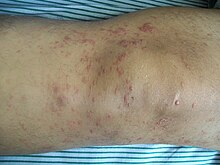
Cutaneous T-cell lymphoma (CTCL) is a class of non-Hodgkin lymphoma, which is a type of cancer of the immune system. Unlike most non-Hodgkin lymphomas, CTCL is caused by a mutation of T cells. The cancerous T cells in the body initially migrate to the skin, causing various lesions to appear. These lesions change shape as the disease progresses, typically beginning as what appears to be a rash which can be very itchy and eventually forming plaques and tumors before spreading to other parts of the body.

<span class="mw-page-title-main">Chlormethine</span> Chemical compound

Chlormethine, also known as mechlorethamine, mustine, HN2, and embikhin (эмбихин), is a nitrogen mustard sold under the brand name Mustargen among others. It is the prototype of alkylating agents, a group of anticancer chemotherapeutic drugs. It works by binding to DNA, crosslinking two strands and preventing cell duplication. It binds to the N7 nitrogen on the DNA base guanine. As the chemical is a blister agent, its use is strongly restricted within the Chemical Weapons Convention where it is classified as a Schedule 1 substance.

<span class="mw-page-title-main">Sézary disease</span> Medical condition

Sézary disease, or Sézary syndrome, is a type of cutaneous T-cell lymphoma that was first described by Albert Sézary. The affected T cells, known as Sézary's cells or Lutzner cells, have pathological quantities of mucopolysaccharides. Sézary disease is sometimes considered a late stage of mycosis fungoides with lymphadenopathy.

<span class="mw-page-title-main">Febrile neutrophilic dermatosis</span> Medical condition

Sweet syndrome (SS), or acute febrile neutrophilic dermatosis, is a skin disease characterized by the sudden onset of fever, an elevated white blood cell count, and tender, red, well-demarcated papules and plaques that show dense infiltrates by neutrophil granulocytes on histologic examination.

<span class="mw-page-title-main">T-cell lymphoma</span> Medical condition

T-cell lymphoma is a rare form of cancerous lymphoma affecting T-cells. Lymphoma arises mainly from the uncontrolled proliferation of T-cells and can become cancerous.

Florid cutaneous papillomatosis (FCP), is an obligate paraneoplastic syndrome.

Large plaque parapsoriasis are skin lesions that may be included in the modern scheme of cutaneous conditions described as parapsoriasis. These lesions, called plaques, may be irregularly round-shaped to oval and are 10 cm (4 in) or larger in diameter. They can be very thin plaques that are asymptomatic or mildly pruritic. Large-plaque parapsoriasis is a common associate of retiform parapsoriasis, can be accompanied by poikiloderma vasculare atrophicans, and can in rare occasions be a precursor to cutaneous T-cell lymphoma.

Pagetoid reticulosis is a cutaneous condition, an uncommon lymphoproliferative disorder, sometimes considered a form of mycosis fungoides.

Granulomatous slack skin (GSS) is a rare cutaneous condition, a variant of lymphoma that typically presents in middle-aged adults.

Secondary cutaneous CD30+ large-cell lymphoma is a cutaneous condition that may arise in cases of mycosis fungoides, and in patients with lymphomatoid papulosis.

Pleomorphic T-cell lymphoma is a cutaneous condition characterized by a 5-year survival rate of 62%.

<span class="mw-page-title-main">Lutzner cells</span>

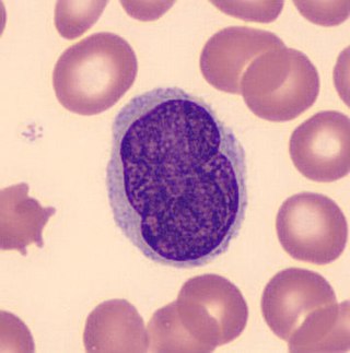
Lutzner cells were discovered by Marvin A. Lutzner, Lucien-Marie Pautrier, and Albert Sézary. These cells are described as the smaller forms of Sézary cells, or Sézary-Lutzner cells, and the two variants are recognised as being morphologically different. Aggregates of these cells in mycosis fungoides are known as a Pautrier's microabscesses. They are a form of T-lymphocytes that has been mutated This atypical form of T-lymphocytes contains T-cell receptors on the surface and is found in both the dermis and epidermis layers of the skin. Since Lutzner cells are a mutated form of T-lymphocytes, they develop in bone marrow and are transported to the thymus is order to mature. The production and maturation stages occur before the cell has developed a mutation. Lutzner cells can form cutaneous T-cell lymphoma, which is a form of skin cancer.

<span class="mw-page-title-main">Poikiloderma vasculare atrophicans</span> Medical condition

Poikiloderma vasculare atrophicans (PVA), is a cutaneous condition characterized by hypo- or hyperpigmentation, telangiectasia and skin atrophy. Other names for the condition include prereticulotic poikiloderma and atrophic parapsoriasis. The condition was first described by pioneer American pediatrician Abraham Jacobi in 1906. PVA causes areas of affected skin to appear speckled red and inflamed, yellowish and/or brown, gray or grayish-black, with scaling and a thinness that may be described as "cigarette paper". On the surface of the skin, these areas may range in size from small patches, to plaques, to neoplasms.

Senile pruritus is one of the most common conditions in the elderly or people over 65 years of age with an emerging itch that may be accompanied with changes in temperature and textural characteristics. In the elderly, xerosis, is the most common cause for an itch due to the degradation of the skin barrier over time. However, the cause of senile pruritus is not clearly known. Diagnosis is based on an elimination criteria during a full body examination that can be done by either a dermatologist or non-dermatologist physician.

Mogamulizumab, sold under the brand name Poteligeo, is a humanized, afucosylated monoclonal antibody targeting CC chemokine receptor 4 (CCR4). The U.S. Food and Drug Administration (FDA) approved it in August 2018 for treatment of relapsed or refractory mycosis fungoides and Sézary disease. It was approved in Japan in 2012, for the treatment of relapsed or refractory CCR4+ adult T-cell leukemia/lymphoma (ATCLL) and in 2014, for relapsed or refractory CCR4+ cutaneous T cell lymphoma (CTCL). The latter approval was based on study with 28 subjects.

Thomas S. Kupper is an American physician, academic, and clinician. His work with clinical and research experience spans dermatology, cutaneous oncology, and immunology. He is the Thomas B. Fitzpatrick Professor at Harvard Medical School, and chairs the Departments of Dermatology at Brigham and Women's Hospital. He also leads the Cutaneous Oncology Disease Center at the Dana Farber Cancer Institute.

Günter Burg is a German dermatologist. Born in Mayen, Germany, he holds German and Swiss citizenship. He has been married to Dr. Doris Burg-Nicklas, a neurologist, since 1968. They have two sons: Andreas and Thomas.

References

  1. synd/98 at Who Named It?
  2. Rapini RP, Bolognia JL, Jorizzo JL (2007). Dermatology: 2-Volume Set. St. Louis: Mosby. p. 1867. ISBN   978-1-4160-2999-1.
  3. Alibert JL (1806). Descriptions des maladies de la peau observées a l'Hôpital Saint-Louis, et exposition des meilleures méthodes suivies pour leur traitement (in French). Paris: Barrois l’ainé. p. 286. Archived from the original on 2012-12-12.
  4. Cerroni, 2007, p.13
  5. 1 2 3 Harvey NT, Spagnolo DV, Wood BA (2015-12-01). "'Could it be mycosis fungoides?': an approach to diagnosing patch stage mycosis fungoides". Journal of Hematopathology. 8 (4): 209–223. doi: 10.1007/s12308-015-0247-2 . ISSN   1865-5785.
  6. 1 2 Hristov AC, Tejasvi T, Wilcox RA (September 2019). "Mycosis fungoides and Sézary syndrome: 2019 update on diagnosis, risk-stratification, and management". American Journal of Hematology. 94 (9): 1027–1041. doi: 10.1002/ajh.25577 . hdl: 2027.42/151292 . PMID   31313347.
  7. 1 2 3 4 Cerroni L (March 2018). "Mycosis fungoides-clinical and histopathologic features, differential diagnosis, and treatment". Seminars in Cutaneous Medicine and Surgery. 37 (1): 2–10. doi:10.12788/j.sder.2018.002. PMID   29719014.
  8. 1 2 3 4 5 6 7 Valipour A, Jäger M, Wu P, Schmitt J, Bunch C, Weberschock T (July 2020). "Interventions for mycosis fungoides". The Cochrane Database of Systematic Reviews. 2020 (7): CD008946. doi:10.1002/14651858.CD008946.pub3. PMC   7389258 . PMID   32632956.
  9. Shinkai K, Fox LP (2022), Papadakis MA, McPhee SJ, Rabow MW, McQuaid KR (eds.), "Cutaneous T-Cell Lymphoma (Mycosis Fungoides)", Current Medical Diagnosis & Treatment 2022, New York, NY: McGraw-Hill Education, retrieved 2021-11-29
  10. Ipenburg NA, Aird W (eds.). "Mycosis Fungoides/Sezary Syndrome". DynaMed. Retrieved 2021-11-29.
  11. Ottevanger R, van Beugen S, Evers AW, Willemze R, Vermeer MH, Quint KD (December 2021). "Quality of life in patients with Mycosis Fungoides and Sézary Syndrome: a systematic review of the literature". Journal of the European Academy of Dermatology and Venereology. 35 (12): 2377–2387. doi:10.1111/jdv.17570. PMC   9291074 . PMID   34331819. S2CID   236637083.
  12. Rangoonwala HI, Cascella M (2021), "Peripheral T-Cell Lymphoma", StatPearls, Treasure Island (FL): StatPearls Publishing, PMID   32965972 , retrieved 2021-11-29
  13. "Mycosis Fungoides (Including Sézary Syndrome) Treatment (PDQ)–Patient Version - National Cancer Institute". www.cancer.gov. 2021-09-03. Retrieved 2021-11-29.
  14. Hodak E, Amitay-Laish I (2019-05-01). "Mycosis fungoides: A great imitator". Clinics in Dermatology. Great ımıtators ın dermatology: Part I. 37 (3): 255–267. doi:10.1016/j.clindermatol.2019.01.004. PMID   31178107. S2CID   81927761.
  15. Rubio-Gonzalez B, Zain J, Rosen ST, Querfeld C (January 2017). "Clinical manifestations and pathogenesis of cutaneous lymphomas: current status and future directions". British Journal of Haematology. 176 (1): 16–36. doi: 10.1111/bjh.14402 . PMID   27782301.
  16. Wilcox, Ryan A. (October 2017). "Cutaneous T-cell lymphoma: 2017 update on diagnosis, risk-stratification, and management: WILCOX". American Journal of Hematology. 92 (10): 1085–1102. doi: 10.1002/ajh.24876 . hdl: 2027.42/141823 . PMID   28872191. S2CID   24447286.
  17. 1 2 Jung, Joon Min; Lim, Dong Jun; Won, Chong Hyun; Chang, Sung Eun; Lee, Mi Woo; Lee, Woo Jin (2021-04-01). "Mycosis Fungoides in Children and Adolescents: A Systematic Review". JAMA Dermatology. 157 (4): 431–438. doi:10.1001/jamadermatol.2021.0083. ISSN   2168-6084. PMID   33656521. S2CID   232103411.
  18. Greer JP, Arber DA, List AF, Foerster J (2014). Greer JP, Arber DA, Glader B, List AF, Means Jr R, Rodgers GM, Foerster J (eds.). Wintrobe's clinical hematology (Thirteenth ed.). Wolters Kluwer, Lippincott Williams & Wilkins Health. p. 1957. ISBN   978-1451172683.
  19. 1 2 Vaidya T, Badri T (2021). "Mycosis Fungoides". StatPearls. Treasure Island (FL): StatPearls Publishing. PMID   30137856 . Retrieved 2021-11-29.
  20. Travaglino A, Russo D, Varricchio S, Pignatiello S, Baldo A, Picardi M, et al. (August 2021). "Prognostic Significance of CD30 in Transformed Mycosis Fungoides". American Journal of Clinical Pathology. 156 (3): 350–355. doi:10.1093/ajcp/aqaa261. PMID   33769436.
  21. O'Connell TX (28 November 2013). USMLE Step 2 Secrets. Elsevier Health Sciences. p. 240. ISBN   9780323225021.
  22. 1 2 3 4 5 6 7 Jawed SI, Myskowski PL, Horwitz S, Moskowitz A, Querfeld C (February 2014). "Primary cutaneous T-cell lymphoma (mycosis fungoides and Sézary syndrome): part II. Prognosis, management, and future directions". Journal of the American Academy of Dermatology. 70 (2): 223.e1–223.e17. doi:10.1016/j.jaad.2013.08.033. PMID   24438970.
  23. Zhang P, Wu MX (January 2018). "A clinical review of phototherapy for psoriasis". Lasers in Medical Science. 33 (1): 173–180. doi:10.1007/s10103-017-2360-1. PMC   5756569 . PMID   29067616.
  24. 1 2 Blackmon AL, Pinter-Brown L (2020-09-16). "Spotlight on Mogamulizumab-Kpkc for Use in Adults with Relapsed or Refractory Mycosis Fungoides or Sézary Syndrome: Efficacy, Safety, and Patient Selection". Drug Design, Development and Therapy. 14: 3747–3754. doi: 10.2147/DDDT.S185896 . PMC   7502391 . PMID   32982179.
  25. "Elorac, Inc. announces orphan drug designation for novel topical treatment for pruritus in cutaneous T-cell lymphoma (CTCL)". Archived from the original on 2010-12-30. Retrieved 2010-11-30.
  26. Wang H, Yosipovitch G (January 2010). "New insights into the pathophysiology and treatment of chronic itch in patients with end-stage renal disease, chronic liver disease, and lymphoma". International Journal of Dermatology. 49 (1): 1–11. doi:10.1111/j.1365-4632.2009.04249.x. PMC   2871329 . PMID   20465602.
  27. 1 2 3 Jung JM, Lim DJ, Won CH, Chang SE, Lee MW, Lee WJ (April 2021). "Mycosis Fungoides in Children and Adolescents: A Systematic Review". JAMA Dermatology. 157 (4): 431–438. doi:10.1001/jamadermatol.2021.0083. PMID   33656521. S2CID   232103411.
  28. 1 2 3 Weinstock MA, Reynes JF (January 1999). "The changing survival of patients with mycosis fungoides: a population-based assessment of trends in the United States". Cancer. 85 (1): 208–212. doi: 10.1002/(SICI)1097-0142(19990101)85:1<208::AID-CNCR28>3.0.CO;2-2 . PMID   9921994.
  29. Kaufman AE, Patel K, Goyal K, O'Leary D, Rubin N, Pearson D, et al. (October 2020). "Mycosis fungoides: developments in incidence, treatment and survival". Journal of the European Academy of Dermatology and Venereology. 34 (10): 2288–2294. doi:10.1111/jdv.16325. PMC   7733543 . PMID   32141115.
  30. Ottevanger R, de Bruin DT, Willemze R, Jansen PM, Bekkenk MW, de Haas ER, et al. (August 2021). "Incidence of mycosis fungoides and Sézary syndrome in the Netherlands between 2000 and 2020". The British Journal of Dermatology. 185 (2): 434–435. doi:10.1111/bjd.20048. PMC   8453738 . PMID   33690948.
  31. Korgavkar K, Xiong M, Weinstock M (November 2013). "Changing incidence trends of cutaneous T-cell lymphoma". JAMA Dermatology. 149 (11): 1295–1299. doi: 10.1001/jamadermatol.2013.5526 . PMID   24005876.
  32. "Mr. T, T cell lymphoma survivor". YouTube . 2013-05-30. Archived from the original on 2021-12-13.
  33. "Mr. T - The Ultimate Tough Guy goes Head-to-Head with Cancer". copingmag.com. March 2000. Archived from the original on 2010-01-02.
  34. "Actor reveals he has rare skin cancer: 'Yes Minister' star refuses to" . Independent.co.uk . 23 October 2011. Archived from the original on 2022-06-18.

Further reading