
Necrosis is a form of cell injury which results in the premature death of cells in living tissue by autolysis. The term "necrosis" came about in the mid-19th century and is commonly attributed to German pathologist Rudolf Virchow, who is often regarded as one of the founders of modern pathology. Necrosis is caused by factors external to the cell or tissue, such as infection, or trauma which result in the unregulated digestion of cell components. In contrast, apoptosis is a naturally occurring programmed and targeted cause of cellular death. While apoptosis often provides beneficial effects to the organism, necrosis is almost always detrimental and can be fatal.

Psoriasis is a long-lasting, noncontagious autoimmune disease characterized by patches of abnormal skin. These areas are red, pink, or purple, dry, itchy, and scaly. Psoriasis varies in severity from small localized patches to complete body coverage. Injury to the skin can trigger psoriatic skin changes at that spot, which is known as the Koebner phenomenon.

An ulcer is a sore on the skin or a mucous membrane, accompanied by the disintegration of tissue. Ulcers can result in complete loss of the epidermis and often portions of the dermis and even subcutaneous fat. Ulcers are most common on the skin of the lower extremities and in the gastrointestinal tract. An ulcer that appears on the skin is often visible as an inflamed tissue with an area of reddened skin. A skin ulcer is often visible in the event of exposure to heat or cold, irritation, or a problem with blood circulation.

A callus is an area of thickened and sometimes hardened skin that forms as a response to repeated friction, pressure, or other irritation. Since repeated contact is required, calluses are most often found on the feet and hands, but they may occur anywhere on the skin. Some degree of callus, such as on the bottom of the foot, is normal.

A skin condition, also known as cutaneous condition, is any medical condition that affects the integumentary system—the organ system that encloses the body and includes skin, nails, and related muscle and glands. The major function of this system is as a barrier against the external environment.

Langerhans cell histiocytosis (LCH) is an abnormal clonal proliferation of Langerhans cells, abnormal cells deriving from bone marrow and capable of migrating from skin to lymph nodes.
Diabetic dermopathy is a type of skin lesion usually seen in people with diabetes mellitus. It is characterized by dull-red papules that progress to well-circumscribed, small, round, atrophic hyperpigmented skin lesions usually on the shins. It is the most common of several diabetic skin conditions, being found in up to 30% of diabetics. Similar lesions can occasionally be found in non-diabetics usually following trauma or injury to the area; however, more than 4 lesions strongly suggests diabetes.

Geographic tongue, also known by several other terms, is a condition of the mucous membrane of the tongue, usually on the dorsal surface. It is a common condition, affecting approximately 2–3% of the general population. It is characterized by areas of smooth, red depapillation which migrate over time. The name comes from the map-like appearance of the tongue, with the patches resembling the islands of an archipelago. The cause is unknown, but the condition is entirely benign, and there is no curative treatment. Uncommonly, geographic tongue may cause a burning sensation on the tongue, for which various treatments have been described with little formal evidence of efficacy.

Granuloma annulare (GA) is a common, sometimes chronic skin condition which presents as reddish bumps on the skin arranged in a circle or ring. It can initially occur at any age, though two-thirds of patients are under 30 years old, and it is seen most often in children and young adults. Females are two times as likely to have it as males.
The Koebner phenomenon or Köbner phenomenon, also called the Koebner response or the isomorphic response, attributed to Heinrich Köbner, is the appearance of skin lesions on lines of trauma. The Koebner phenomenon may result from either a linear exposure or irritation. Conditions demonstrating linear lesions after a linear exposure to a causative agent include: molluscum contagiosum, warts and toxicodendron dermatitis. Warts and molluscum contagiosum lesions can be spread in linear patterns by self-scratching ("auto-inoculation"). Toxicodendron dermatitis lesions are often linear from brushing up against the plant. Causes of the Koebner phenomenon that are secondary to scratching rather than an infective or chemical cause include vitiligo, psoriasis, lichen planus, lichen nitidus, pityriasis rubra pilaris, and keratosis follicularis.
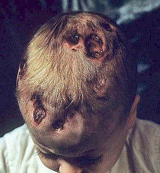
Chronic multifocal Langerhans cell histiocytosis, previously known as Hand–Schüller–Christian disease, is a type of Langerhans cell histiocytosis (LCH), which can affect multiple organs. The condition is traditionally associated with a combination of three features; bulging eyes, breakdown of bone, and diabetes insipidus, although around 75% of cases do not have all three features. Other features may include a fever and weight loss, and depending on the organs involved there may be rashes, asymmetry of the face, ear infections, signs in the mouth and the appearance of advanced gum disease. Features relating to lung and liver disease may occur.

Discoid lupus erythematosus is the most common type of chronic cutaneous lupus (CCLE), an autoimmune skin condition on the lupus erythematosus spectrum of illnesses. It presents with red, painful, inflamed and coin-shaped patches of skin with a scaly and crusty appearance, most often on the scalp, cheeks, and ears. Hair loss may occur if the lesions are on the scalp. The lesions can then develop severe scarring, and the centre areas may appear lighter in color with a rim darker than the normal skin. These lesions can last for years without treatment.

Rosai–Dorfman disease, also known as sinus histiocytosis with massive lymphadenopathy or sometimes as Destombes–Rosai–Dorfman disease, is a rare disorder of unknown cause that is characterized by abundant histiocytes in the lymph nodes or other locations throughout the body.
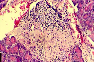
Insulitis is an inflammation of the islets of Langerhans, a collection of endocrine tissue located in the pancreas that helps regulate glucose levels, and is classified by specific targeting of immune cell infiltration in the islets of Langerhans. This immune cell infiltration can result in the destruction of insulin-producing beta cells of the islets, which plays a major role in the pathogenesis, the disease development, of type 1 and type 2 diabetes. Insulitis is present in 19% of individuals with type 1 diabetes and 28% of individuals with type 2 diabetes. It is known that genetic and environmental factors contribute to insulitis initiation, however, the exact process that causes it is unknown. Insulitis is often studied using the non-obese diabetic (NOD) mouse model of type 1 diabetes. The chemokine family of proteins may play a key role in promoting leukocytic infiltration into the pancreas prior to pancreatic beta-cell destruction.

Inflammatory Linear Verrucous Epidermal Nevus is a rare disease of the skin that presents as multiple, discrete, red papules that tend to coalesce into linear plaques that follow the Lines of Blaschko. The plaques can be slightly warty (psoriaform) or scaly (eczema-like). ILVEN is caused by somatic mutations that result in genetic mosaicism. There is no cure, but different medical treatments can alleviate the symptoms.
Actinic granuloma (AG) was first described by O'Brien in 1975 as a rare granulomatous disease. Lesions appear on sun-exposed areas, usually on the face, neck, and scalp, with a slight preference for middle-aged women. They are typically asymptomatic, single or multiple, annular or polycyclic lesions measuring up to 6 cm in diameter, with slow centrifugal expansion, an erythematous elevated edge, and a hypopigmented, atrophic center.
Diabetic cheiroarthropathy, also known as Diabetic stiff hand syndrome or limited joint mobility syndrome, is a cutaneous condition characterized by waxy, thickened skin and limited joint mobility of the hands and fingers, leading to flexion contractures, a condition associated with diabetes mellitus and it is observed in roughly 30% of diabetic patients with longstanding disease. It can be a predictor for other diabetes-related complications and was one of the earliest known complications of diabetes, first documented in 1974.
A coma blister, or coma bullae, is a skin lesion or blister that typically arises due to pressure in an individual with impaired consciousness. They vary in size, ranging from 4 to 5 centimeters in diameter, and may appear hemorrhagic or blood filled. Coma blisters are usually found in the extremities and trunk. These types of blisters have been associated with the overdose of central nervous system (CNS) depressants especially barbiturates, but also tricyclic antidepressants, hypnotics, benzodiazepines, opiates, antipsychotics, and alcohol. However, studies have found that coma blisters are not caused by the toxicity of these drugs, but due to hypoxia and external pressure on the comatose individual's skin from being immobilized. Coma blisters have been frequently found on individuals who have overdosed on drugs, but have also been found on individuals with chronic kidney failure, hypercalcemia, diabetic ketoacidosis, and a variety of neurologic conditions. Coma blisters are more frequent in adults and less common among children as demonstrated by the few cases published in literature.
Histiocytic diseases in dogs are a group of diseases in dogs which may involve the skin, and which can be difficult to differentiate from granulomatous, reactive inflammatory or lymphoproliferative diseases. The clinical presentation and behaviour as well as response to therapy vary greatly among the syndromes.
T-cell/histiocyte-rich large B-cell lymphoma (THRLBCL) is a malignancy of B cells. B-cells are lymphocytes that normally function in the humoral immunity component of the adaptive immune system by secreting antibodies that, for example, bind to and neutralize invasive pathogens. Among the various forms of B-cell lymphomas, THRLBCL is a rarely occurring subtype of the diffuse large B-cell lymphomas (DLBCL). DLBCL are a large group of lymphomas that account for ~25% of all non-Hodgkin lymphomas worldwide. THRLBCL is distinguished from the other DLBCL subtypes by the predominance of non-malignant T-cell lymphocytes and histiocytes over malignant B-cells in its tumors and tissue infiltrates.

