
Exocrine glands are glands that secrete substances onto an epithelial surface by way of a duct. Examples of exocrine glands include sweat, salivary, mammary, ceruminous, lacrimal, sebaceous, prostate and mucous. Exocrine glands are one of two types of glands in the human body, the other being endocrine glands, which secrete their products directly into the bloodstream. The liver and pancreas are both exocrine and endocrine glands; they are exocrine glands because they secrete products—bile and pancreatic juice—into the gastrointestinal tract through a series of ducts, and endocrine because they secrete other substances directly into the bloodstream.

The salivary glands in mammals are exocrine glands that produce saliva through a system of ducts. Humans have three paired major salivary glands, as well as hundreds of minor salivary glands. Salivary glands can be classified as serous, mucous or seromucous (mixed).

Epithelium is one of the four basic types of animal tissue, along with connective tissue, muscle tissue and nervous tissue. It is a thin, continuous, protective layer of compactly packed cells with little intercellular matrix. Epithelial tissues line the outer surfaces of organs and blood vessels throughout the body, as well as the inner surfaces of cavities in many internal organs. An example is the epidermis, the outermost layer of the skin.

The seminal vesicles, are a pair of two convoluted tubular glands that lie behind the urinary bladder of some male mammals. They secrete fluid that partly composes the semen.

The nasal cavity is a large, air-filled space above and behind the nose in the middle of the face. The nasal septum divides the cavity into two cavities, also known as fossae. Each cavity is the continuation of one of the two nostrils. The nasal cavity is the uppermost part of the respiratory system and provides the nasal passage for inhaled air from the nostrils to the nasopharynx and rest of the respiratory tract.

Thyroid follicular cells are the major cell type in the thyroid gland, and are responsible for the production and secretion of the thyroid hormones thyroxine (T4) and triiodothyronine (T3). They form the single layer of cuboidal epithelium that makes up the outer structure of the almost spherical thyroid follicle.
The paired submandibular glands are major salivary glands located beneath the floor of the mouth. They each weigh about 15 grams and contribute some 60–67% of unstimulated saliva secretion; on stimulation their contribution decreases in proportion as the parotid secretion rises to 50%. The average length of the normal human submandibular salivary gland is approximately 27mm, while the average width is approximately 14.3mm.

The lacrimal glands are paired exocrine glands, one for each eye, found in most terrestrial vertebrates and some marine mammals, that secrete the aqueous layer of the tear film. In humans, they are situated in the upper lateral region of each orbit, in the lacrimal fossa of the orbit formed by the frontal bone. Inflammation of the lacrimal glands is called dacryoadenitis. The lacrimal gland produces tears which are secreted by the lacrimal ducts, and flow over the ocular surface, and then into canals that connect to the lacrimal sac. From that sac, the tears drain through the lacrimal duct into the nose.

In anatomy, serous membrane is a smooth tissue membrane of mesothelium lining the contents and inside wall of body cavities, which secrete serous fluid to allow lubricated sliding movements between opposing surfaces. The serous membrane that covers internal organs is called a visceral membrane; while the one that covers the cavity wall is called the parietal membrane. Between the two opposing serosal surfaces is often a potential space, mostly empty except for the small amount of serous fluid.
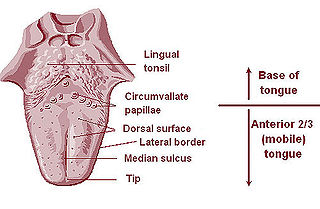
The lingual tonsils are a collection of lymphatic tissue located in the lamina propria of the root of the tongue. This lymphatic tissue consists of the lymphatic nodules rich in cells of the immune system (immunocytes). The immunocytes initiate the immune response when the lingual tonsils get in contact with invading microorganisms.
In physiology, the term serous fluid or serosal fluid is any of various body fluids resembling serum, that are typically pale yellow and transparent and of a benign nature. The fluid fills the inside of body cavities. Serous fluid originates from serous glands, with secretions enriched with proteins and water. Serous fluid may also originate from mixed glands, which contain both mucous and serous cells. A common trait of serous fluids is their role in assisting digestion, excretion, and respiration.
Theca interna cells express receptors for luteinizing hormone (LH) to produce androstenedione, which via a few steps, gives the granulosa the precursor for estrogen manufacturing.
The lacrimal apparatus is the physiological system containing the orbital structures for tear production and drainage.
It consists of:

The esophageal glands are glands that are part of the digestive system of various animals, including humans.

Olfactory glands, also known as Bowman's glands, are a type of nasal gland situated in the olfactory mucosa, beneath the olfactory epithelium, in the lamina propria, a connective tissue also containing fibroblasts, blood vessels and bundles of fine axons from the olfactory neurons.
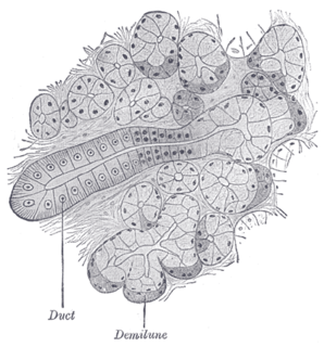
Serous demilunes, also known as Crescents of Giannuzzi or Demilunes of Heidenhain, are cellular formations in the shape of a half-moon on some salivary glands.
An apocrine sweat gland is composed of a coiled secretory portion located at the junction of the dermis and subcutaneous fat, from which a straight portion inserts and secretes into the infundibular portion of the hair follicle. In humans, apocrine sweat glands are found only in certain locations of the body: the axillae (armpits), areola and nipples of the breast, ear canal, eyelids, wings of the nostril, perineal region, and some parts of the external genitalia. Modified apocrine glands include the ciliary glands in the eyelids; the ceruminous glands, which produce ear wax; and the mammary glands, which produce milk. The rest of the body is covered by eccrine sweat glands.
Mucous gland, also known as muciparous glands, are found in several different parts of the body, and they typically stain lighter than serous glands during standard histological preparation. Most are multicellular, but goblet cells are single-celled glands.
Centroacinar cells are spindle-shaped cells in the exocrine pancreas. They represent an extension of the intercalated duct into each pancreatic acinus. These cells are commonly known as duct cells, and secrete an aqueous bicarbonate solution under stimulation by the hormone secretin. They also secrete mucin.
The theca folliculi comprise a layer of the ovarian follicles. They appear as the follicles become secondary follicles.

