Sebaceous gland

Last updated

Hair follicle-en.svg
Schematic view of hair follicle and sebaceous gland
Skin.png
Cross-section of all skin layers. A hair follicle with associated structures. (Sebaceous glands labeled at center left.)
Identifiers
MeSH D012627
TA98 A16.0.00.030
A15.2.07.044
TA2 7082
FMA 59160
Anatomical terminology

A sebaceous gland or oil gland [1] is a microscopic exocrine gland in the skin that opens into a hair follicle to secrete an oily or waxy matter, called sebum, which lubricates the hair and skin of mammals. [2] In humans, sebaceous glands occur in the greatest number on the face and scalp, but also on all parts of the skin except the palms of the hands and soles of the feet. In the eyelids, meibomian glands, also called tarsal glands, are a type of sebaceous gland that secrete a special type of sebum into tears. Surrounding the female nipple, areolar glands are specialized sebaceous glands for lubricating the nipple. Fordyce spots are benign, visible, sebaceous glands found usually on the lips, gums and inner cheeks, and genitals.

Contents

Structure

Location

Sebaceous glands are found throughout all areas of the skin, except the palms of the hands and soles of the feet. [3] There are two types of sebaceous glands: those connected to hair follicles and those that exist independently. [4]

Sebaceous glands are found in hair-covered areas, where they are connected to hair follicles. One or more glands may surround each hair follicle, and the glands themselves are surrounded by arrector pili muscles, forming a pilosebaceous unit. The glands have an acinar structure (like a many-lobed berry), in which multiple glands branch off a central duct. The glands deposit sebum on the hairs and bring it to the skin surface along the hair shaft. The structure, consisting of hair, hair follicle, arrector pili muscles, and sebaceous gland, is an epidermal invagination known as a pilosebaceous unit. [4]

Sebaceous glands are also found in hairless areas (glabrous skin) of the eyelids, nose, penis, labia minora, the inner mucosal membrane of the cheek, and nipples. [4] Some sebaceous glands have unique names. Sebaceous glands on the lip and mucosa of the cheek, and on the genitalia, are known as Fordyce spots, and glands on the eyelids are known as meibomian glands. Sebaceous glands of the breast are also known as Montgomery's glands. [5]

Development

Sebaceous glands are first visible from the 13th to the 16th week of fetal development, as bulgings off hair follicles. [6] Sebaceous glands develop from the same tissue that gives rise to the epidermis of the skin. Overexpression of the signalling factors Wnt, Myc and SHH all increase the likelihood of sebaceous gland presence. [5]

The sebaceous glands of a human fetus secrete a substance called vernix caseosa, a waxy, translucent white substance coating the skin of newborns. [7] After birth, activity of the glands decreases until there is almost no activity during ages two–six years, and then increases to a peak of activity during puberty, due to heightened levels of androgens. [6]

Function

Relative to keratinocytes that make up the hair follicle, sebaceous glands are composed of huge cells with many large vesicles that contain the sebum. [8] These cells express Na+ and Cl ion channels, ENaC and CFTR (see Fig. 6 and Fig. 7 in reference [8] ).

Sebaceous glands secrete the oily, waxy substance called sebum (Latin : fat, tallow ) that is made of triglycerides, wax esters, squalene, and metabolites of fat-producing cells. Sebum lubricates the skin and hair of mammals. [9] Sebaceous secretions in conjunction with apocrine glands also play an important thermoregulatory role. In hot conditions, the secretions emulsify the sweat produced by the eccrine sweat glands and this produces a sheet of sweat that is not readily lost in drops of sweat. This is of importance in delaying dehydration. In colder conditions, the nature of sebum becomes more lipid, and in coating the hair and skin, rain is effectively repelled. [10] [11]

Sebum is produced in a holocrine process, in which cells within the sebaceous gland rupture and disintegrate as they release the sebum and the cell remnants are secreted together with the sebum. [12] [13] The cells are constantly replaced by mitosis at the base of the duct. [4]

Sebum

Sebum is secreted by the sebaceous gland in humans. It is primarily composed of triglycerides (≈41%), wax esters (≈26%), squalene (≈12%), and free fatty acids (≈16%). [7] [14] The composition of sebum varies across species. [14] Wax esters and squalene are unique to sebum and not produced as final products anywhere else in the body. [5] Sapienic acid is a sebum fatty acid that is unique to humans, and is implicated in the development of acne. [15] Sebum is odorless, but its breakdown by bacteria can produce strong odors. [16]

Sex hormones are known to affect the rate of sebum secretion; androgens such as testosterone have been shown to stimulate secretion, and estrogens have been shown to inhibit secretion. [17] Dihydrotestosterone acts as the primary androgen in the prostate and in hair follicles. [18] [19]

Immune function and nutrition

Sebaceous glands are part of the body's integumentary system and serve to protect the body against microorganisms. Sebaceous glands secrete acids that form the acid mantle. This is a thin, slightly acidic film on the surface of the skin that acts as a barrier to microbes that might penetrate the skin. [20] The pH of the skin is between 4.5 and 6.2, [21] an acidity that helps to neutralize the alkaline nature of contaminants. [22] Sebaceous lipids help maintain the integrity of the skin barrier [10] [23] [24] and supply vitamin E to the skin. [25]

Unique sebaceous glands

During the last three months of fetal development, the sebaceous glands of the fetus produce vernix caseosa, a waxy white substance that coats the skin to protect it from amniotic fluid. [26]

The areolar glands are in the areola that surrounds the nipple in the female breast. These glands secrete an oily fluid that lubricates the nipple, and also secrete volatile compounds that are thought to serve as an olfactory stimulus for the newborn. During pregnancy and lactation these glands, also called Montgomery's glands, become enlarged. [27]

Meibomian glands, in the eyelids, secrete a form of sebum called meibum onto the eye, that slows the evaporation of tears. [28] They also serve to create an airtight seal when the eyes are closed, and their lipid quality also prevents the eyelids from sticking together. They attach directly to the follicles of the eyelashes, which are arranged vertically within the tarsal plates of the eyelids.

Fordyce spots, or Fordyce granules, are ectopic sebaceous glands found on the genitals and oral mucosa. They show themselves as yellowish-white milia (milk spots). [29]

Earwax is partly composed of sebum produced by glands in the ear canal. These secretions are viscous and have a high lipid content, which provides good lubrication. [30]

Clinical significance

Conditions of sebaceous glands Blausen 0811 SkinPores.png
Conditions of sebaceous glands

Sebaceous glands are involved in skin problems such as acne and keratosis pilaris. In the skin pores, sebum and keratin can create a hyperkeratotic plug called a comedo.

Acne

Acne is a common occurrence, particularly during puberty in teenagers, and is thought to relate to an increased production of sebum due to hormonal factors. The increased production of sebum can lead to a blockage of the sebaceous gland duct. This can cause a comedo (commonly called a blackhead or a whitehead), which can lead to infection, particularly by the bacteria Cutibacterium acnes . This can inflame the comedones, which then change into the characteristic acne lesions. Comedones generally occur on the areas with more sebaceous glands, particularly the face, shoulders, upper chest and back. Comedones may be "black" or "white" depending on whether the entire pilosebaceous unit, or just the sebaceous duct, is blocked. [31] Sebaceous filaments—innocuous build-ups of sebum—are often mistaken for whiteheads.

There are many treatments available for acne from reducing sugars in the diet, to medications that include antibiotics, benzoyl peroxide, retinoids, and hormonal treatments. [31] Retinoids reduce the amount of sebum produced by the sebaceous glands. [32] Should the usual treatments fail, the presence of the Demodex mite could be looked for as the possible cause. [33]

Other

Other conditions that involve the sebaceous glands include:

History

The word sebaceous, meaning 'consisting of sebum', was first termed in 1728 and comes from the Latin for 'tallow'. [40] Sebaceous glands have been documented since at least 1746 by Jean Astruc, who defined them as "...the glands which separate the fat." [41] :viii He describes them in the oral cavity and on the head, eyelids, and ears, as "universally" acknowledged. [41] :22–25 viii Astruc describes them being blocked by "small animals" that are "implanted" in the excretory ducts [41] :64 and attributes their presence in the oral cavity to apthous ulcers, noting that "these glands naturally [secrete] a viscous humour, which puts on various colours and consistencies... in its natural state is very mild, balsamic, and intended to wet and lubricate the mouth". [41] :85–86 In The Principles of Physiology 1834, Andrew Combe noted that the glands were not present in the palms of the hands or soles of the feet. [42]

Other animals

Cheiromeles torquatus.jpg
Example of a gular gland in a male black bonneted bat [44]

The preputial glands of mice and rats are large modified sebaceous glands that produce pheromones used for territorial marking. [5] These and the scent glands in the flanks of hamsters have a similar composition to human sebaceous glands, are androgen responsive, and have been used as a basis for study. [5] Some species of bat, including the Mexican free-tailed, have a specialized sebaceous gland occurring on the throat called a "gular gland". [45] This gland is present more frequently in males than females, and it is hypothesized that the secretions of the gland are used for scent-marking. [46]

Sebaceous adenitis is an autoimmune disease that affects sebaceous glands. It is mainly known to occur in dogs, particularly poodles and akitas, where it is thought to be generally autosomal recessively inherited. It has also been described in cats, and one report describes this condition in a rabbit. In these animals, it causes hair loss, though the nature and distribution of the hair loss differs greatly. [47]

See also

Related Research Articles

<span class="mw-page-title-main">Exocrine gland</span> Gland that secretes substances onto an epithelial surface by way of a duct

Exocrine glands are glands that secrete substances onto an epithelial surface by way of a duct. Examples of exocrine glands include sweat, salivary, mammary, ceruminous, lacrimal, sebaceous, prostate and mucous. Exocrine glands are one of two types of glands in the human body, the other being endocrine glands, which secrete their products directly into the bloodstream. The liver and pancreas are both exocrine and endocrine glands; they are exocrine glands because they secrete products—bile and pancreatic juice—into the gastrointestinal tract through a series of ducts, and endocrine because they secrete other substances directly into the bloodstream. Exocrine sweat glands are part of the integumentary system; they have eccrine and apocrine types.

<span class="mw-page-title-main">Acne</span> Skin condition characterized by pimples

Acne, also known as acne vulgaris, is a long-term skin condition that occurs when dead skin cells and oil from the skin clog hair follicles. Typical features of the condition include blackheads or whiteheads, pimples, oily skin, and possible scarring. It primarily affects skin with a relatively high number of oil glands, including the face, upper part of the chest, and back. The resulting appearance can lead to lack of confidence, anxiety, reduced self-esteem, and, in extreme cases, depression or thoughts of suicide.

<span class="mw-page-title-main">Isotretinoin</span> Medication primarily used to treat severe acne

Isotretinoin, also known as 13-cis-retinoic acid and sold under the brand name Accutane among others, is a medication primarily used to treat severe acne. It is also used to prevent certain skin cancers, and in the treatment of other cancers. It is used to treat harlequin-type ichthyosis, a usually lethal skin disease, and lamellar ichthyosis. It is a retinoid, meaning it is related to vitamin A, and is found in small quantities naturally in the body. Its isomer, tretinoin, is also an acne drug.

<span class="mw-page-title-main">Sebaceous hyperplasia</span> Medical condition

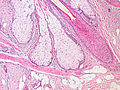
Sebaceous hyperplasia is a disorder of the sebaceous glands in which they become enlarged, producing flesh-colored or yellowish, shiny, often umbilicated bumps on the face. Newly formed nodules often swell with sweating, but this diminishes over time.

<span class="mw-page-title-main">Seborrhoeic dermatitis</span> Skin disease

Seborrhoeic dermatitis is a long-term skin disorder. Symptoms include flaky, scaly, greasy, and occasionally itchy and inflamed skin. Areas of the skin rich in oil-producing glands are often affected including the scalp, face, and chest. It can result in social or self-esteem problems. In babies, when the scalp is primarily involved, it is called cradle cap. Seborrhoeic dermatitis of the scalp may be described in lay terms as dandruff due to the dry, flaky character of the skin. However, as dandruff may refer to any dryness or scaling of the scalp, not all dandruff is seborrhoeic dermatitis. Seborrhoeic dermatitis is sometimes inaccurately referred to as seborrhoea.

<i>Cutibacterium acnes</i> Species of bacterium

Cutibacterium acnes is the relatively slow-growing, typically aerotolerant anaerobic, gram-positive bacterium (rod) linked to the skin condition of acne; it can also cause chronic blepharitis and endophthalmitis, the latter particularly following intraocular surgery. Its genome has been sequenced and a study has shown several genes can generate enzymes for degrading skin and proteins that may be immunogenic.

<span class="mw-page-title-main">Sebacic acid</span> Chemical compound

Sebacic acid is a naturally occurring dicarboxylic acid with the chemical formula HO2C(CH2)8CO2H. It is a white flake or powdered solid. Sebaceus is Latin for tallow candle, sebum is Latin for tallow, and refers to its use in the manufacture of candles. Sebacic acid is a derivative of castor oil.

<span class="mw-page-title-main">Sweat gland</span> Small sweat-producing tubular skin structures

Sweat glands, also known as sudoriferous or sudoriparous glands, from Latin sudor 'sweat', are small tubular structures of the skin that produce sweat. Sweat glands are a type of exocrine gland, which are glands that produce and secrete substances onto an epithelial surface by way of a duct. There are two main types of sweat glands that differ in their structure, function, secretory product, mechanism of excretion, anatomic distribution, and distribution across species:

The arrector pili muscles, also known as hair erector muscles, are small muscles attached to hair follicles in mammals. Contraction of these muscles causes the hairs to stand on end, known colloquially as goose bumps (piloerection).

<span class="mw-page-title-main">Meibomian gland</span> Sebaceous glands along the rims of the eyelid

Meibomian glands are sebaceous glands along the rims of the eyelid inside the tarsal plate. They produce meibum, an oily substance that prevents evaporation of the eye's tear film. Meibum prevents tears from spilling onto the cheek, traps them between the oiled edge and the eyeball, and makes the closed lids airtight. There are about 25 such glands on the upper eyelid, and 20 on the lower eyelid.

<span class="mw-page-title-main">Comedo</span> Medical condition

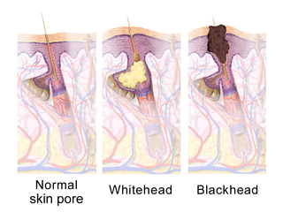
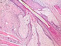
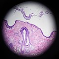
A comedo is a clogged hair follicle (pore) in the skin. Keratin combines with oil to block the follicle. A comedo can be open (blackhead) or closed by skin (whitehead) and occur with or without acne. The word "comedo" comes from the Latin comedere, meaning "to eat up", and was historically used to describe parasitic worms; in modern medical terminology, it is used to suggest the worm-like appearance of the expressed material.

<span class="mw-page-title-main">Holocrine</span> Term used to classify the mode of secretion in exocrine glands in the study of histology

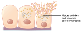
Holocrine is a term used to classify the mode of secretion in exocrine glands in the study of histology. Holocrine secretions are produced in the cytoplasm of the cell and released by the rupture of the plasma membrane, which destroys the cell and results in the secretion of the product into the lumen.

An apocrine sweat gland is composed of a coiled secretory portion located at the junction of the dermis and subcutaneous fat, from which a straight portion inserts and secretes into the infundibular portion of the hair follicle. In humans, apocrine sweat glands are found only in certain locations of the body: the axillae (armpits), areola and nipples of the breast, ear canal, eyelids, wings of the nostril, perineal region, and some parts of the external genitalia. Modified apocrine glands include the ciliary glands in the eyelids; the ceruminous glands, which produce ear wax; and the mammary glands, which produce milk. They are distinct from eccrine sweat glands which cover the whole body.

<span class="mw-page-title-main">Trichilemmal cyst</span> Common cyst that forms from a hair follicle

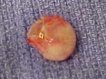
A trichilemmal cyst is a common cyst that forms from a hair follicle, most often on the scalp, and is smooth, mobile, and filled with keratin, a protein component found in hair, nails, skin, and horns. Trichilemmal cysts are clinically and histologically distinct from trichilemmal horns, hard tissue that is much rarer and not limited to the scalp. Rarely, these cysts may grow more extensively and form rapidly multiplying trichilemmal tumors, also called proliferating trichilemmal cysts, which are benign, but may grow aggressively at the cyst site. Very rarely, trichilemmal cysts can become cancerous.

Skin appendages are anatomical skin-associated structures that serve a particular function including sensation, contractility, lubrication and heat loss in animals. In humans, some of the more common skin appendages are hairs, arrector pilli, sebaceous glands, sweat glands (can secrete sweat with strong odour or with a faint odour, and nails.

Madarosis is a condition that results in the loss of eyelashes, and sometimes eyebrows. The term "madarosis" is derived from the ancient Greek "madaros", meaning "bald". It originally was a disease of only losing eyelashes but it currently is the loss of both eyelashes and eyebrows. Eyebrows and eyelashes are both important in the prevention of bacteria and other foreign objects from entering the eye. A majority of patients with madarosis have leprosy, and it was reported that 76% of patients with varying types of leprosy had madarosis.

<span class="mw-page-title-main">Sebaceous carcinoma</span> Medical condition

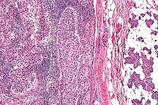
Sebaceous carcinoma, also known as sebaceous gland carcinoma (SGc), sebaceous cell carcinoma, and meibomian gland carcinoma is an uncommon malignant cutaneous tumor. Most are typically about 1.4 cm at presentation. SGc originates from sebaceous glands in the skin and, therefore, may originate anywhere in the body where these glands are found. SGc can be divided into 2 types: periocular and extraocular. The periocular region is rich in sebaceous glands making it a common site of origin. The cause of these lesions in the vast majority of cases is unknown. Occasional cases may be associated with Muir-Torre syndrome. SGc accounts for approximately 0.7% of all skin cancers, and the incidence of SGc is highest in Caucasian, Asian, and Indian populations. Due to the rarity of this tumor and variability in clinical and histological presentation, SGc is often misdiagnosed as an inflammatory condition or a more common neoplasm. SGc is commonly treated with wide local excision or Mohs micrographic surgery, and the relative survival rates at 5 and 10 years are 92.72 and 86.98%, respectively.

<span class="mw-page-title-main">Pimple</span> Type of comedo

A pimple or zit is a kind of comedo that results from excess sebum and dead skin cells getting trapped in the pores of the skin. In its aggravated state, it may evolve into a pustule or papule. Pimples can be treated by acne medications, antibiotics, and anti-inflammatories prescribed by a physician, or various over the counter remedies purchased at a pharmacy.

Infantile acne is a form of acne that begins in very young children. Typical symptoms include inflammatory and noninflammatory lesions, papules and pustules most commonly present on the face. No cause of infantile acne has been established but it may be caused by increased sebaceous gland secretions due to elevated androgens, genetics and the fetal adrenal gland causing increased sebum production. Infantile acne can resolve by itself by age 1 or 2. However, treatment options include topical benzyl peroxide, topical retinoids and topical antibiotics in most cases.

Sapienic acid is a fatty acid that is a major component of human sebum. Unique to humans, it takes its scientific name from the root sapiens. The equivalent fatty acid in mouse sebum is palmitoleic acid. Sapienic acid salts, esters, anion, and conjugate base are known as sapienates.

References

  1. "Hair follicle sebaceous gland: MedlinePlus Medical Encyclopedia Image". medlineplus.gov. Archived from the original on 6 June 2023. Retrieved 6 June 2023.
  2. Lovászi, Marianna; Szegedi, Andrea; Zouboulis, Christos C.; Törőcsik, Dániel (17 October 2017). "Sebaceous-immunobiology is orchestrated by sebum lipids". Dermato-endocrinology. 9 (1). Informa UK Limited: e1375636. doi:10.1080/19381980.2017.1375636. ISSN   1938-1980. PMC   5821166 . PMID   29484100.
  3. 1 2 James, William D.; Berger, Timothy; Elston, Dirk M. (2006). Andrews' Diseases of the Skin: Clinical dermatology. Saunders Elsevier. p. 7. ISBN   978-0-7216-2921-6.
  4. 1 2 3 4 Young, Barbra; Lowe, James S; Stevens, Alan; Heath, John W; Deakin, Philip J (March 2006). Wheater's Functional Histology (5 ed.). Elsevier Health Sciences. pp. 175–178. ISBN   978-0-443-06850-8.
  5. 1 2 3 4 5 6 7 Smith, K. R.; Thiboutot, D. M. (2007). "Thematic Review Series: Skin Lipids. Sebaceous Gland Lipids: Friend Or Foe?". Journal of Lipid Research. 49 (2): 271–281. doi: 10.1194/jlr.R700015-JLR200 . PMID   17975220.
  6. 1 2 Thiboutot, D (July 2004). "Regulation of human sebaceous glands". The Journal of Investigative Dermatology. 123 (1): 1–12. doi: 10.1111/j.1523-1747.2004.t01-2-.x . PMID   15191536.
  7. 1 2 Thody, A. J.; Shuster, S. (1989). "Control and Function of Sebaceous Glands". Physiological Reviews. 69 (2): 383–416. doi:10.1152/physrev.1989.69.2.383. PMID   2648418.
  8. 1 2 Hanukoglu I, Boggula VR, Vaknine H, Sharma S, Kleyman T, Hanukoglu A (January 2017). "Expression of epithelial sodium channel (ENaC) and CFTR in the human epidermis and epidermal appendages". Histochemistry and Cell Biology. 147 (6): 733–748. doi:10.1007/s00418-016-1535-3. PMID   28130590. S2CID   8504408. Archived from the original on 17 November 2019. Retrieved 29 June 2019.
  9. Dellmann's textbook of veterinary histology (405 pages), Jo Ann Coers Eurell, Brian L. Frappier, 2006, p.29, weblink: Books-Google-RTOC Archived 24 February 2024 at the Wayback Machine .
  10. 1 2 Zouboulis CC (2004). "Acne and Sebaceous Gland Function". Clinics in Dermatology. 22 (5): 360–366. doi:10.1016/j.clindermatol.2004.03.004. PMID   15556719.
  11. Porter AM (2001). "Why do we have apocrine and sebaceous glands?". J R Soc Med. 94 (5): 236–7. doi:10.1177/014107680109400509. PMC   1281456 . PMID   11385091.
  12. Victor Eroschenko, diFiore's Atlas of Histology with functional correlations, Lippincott Williams & Wilkins, 10th edition, 2005. p. 41
  13. Dorland's (2012). Dorland's Illustrated Medical Dictionary (32nd ed.). Elsevier Saunders. p. 866. ISBN   978-0-19-856878-0.
  14. 1 2 Cheng JB, Russell DW (September 2004). "Mammalian Wax Biosynthesis II: Expression cloning of wax synthase cDNAs encoding a member of the acyltransferase enzyme family". The Journal of Biological Chemistry. 279 (36): 37798–807. doi: 10.1074/jbc.M406226200 . PMC   2743083 . PMID   15220349.
  15. Webster, Guy F.; Anthony V. Rawlings (2007). Acne and Its Therapy. Basic and clinical dermatology. Vol. 40. CRC Press. p. 311. ISBN   978-0-8247-2971-4.
  16. 1 2 Draelos, Zoe Diana (2005). Hair care: an illustrated dermatologic handbook. London; New York: Taylor & Francis. p. 26. ISBN   978-1-84184-194-6. Archived from the original on 24 February 2024. Retrieved 7 December 2016.
  17. Sweeney TM (December 1968). "The Effect of Estrogen and Androgen on the Sebaceous Gland Turnover Time". The Journal of Investigative Dermatology. 53 (1): 8–10. doi: 10.1038/jid.1969.100 . PMID   5793140.
  18. Amory JK, Anawalt BD, Matsumoto AM, Page ST, Bremner WJ, Wang C, Swerdloff RS, Clark RV (June 2008). "The effect of 5alpha-reductase inhibition with dutasteride and finasteride on bone mineral density, serum lipoproteins, hemoglobin, prostate specific antigen and sexual function in healthy young men". J. Urol. 179 (6): 2333–8. doi:10.1016/j.juro.2008.01.145. PMC   2684818 . PMID   18423697.
  19. Wilkinson, P.F.; Millington, R. (1983). Skin (Digitally printed ed.). Cambridge: Cambridge university press. p. 151. ISBN   978-0-521-24122-9.
  20. Monika-Hildegard Schmid-Wendtner; Korting Schmid-Wendtner (2007). Ph and Skin Care. ABW Wissenschaftsverlag. pp. 31–. ISBN   978-3-936072-64-8 . Retrieved 19 June 2012.
  21. Zlotogorski A (1987). "Distribution of skin surface pH on the forehead and cheek of adults". Arch. Dermatol. Res. 279 (6): 398–401. doi:10.1007/bf00412626. PMID   3674963. S2CID   3065931.
  22. Schmid MH, Korting HC (1995). "The concept of the acid mantle of the skin: its relevance for the choice of skin cleansers" (PDF). Dermatology. 191 (4): 276–80. doi:10.1159/000246568. PMID   8573921. Archived from the original (PDF) on 1 March 2011.
  23. Youn, S. W. (2010). "The Role of Facial Sebum Secretion in Acne Pathogenesis: Facts and Controversies". Clinics in Dermatology. 28 (1): 8–11. doi:10.1016/j.clindermatol.2009.03.011. PMID   20082943.
  24. Drake, David R.; Brogden, Kim A.; Dawson, Deborah V.; Wertz, Philip W. (10 May 2011). "Thematic Review Series: Skin Lipids. Antimicrobial lipids at the skin surface". Journal of Lipid Research. 49 (1): 4–11. doi: 10.1194/jlr.R700016-JLR200 . PMID   17906220. S2CID   10119536. Archived from the original on 9 January 2020. Retrieved 17 October 2011.
  25. Thiele, Jens J.; Weber, Stefan U.; Packer, Lester (1999). "Sebaceous Gland Secretion is a Major Physiologic Route of Vitamin E Delivery to Skin". Journal of Investigative Dermatology. 113 (6): 1006–1010. doi: 10.1046/j.1523-1747.1999.00794.x . PMID   10594744.
  26. Zouboulis, Christos C.; Baron, Jens Malte; Böhm, Markus; Kippenberger, Stefan; Kurzen, Hjalmar; Reichrath, Jörg; Thielitz, Anja (2008). "Frontiers in Sebaceous Gland Biology and Pathology". Experimental Dermatology. 17 (6): 542–551. doi: 10.1111/j.1600-0625.2008.00725.x . PMID   18474083.
  27. Doucet, Sébastien; Soussignan, Robert; Sagot, Paul; Schaal, Benoist (2009). Hausberger, Martine (ed.). "The Secretion of Areolar (Montgomery's) Glands from Lactating Women Elicits Selective, Unconditional Responses in Neonates". PLOS ONE. 4 (10): e7579. Bibcode:2009PLoSO...4.7579D. doi: 10.1371/journal.pone.0007579 . PMC   2761488 . PMID   19851461.
  28. McCulley, JP; Shine, WE (March 2004). "The lipid layer of tears: dependent on meibomian gland function". Experimental Eye Research. 78 (3): 361–5. doi:10.1016/s0014-4835(03)00203-3. PMID   15106913.
  29. Dorland's (2012). Dorland's Illustrated Medical Dictionary (32nd ed.). Elsevier Saunders. p. 802. ISBN   978-0-19-856878-0.
  30. Roeser, RJ; Ballachanda, BB (December 1997). "Physiology, pathophysiology, and anthropology/epidemiology of human earcanal secretions". Journal of the American Academy of Audiology. 8 (6): 391–400. PMID   9433685.
  31. 1 2 Colledge N, Walker B, Ralston S, eds. (2010). Davidson's principles and practice of medicine (21st ed.). Edinburgh: Churchill Livingstone/Elsevier. pp. 1267–1268. ISBN   978-0-7020-3085-7.
  32. Farrell LN, Strauss JS, Stranieri AM (December 1980). "The treatment of severe cystic acne with 13-cis-retinoic acid. Evaluation of sebum production and the clinical response in a multiple-dose trial". Journal of the American Academy of Dermatology. 3 (6): 602–11. doi:10.1016/S0190-9622(80)80074-0. PMID   6451637.
  33. Zhao YE, Peng Y, Wang XL, Wu LP, Wang M, Yan HL, Xiao SX (2011). "Facial dermatosis associated with Demodex: a case-control study". J Zhejiang Univ Sci B. 12 (12): 1008–15. doi:10.1631/jzus.B1100179. PMC   3232434 . PMID   22135150.
  34. James, William D.; Berger, Timothy G. (2006). Andrews' Diseases of the Skin: clinical Dermatology. Saunders Elsevier. p. 662. ISBN   978-0-7216-2921-6.
  35. Dessinioti, C.; Katsambas, A. (2013). "Seborrheic dermatitis: etiology, risk factors, and treatments: facts and controversies". Clin Dermatol. 31 (4): 343–51. doi:10.1016/j.clindermatol.2013.01.001. PMID   23806151.
  36. 1 2 Rapini, Ronald P.; Bolognia, Jean L.; Jorizzo, Joseph L. (2007). Dermatology: 2-Volume Set. St. Louis: Mosby. ISBN   978-1-4160-2999-1.
  37. Nelson BR, Hamlet KR, Gillard M, Railan D, Johnson TM (July 1995). "Sebaceous carcinoma". J. Am. Acad. Dermatol. 33 (1): 1–15, quiz 16–8. doi:10.1016/0190-9622(95)90001-2. PMID   7601925.
  38. Neville BW, Damm DD, Allen CA, Bouquot JE (2002). Oral & maxillofacial pathology (2nd ed.). Philadelphia: W. B. Saunders. p. 31. ISBN   978-0-7216-9003-2.
  39. Kovich O, Hale E (2005). "Nevus sebaceus". Dermatology Online Journal. 11 (4): 16. doi:10.5070/D33BQ5524C. PMID   16403388. Archived from the original on 2 October 2012. Retrieved 21 November 2019.
  40. Harper, Douglas. "Sebaceous". Etymology Online. Archived from the original on 1 March 2017. Retrieved 5 April 2014.
  41. 1 2 3 4 Astruc, Jean (1746). A General and Compleat Treatise on All the Diseases Incident to Children. J. Nourse. p.  3. Sebaceous glands.
  42. Rosenthal, Stanley A; Furnari, Domenica (1958). "Slide Agglutination as a Presumptive Test in the Laboratory Diagnosis of Candida Albicans1". The Journal of Investigative Dermatology. 31 (5): 251–253. doi: 10.1038/jid.1958.50 . PMID   13598929.
  43. Dobson, G. E. (1878). Catalogue of the Chiroptera in the collection of the British Museum. Order of the Trustees.
  44. Dobson, G. E. (1878). Catalogue of the Chiroptera in the collection of the British Museum. Order of the Trustees.
  45. Gutierrez, Mercedes; Aoki, Agustin (1973). "Fine structure of the gular gland of the free-tailed bat Tadarida brasiliensis". Journal of Morphology. 141 (3): 293–305. doi:10.1002/jmor.1051410305. PMID   4753444. S2CID   3093610.
  46. Heideman, P. D., Erickson, K. R., & Bowles, J. B. (1990). Notes on the breeding biology, gular gland and roost habits of Molossus sinaloae (Chiroptera, Molossidae) Archived 21 October 2021 at the Wayback Machine . Zeitschrift für Säugetierkunde, 55(5), 303-307.
  47. Lars Mecklenburg; Monika Linek; Desmond J. Tobin (15 September 2009). Hair Loss Disorders in Domestic Animals. John Wiley & Sons. pp. 269–. ISBN   978-0-8138-1934-1. Archived from the original on 24 February 2024. Retrieved 25 September 2016.