Respiratory epithelium

Last updated
Respiratory epithelium
Blausen 0766 RespiratoryEpithelium.png
Illustration depicting the respiratory epithelium. Basal cells labelled as stem cells.
Details
System Respiratory system
Identifiers
MeSH D020545
TH H3.05.00.0.00003
Anatomical terms of microanatomy

Respiratory epithelium, or airway epithelium, [1] is a type of ciliated columnar epithelium found lining most of the respiratory tract as respiratory mucosa, [2] where it serves to moisten and protect the airways. It is not present in the vocal cords of the larynx, or the oropharynx and laryngopharynx, where instead the epithelium is stratified squamous. [3] It also functions as a barrier to potential pathogens and foreign particles, preventing infection and tissue injury by the secretion of mucus and the action of mucociliary clearance.

Contents

Structure

Pseudostratified columnar epithelium, animated image highlights the epithelial cells, goblet cells, then underlying connective tissue Psuedostratified epithelium animated.gif
Pseudostratified columnar epithelium, animated image highlights the epithelial cells, goblet cells, then underlying connective tissue
Cells of the respiratory epithelium. Basal cells shown in purple, ciliated cells shown in brown, goblet cells shown in green, and submucosal gland shown in blue. Respiratory Tract Cells.png
Cells of the respiratory epithelium. Basal cells shown in purple, ciliated cells shown in brown, goblet cells shown in green, and submucosal gland shown in blue.

The respiratory epithelium lining the upper respiratory airways is classified as ciliated pseudostratified columnar epithelium. [4] This designation is due to the arrangement of the multiple cell types composing the respiratory epithelium. While all cells make contact with the basement membrane and are, therefore, a single layer of cells, their nuclei are not aligned in the same plane. Hence, it appears as though several layers of cells are present and the epithelium is called pseudostratified (falsely layered). The respiratory mucosa transitions to simple ciliated cuboidal epithelium and finally to simple squamous epithelium in the alveolar ducts and alveoli. [5]

Cells

The cells in the respiratory epithelium are of five main types: a) ciliated cells, b) goblet cells, c) brush cells, d) airway basal cells, and e) small granule cells (NDES) [6] Goblet cells become increasingly fewer further down the respiratory tree until they are absent in the terminal bronchioles; club cells take over their role to some extent here. [7] Another important cell type is the pulmonary neuroendocrine cell. These are innervated cells that only make up around 0.5% of the respiratory epithelial cells. [7] The ciliated cells are columnar epithelial cells with specialized ciliary modifications. The ciliated cells make up between 50 and 80 per cent of the epithelium. [8]

Between the ciliated cells are numerous microvilli, attached as tufts to brush cells sometimes referred to as pulmonary brush cells; [9] these are also known as the tuft cells of the gastrointestinal tract, or intestinal tuft cells, [10] although there is a difference between the two types: the brush cells lack the terminal web that lies under the microvilli of the tuft cells. [9] Although their function is not yet fully understood, it has been suggested that they exhibit a virulence associated clearance role, activating mucociliary clearance by releasing acetylcholine. [11]

Function

The respiratory epithelium functions to moisten and protect the airways. It acts as a physical barrier to pathogens, as well as their removal in the mechanism of mucociliary clearance.

The ciliated cells are the primary components in the mucociliary clearance mechanism. Each epithelial cell has around 200 cilia that beat constantly at a rate of between 10 and 20 times per second. The direction of their beat is targeted towards the pharynx, either upwards from the lower respiratory tract or downwards from the nasal structures. [12]

Goblet cells, so named because they are shaped like a wine goblet, are columnar epithelial cells that contain membrane-bound mucous granules and secrete mucus as part of the airway surface liquid (ASL), also known as the epithelial lining fluid, the composition of which is tightly regulated; the mucus helps maintain epithelial moisture and traps particulate material and pathogens moving through the airway. and determines how well mucociliary clearance works. [13] [14]

The basal cells are small, nearly cuboidal that differentiate into the other cell types found within the epithelium. Basal cells respond to injury of the airway epithelium, migrating to cover a site denuded of differentiated epithelial cells, and subsequently differentiating to restore a healthy epithelial cell layer. The differentiated epithelial cells can also dedifferentiate into stem cells and contribute to the repairing of the barrier. [15]

Club cells carry out similar functions in the more distal airways.

Certain parts of the respiratory tract, such as the oropharynx, are also subject to the abrasive swallowing of food. To prevent the destruction of the epithelium in these areas, it changes to stratified squamous epithelium, which is better suited to the constant sloughing and abrasion. The squamous layer of the oropharynx is continuous with the esophagus.[ citation needed ]

The respiratory epithelium has a further role of immunity for the lungs - that of glucose homeostasis. [16] The glucose concentration in the airway surface liquid is held at a level of around 12 times lower than that of the blood sugar concentration. [16] The tight junctions act as a barrier that restricts the passage of glucose across the epithelium into the airway lumen. Some glucose passes through, where it diffuses into the airway surface liquid to be kept at its reduced level by pulmonary glucose transport, and metabolism. [17] However, airway inflammation decreases the effectiveness of the tight junctions making the barrier more permeable to glucose. Higher levels of glucose promote the proliferation of bacteria by providing glucose as a source for carbon for them. [16] Increased levels of glucose in the airway surface liquid is associated with respiratory diseases, and hyperglycemia. [17]

Clinical significance

Long-term irritation of the epithelial cells can cause the overproduction of mucus, known as mucus hypersecretion. Mucus hypersecretion results in the productive cough of chronic bronchitis. [18]

Pulmonary neuroendocrine cells have been associated with a range of chronic lung disorders. They are also the originating cells of small-cell lung cancer. [19]

Related Research Articles

<span class="mw-page-title-main">Lung</span> Primary organ of the respiratory system

The lungs are the central organs of the respiratory system in humans and most other animals, including some snails and a small number of fish. In mammals and most other vertebrates, two lungs are located near the backbone on either side of the heart. Their function in the respiratory system is to extract oxygen from the air and transfer it into the bloodstream, and to release carbon dioxide from the bloodstream into the atmosphere, in a process of gas exchange. The pleurae, which are thin, smooth, and moist, serve to reduce friction between the lungs and chest wall during breathing, allowing for easy and effortless movements of the lungs.

<span class="mw-page-title-main">Pulmonary alveolus</span> Hollow cavity found in the lungs

A pulmonary alveolus, also known as an air sac or air space, is one of millions of hollow, distensible cup-shaped cavities in the lungs where pulmonary gas exchange takes place. Oxygen is exchanged for carbon dioxide at the blood–air barrier between the alveolar air and the pulmonary capillary. Alveoli make up the functional tissue of the mammalian lungs known as the lung parenchyma, which takes up 90 percent of the total lung volume.

<span class="mw-page-title-main">Respiratory tract</span> Organs involved in transmission of air to and from the point where gases diffuse into tissue

The respiratory tract is the subdivision of the respiratory system involved with the process of respiration in mammals. The respiratory tract is lined with respiratory epithelium as respiratory mucosa.

<span class="mw-page-title-main">Epithelium</span> Tissue lining the surfaces of organs in animals

Epithelium or epithelial tissue is a thin, continuous, protective layer of compactly packed cells with little intercellular matrix. Epithelial tissues line the outer surfaces of organs and blood vessels throughout the body, as well as the inner surfaces of cavities in many internal organs. An example is the epidermis, the outermost layer of the skin. Epithelial tissue is one of the four basic types of animal tissue, along with connective tissue, muscle tissue and nervous tissue. These tissues also lack blood or lymph supply. The tissue is supplied by nerves.

<span class="mw-page-title-main">Bronchus</span> Airway in the respiratory tract

A bronchus is a passage or airway in the lower respiratory tract that conducts air into the lungs. The first or primary bronchi to branch from the trachea at the carina are the right main bronchus and the left main bronchus. These are the widest bronchi, and enter the right lung, and the left lung at each hilum. The main bronchi branch into narrower secondary bronchi or lobar bronchi, and these branch into narrower tertiary bronchi or segmental bronchi. Further divisions of the segmental bronchi are known as 4th order, 5th order, and 6th order segmental bronchi, or grouped together as subsegmental bronchi. The bronchi, when too narrow to be supported by cartilage, are known as bronchioles. No gas exchange takes place in the bronchi.

<span class="mw-page-title-main">Bronchiole</span> Passageways by which air passes through the nose or mouth to the alveoli of the lungs

The bronchioles or bronchioli are the smaller branches of the bronchial airways in the lower respiratory tract. They include the terminal bronchioles, and finally the respiratory bronchioles that mark the start of the respiratory zone delivering air to the gas exchanging units of the alveoli. The bronchioles no longer contain the cartilage that is found in the bronchi, or glands in their submucosa.

<span class="mw-page-title-main">Mucus</span> Secretion produced by mucous membranes

Mucus is a slippery aqueous secretion produced by, and covering, mucous membranes. It is typically produced from cells found in mucous glands, although it may also originate from mixed glands, which contain both serous and mucous cells. It is a viscous colloid containing inorganic salts, antimicrobial enzymes, immunoglobulins, and glycoproteins such as lactoferrin and mucins, which are produced by goblet cells in the mucous membranes and submucosal glands. Mucus serves to protect epithelial cells in the linings of the respiratory, digestive, and urogenital systems, and structures in the visual and auditory systems from pathogenic fungi, bacteria and viruses. Most of the mucus in the body is produced in the gastrointestinal tract.

Club cells, also known as bronchiolar exocrine cells, are low columnar/cuboidal cells with short microvilli, found in the small airways (bronchioles) of the lungs. They were formerly known as Clara cells.

<span class="mw-page-title-main">Metaplasia</span> Medical condition

Metaplasia is the transformation of a cell type to another cell type. The change from one type of cell to another may be part of a normal maturation process, or caused by some sort of abnormal stimulus. In simplistic terms, it is as if the original cells are not robust enough to withstand their environment, so they transform into another cell type better suited to their environment. If the stimulus causing metaplasia is removed or ceases, tissues return to their normal pattern of differentiation. Metaplasia is not synonymous with dysplasia, and is not considered to be an actual cancer. It is also contrasted with heteroplasia, which is the spontaneous abnormal growth of cytologic and histologic elements. Today, metaplastic changes are usually considered to be an early phase of carcinogenesis, specifically for those with a history of cancers or who are known to be susceptible to carcinogenic changes. Metaplastic change is thus often viewed as a premalignant condition that requires immediate intervention, either surgical or medical, lest it lead to cancer via malignant transformation.

<span class="mw-page-title-main">Goblet cell</span> Epithelial cells that secrete mucins

Goblet cells are simple columnar epithelial cells that secrete gel-forming mucins, like mucin 5AC. The goblet cells mainly use the merocrine method of secretion, secreting vesicles into a duct, but may use apocrine methods, budding off their secretions, when under stress. The term goblet refers to the cell's goblet-like shape. The apical portion is shaped like a cup, as it is distended by abundant mucus laden granules; its basal portion lacks these granules and is shaped like a stem.

<span class="mw-page-title-main">Simple columnar epithelium</span> Tissue type

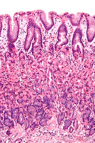
Simple columnar epithelium is a single layer of columnar epithelial cells which are tall and slender with oval-shaped nuclei located in the basal region, attached to the basement membrane. In humans, simple columnar epithelium lines most organs of the digestive tract including the stomach, and intestines. Simple columnar epithelium also lines the uterus.

<span class="mw-page-title-main">Pseudostratified columnar epithelium</span> Tissue type

Pseudostratified columnar epithelium is a type of epithelium that, though comprising only a single layer of cells, has its cell nuclei positioned in a manner suggestive of stratified columnar epithelium. A stratified epithelium rarely occurs as squamous or cuboidal.

<span class="mw-page-title-main">Intestinal epithelium</span> Single-cell layer lining the intestines

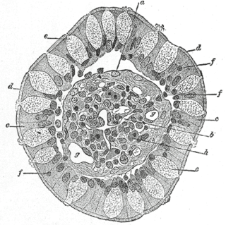
The intestinal epithelium is the single cell layer that forms the luminal surface (lining) of both the small and large intestine (colon) of the gastrointestinal tract. Composed of simple columnar epithelium its main functions are absorption, and secretion. Useful substances are absorbed into the body, and the entry of harmful substances is restricted. Secretions include mucins, and peptides.

<span class="mw-page-title-main">Mucociliary clearance</span>

Mucociliary clearance (MCC), mucociliary transport, or the mucociliary escalator describes the self-clearing mechanism of the airways in the respiratory system. It is one of the two protective processes for the lungs in removing inhaled particles including pathogens before they can reach the delicate tissue of the lungs. The other clearance mechanism is provided by the cough reflex. Mucociliary clearance has a major role in pulmonary hygiene.

Histology is the study of the minute structure, composition, and function of tissues. Mature human vocal cords are composed of layered structures which are quite different at the histological level.

<span class="mw-page-title-main">Nasal mucosa</span> Part of the mucus membrane lining the nasal cavity

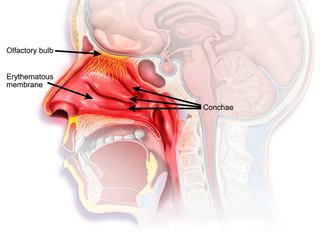
The nasal mucosa lines the nasal cavity. It is part of the respiratory mucosa, the mucous membrane lining the respiratory tract. The nasal mucosa is intimately adherent to the periosteum or perichondrium of the nasal conchae. It is continuous with the skin through the nostrils, and with the mucous membrane of the nasal part of the pharynx through the choanae. From the nasal cavity its continuity with the conjunctiva may be traced, through the nasolacrimal and lacrimal ducts; and with the frontal, ethmoidal, sphenoidal, and maxillary sinuses, through the several openings in the nasal meatuses. The mucous membrane is thickest, and most vascular, over the nasal conchae. It is also thick over the nasal septum where increased numbers of goblet cells produce a greater amount of nasal mucus. It is very thin in the meatuses on the floor of the nasal cavities, and in the various sinuses. It is one of the most commonly infected tissues in adults and children. Inflammation of this tissue may cause significant impairment of daily activities, with symptoms such as stuffy nose, headache, mouth breathing, etc.

<span class="mw-page-title-main">Tuft cell</span>

Tuft cells are chemosensory cells in the epithelial lining of the intestines. Similar tufted cells are found in the respiratory epithelium where they are known as brush cells. The name "tuft" refers to the brush-like microvilli projecting from the cells. Ordinarily there are very few tuft cells present but they have been shown to greatly increase at times of a parasitic infection. Several studies have proposed a role for tuft cells in defense against parasitic infection. In the intestine, tuft cells are the sole source of secreted interleukin 25 (IL-25).

<span class="mw-page-title-main">Anatomical terms of microanatomy</span> Anatomical terminology is used to describe microanatomical (or histological) structures

Anatomical terminology is used to describe microanatomical structures. This helps describe precisely the structure, layout and position of an object, and minimises ambiguity. An internationally accepted lexicon is Terminologia Histologica.

<span class="mw-page-title-main">Air-liquid interface cell culture</span>

Air liquid interface cell culture (ALI) is a method of cell culture by which basal stem cells are grown with their basal surfaces in contact with media, and the top of the cellular layer is exposed to the air. The cells are then lifted and media is changed until the development of a mucociliary phenotype of a pseudostratified epithelium, similar to the tracheal epithelium.

Airway basal cells are found deep in the respiratory epithelium, attached to, and lining the basement membrane.

References

  1. Crystal, R (September 2008). "Airway epithelial cells: current concepts and challenges". Proc Am Thorac Soc. 15 (7): 772–777. doi:10.1513/pats.200805-041HR. PMC   5820806 . PMID   18757316.
  2. "Respiratory mucosa". meshb.nlm.nih.gov. Retrieved 26 July 2019.
  3. Saladin, K (2012). Anatomy & physiology: the unity of form and function (6th ed.). McGraw-Hill. pp. 857–859. ISBN   9780073378251.
  4. Mescher AL, "Chapter 17. The Respiratory System" (Chapter). Mescher AL: Junqueira's Basic Histology: Text & Atlas, 12e: "AccessMedicine | the Respiratory System: Introduction". Archived from the original on 2013-06-03. Retrieved 2015-02-24..
  5. "Bronchi, Bronchial Tree & Lungs". nih.gov. Retrieved 17 September 2019.
  6. Mescher, Anthony L. (2018). Junqueira's basic histology: text and atlas (Fifteenth ed.). [New York]. p. 350. ISBN   978-1-26-002618-4.{{cite book}}: CS1 maint: location missing publisher (link)
  7. 1 2 Weinberger, Steven; Cockrill, Barbara; Mandel, Jess (2019). Principles of pulmonary medicine (Seventh ed.). Elsevier. p. 67. ISBN   9780323523714.
  8. Yaghi, A; Dolovich, MB (11 November 2016). "Airway Epithelial Cell Cilia and Obstructive Lung Disease". Cells. 5 (4): 40. doi: 10.3390/cells5040040 . PMC   5187524 . PMID   27845721.
  9. 1 2 Reid, L; Meyrick, B; Antony, VB; Chang, LY; Crapo, JD; Reynolds, HY (1 July 2005). "The mysterious pulmonary brush cell: a cell in search of a function". American Journal of Respiratory and Critical Care Medicine. 172 (1): 136–9. doi:10.1164/rccm.200502-203WS. PMC   2718446 . PMID   15817800.
  10. Hasleton, Philip (1996). Spencer's Pathology of the Lung. McGraw-Hill. pp.  10. ISBN   0071054480.
  11. Perniss, Alexander; Lui, Shuya; Boonen, Brett; Zufall, Frank; Buffe, Bernd; Kummer, Wolfgang (2020-04-14). "Chemosensory Cell-Derived Acetylcholine Drives Tracheal Mucociliary Clearance in Response to Virulence-Associated Formyl Peptides". Cell Immunity. 52 (2): 683–699.e11. doi: 10.1016/j.immuni.2020.03.005 . PMID   32294408.
  12. Hall, John (2011). Guyton and Hall Textbook of Medical Physiology. Saunders/Elsevier. p. 473. ISBN   9781416045748.
  13. Stanke F The Contribution of the Airway Epithelial Cell to Host Defense. Mediators Inflamm. 2015;2015:463016. PMID   26185361 PMC 4491388
  14. U.S. EPA. Integrated Science Assessment for Oxides of Nitrogen – Health Criteria (2016 Final Report). U.S. Environmental Protection Agency, Washington, DC, EPA/600/R-15/068, 2016. Federal Register Notice Jan 28, 2016 Free download available at Report page at EPA website.
  15. Hiemstra, PS; McCray PB, Jr; Bals, R (April 2015). "The innate immune function of airway epithelial cells in inflammatory lung disease". The European Respiratory Journal. 45 (4): 1150–62. doi:10.1183/09031936.00141514. PMC   4719567 . PMID   25700381.
  16. 1 2 3 Baker, EH; Baines, DL (February 2018). "Airway Glucose Homeostasis: A New Target in the Prevention and Treatment of Pulmonary Infection". Chest. 153 (2): 507–514. doi:10.1016/j.chest.2017.05.031. PMID   28610911. S2CID   13733461.
  17. 1 2 Garnett, JP; Baker, EH; Baines, DL (November 2012). "Sweet talk: insights into the nature and importance of glucose transport in lung epithelium". The European Respiratory Journal. 40 (5): 1269–76. doi: 10.1183/09031936.00052612 . PMID   22878875.
  18. Global Initiative for Chronic Obstructive Lung Disease - GOLD (PDF). 2018. p. 15. Retrieved 10 November 2019.
  19. Garg, A; Sui, P; Verheyden, JM; Young, LR; Sun, X (2019). "Consider the lung as a sensory organ: A tip from pulmonary neuroendocrine cells". Current Topics in Developmental Biology. 132: 67–89. doi:10.1016/bs.ctdb.2018.12.002. ISBN   9780128104897. PMID   30797518. S2CID   73489416.

Additional images