Intestinal epithelium

Last updated
Intestinal epithelium
Simple columnar epithelial cells.png
Simple columnar epithelial cells
Bischoff fig 2 adapted 01.png
Cell types of the intestinal epithelium
Identifiers
MeSH D007413
FMA 15695 17229, 15695
Anatomical terminology

The intestinal epithelium is the single cell layer that forms the luminal surface (lining) of both the small and large intestine (colon) of the gastrointestinal tract. Composed of simple columnar epithelium its main functions are absorption, and secretion. Useful substances are absorbed into the body, and the entry of harmful substances is restricted. Secretions include mucins, and peptides.

Contents

Absorptive cells in the small intestine are known as enterocytes, and in the colon they are known as colonocytes. The other cell types are the secretory cells – goblet cells, Paneth cells, enteroendocrine cells, and Tuft cells. Paneth cells are absent in the colon. [1] [2]

As part of its protective role, the intestinal epithelium forms an important component of the intestinal mucosal barrier. Certain diseases and conditions are caused by functional defects in the intestinal epithelium. On the other hand, various diseases and conditions can lead to its dysfunction which, in turn, can lead to further complications.

Structure

Proliferative stem cells residing at the base of the intestinal glands produce new epithelial cells which migrate upwards and out of the crypt. Eventually, they are shed into the intestinal lumen Epithelial cell migration.tif
Proliferative stem cells residing at the base of the intestinal glands produce new epithelial cells which migrate upwards and out of the crypt. Eventually, they are shed into the intestinal lumen

The intestinal epithelium is part of the mucosal lining. The epithelium is simple cuboidal epithelium composed of a single layer of cells, while the other two layers of the mucosa, the lamina propria and the muscularis mucosae, support and communicate with the epithelial layer. To securely contain the contents of the intestinal lumen, the cells of the epithelial layer are joined together by tight junctions, thus forming a contiguous and relatively impermeable membrane.

Drawing showing the relationship between villi and microvilli of the small intestine. The luminal surface of the enterocytes have microvilli (1 micrometer long) while the cell layer itself is folded to form villi (0.5-1.6 millimeters long) and crypts. Both serve to increase the total absorption surface of the intestine. Villi & microvilli of small intestine.svg
Drawing showing the relationship between villi and microvilli of the small intestine. The luminal surface of the enterocytes have microvilli (1 micrometer long) while the cell layer itself is folded to form villi (0.5-1.6 millimeters long) and crypts. Both serve to increase the total absorption surface of the intestine.

Epithelial cells are continuously renewed every 4–5 days through a process of cell division, maturation, and migration. Renewal relies on proliferative cells (stem cells) that reside at the crypt (base) of the intestinal glands (epithelial invaginations into the underlying connective tissue). [3] After being formed at the base, the new cells migrate upwards and out of the crypt, maturing along the way. Eventually, they undergo apoptosis and are shed off into the intestinal lumen. [4] In this way, the lining of the intestine is constantly renewed while the number of cells making up the epithelial layer remains constant. [5]

In the small intestine, the mucosal layer is specially adapted to provide a large surface area in order to maximize the absorption of nutrients. The expansion of the absorptive surface, 600 times beyond that of a simple cylindrical tube, is achieved by three anatomical features: [6]

The brush border on the apical surface of the epithelial cells is covered with glycocalyx, which is composed of oligosaccharides attached to membrane glycoproteins and glycolipids. [7]

TEM image of a thin section cut through an epithelial cell showing the luminal surface (apical end) of the cell packed with microvilli that make up the absorbing surface. Each microvillus is approximately 1 micrometers long and 0.1 micrometer in diameter Microvilli.jpg
TEM image of a thin section cut through an epithelial cell showing the luminal surface (apical end) of the cell packed with microvilli that make up the absorbing surface. Each microvillus is approximately 1 micrometers long and 0.1 micrometer in diameter

Cell types

Different cell types are produced by the stem cells that reside at the base of the crypts. [8] Each type matures according to its specific differentiation program as it migrates up and out of the crypt. Many of the genes necessary for differentiation into the different epithelial cell types have been identified and characterized. The cell types produced are: enterocytes (small intestine) (known as colonocytes in colon), Goblet cells, enteroendocrine cells, Paneth cells, microfold cells, cup cells and tuft cells. Their functions are listed here: [9]

Throughout the digestive tract, the distribution of the different types of epithelial cells varies according to the function of that region. [5]

Structural components of cellular junctions

Types of cell junctions (click to enlarge). 402 Types of Cell Junctions new.jpg
Types of cell junctions (click to enlarge).

Important for the barrier function of intestinal epithelium, its cells are joined securely together by four types of cell junction which can be identified at the ultrastructural level: [16] [17]

Gap junctions

Gap junctions bring the adjacent cells within 2 nanometers of each other. They are formed by several homologous proteins encoded by the connexin gene family coming together to form a multiprotein complex. The molecular structure of this complex is in the form of a hexamer. The complex, which is embedded in the cell membranes of the two joined cells, forms a gap or channel in the middle of the six proteins. This channel allows various molecules, ions and electrical impulses to pass between the two cells. [18]

Desmosomes

These complexes, consisting of transmembrane adhesion proteins of the cadherin family, link adjacent cells together through their cytoskeletons. [19] Desmosomes leave a gap of 30 nanometers between cells. [18]

Adherens junctions

Adherens junctions, also called zonula adherens, are multiprotein complexes formed by proteins of the catenin and cadherin families. They are located in the membrane at the contact points between the cells. They are formed by interactions between intracellular adapter proteins, transmembrane proteins and the actin cytoskeletons of the cells . Besides their role in linking adjacent cells, these complexes are important for regulating epithelial migration, cell polarity, and the formation of other cell junction complexes. [17]

Tight junctions

Tight junctions, also called zonula occludens, are the most important components of the intestinal epithelium for its barrier function. [20] These complexes, formed primarily of members of the claudin and the occludin families, consist of about 35 different proteins, [16] form a ring shaped continuous ribbon around the cells, and are located near the borders of the lateral and apical membranes. [17]

The extracellular domains of the transmembrane proteins in adjacent cells cross connect to form a tight seal. These interactions include those between proteins in the same membrane ("cis") and proteins in adjacent cells ("trans"). In addition, interactions can be homophilic (between identical proteins) or heterophilic (between different proteins). [17]

Similar to adherens junctions, the intracellular domains of tight junctions interact with different scaffold proteins, adapter proteins and signaling complexes to regulate cytoskeletal linking, cell polarity, cell signaling and vesical trafficking. [17]

Tight junctions provide a narrow but modifiable seal between adjacent cells in the epithelial layer and thereby provide selective paracellular transport of solutes. [17] While previously thought to be static structures, tight junctions are now known to be dynamic and can change the size of the opening between cells and thereby adapt to the different states of development, physiologies and pathologies. [20] They function as a selective and semipermeable paracellular barrier between apical and basolateral compartments of the epithelial layer. They function to facilitate the passage of small ions and water-soluble solutes through the paracellular space while preventing the passage of luminal antigens, microorganisms and their toxins. [17]

Physiology

The intestinal epithelium has a complex anatomical structure which facilitates motility and coordinated digestive, absorptive, immunological and neuroendocrine functions. [21]

The mucus secreted by goblet cells acts as a lubricant and protects the epithelial cell layer against irritation from mucosal contents. [22]

Traditionally, crypt cells were considered primarily as secretory cells while enterocytes are considered principally absorptive. However, recent studies have challenged this classical functional partitioning and have shown that both the surface and crypt cells can perform both secretory and absorptive functions and that, in fact, these functions can occur simultaneously. [23] [24]

Nutrient uptake

Overlaying the brush border of the apical surface of the enterocytes is the glycocalyx, which is a loose network composed of the oligosaccharide side chains of integral membrane hydrolases and other enzymes essential for the digestion of proteins and carbohydrates. These glycoproteins, glycolipids, and enzymes catalyze the final digestive stages of luminal carbohydrates and proteins. The monosaccharides and amino acids thus produced are subsequently transported across the intestinal epithelium and eventually into the bloodstream. [7]

The absorption of electrolytes and water is one of the most important functions of the digestive tract. Water absorption is passive and isotonic - depending on the speed and direction of solute flow. Other factors influencing fluid absorption are osmolarity and the specific intestinal region. [21] Regulated selective permeability is performed through two major routes: the transcellular (transepithelial) route and the paracellular route. [17]

Transcellular permeability

Scheme of selective permeability routes of epithelial cells (red arrows). The transcellular (through the cells) and paracellular (between the cells) routes control the passage of substances between the intestinal lumen and blood. Selective permeability routes in epithelium.png
Scheme of selective permeability routes of epithelial cells (red arrows). The transcellular (through the cells) and paracellular (between the cells) routes control the passage of substances between the intestinal lumen and blood.

This consists of specific transport of solutes across the epithelial cells. It is predominantly regulated by the activities of specialised transporters that translocate specific electrolytes, amino acids, sugars, short chain fatty acids and other molecules into or out of the cell. [17]

Paracellular permeability

Paracellular permeability depends on transport through the spaces that exist between epithelial cells. It is regulated by cellular junctions that are localized in the laminal membranes of the cells. [17] This is the main route of passive flow of water and solutes across the intestinal epithelium. Regulation depends on the intercellular tight junctions which have the most influence on paracellular transport. [25] Studies using the electron microscope showed that the electrical resistance of epithelial layers depends on the complexity and number of filaments within the tight junction transmembrane protein complexes. [21] Also, the plasma membrane resistance and variable transmembrane conductance of the epithelial cells can also modulate paracellular pathway function. [21]

Functions

The barrier formed by the intestinal epithelium separates the external environment (the contents of the intestinal lumen) from the body [17] and is the most extensive and important mucosal surface of body. [20]

The intestinal epithelium serves several crucial functions, exhibiting both innate and adaptive immune features. It closely monitors its intracellular and extracellular environment, communicates messages to neighbouring cells and rapidly initiates active defensive and repair measures, if necessary. [26] On the one hand, it acts as a barrier, preventing the entry of harmful substances such as foreign antigens, toxins and microorganisms. [16] [17] On the other hand, it acts as a selective filter which facilitates the uptake of dietary nutrients, electrolytes, water and various other beneficial substances from the intestinal lumen. [17]

When barrier integrity is lost, intestinal permeability increases and uncontrolled passage of harmful substances can occur. This can lead to, depending on the genetic predisposition of the individual, the development of inflammation, infection, allergies, autoimmune diseases or cancer - within the intestine itself or other organs. [21]

Although they primarily function as part of the digestive system, enterocytes of the intestinal epithelium also express toll-like receptors and nucleotide oligomerization domain proteins that recognize diverse types of microbes and contribute to immune system function. [27] [28] Thus the intestinal epithelium not only serves as a physical barrier separating the intestinal lumen from the body proper but also carries out pathogen recognition functions as part of the intrinsic immune system.

Importance for human health

Loss of integrity of the intestinal epithelium plays a key pathogenic role in inflammatory bowel disease (IBD). [29] Changes in the composition of the intestinal microbiota are an important environmental factor in the development of IBD. Detrimental changes in the intestinal microbiota induce an inappropriate (uncontrolled) immune response that results in damage to the intestinal epithelium. Breaches in this critical barrier (the intestinal epithelium) allow further infiltration of microbiota that, in turn, elicit further immune responses. IBD is a multifactorial disease that is nonetheless driven in part by an exaggerated immune response to gut microbiota that causes defects in epithelial barrier function. [30]

Bile acids are normal components of the luminal contents of the gastrointestinal tract where they can act as physiologic detergents and regulators of intestinal epithelial homeostasis. [31] Excessive long term exposure of intestinal epithelial cells to bile acids may cause oxidative stress leading to oxidative DNA damage and carcinogenic mutation. [32]

See also

Related Research Articles

<span class="mw-page-title-main">Ileum</span> Final section of the small intestine

The ileum is the final section of the small intestine in most higher vertebrates, including mammals, reptiles, and birds. In fish, the divisions of the small intestine are not as clear and the terms posterior intestine or distal intestine may be used instead of ileum. Its main function is to absorb vitamin B12, bile salts, and whatever products of digestion that were not absorbed by the jejunum.

<span class="mw-page-title-main">Microvillus</span> Microscopic protrusion of a cell membrane that increases surface area substantially

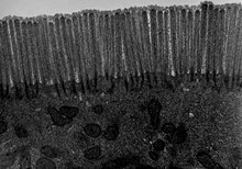
Microvilli are microscopic cellular membrane protrusions that increase the surface area for diffusion and minimize any increase in volume, and are involved in a wide variety of functions, including absorption, secretion, cellular adhesion, and mechanotransduction.

<span class="mw-page-title-main">Epithelium</span> Tissue lining the surfaces of organs in animals

Epithelium or epithelial tissue is a thin, continuous, protective layer of compactly packed cells with little intercellular matrix. Epithelial tissues line the outer surfaces of organs and blood vessels throughout the body, as well as the inner surfaces of cavities in many internal organs. An example is the epidermis, the outermost layer of the skin. Epithelial tissue is one of the four basic types of animal tissue, along with connective tissue, muscle tissue and nervous tissue. These tissues also lack blood or lymph supply. The tissue is supplied by nerves.

<span class="mw-page-title-main">Tight junction</span> Structure preventing inter-cell leakage

Tight junctions, also known as occluding junctions or zonulae occludentes, are multiprotein junctional complexes whose canonical function is to prevent leakage of solutes and water and seals between the epithelial cells. They also play a critical role maintaining the structure and permeability of endothelial cells. Tight junctions may also serve as leaky pathways by forming selective channels for small cations, anions, or water. The corresponding junctions that occur in invertebrates are septate junctions.

<span class="mw-page-title-main">Caco-2</span>

Caco-2 is an immortalized cell line of human colorectal adenocarcinoma cells. It is primarily used as a model of the intestinal epithelial barrier. In culture, Caco-2 cells spontaneously differentiate into a heterogeneous mixture of intestinal epithelial cells. It was developed in 1977 by Jorgen Fogh at the Sloan-Kettering Institute for Cancer Research.

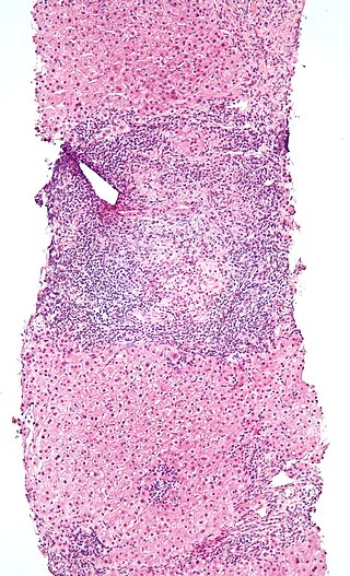
Gut-associated lymphoid tissue (GALT) is a component of the mucosa-associated lymphoid tissue (MALT) which works in the immune system to protect the body from invasion in the gut.

<span class="mw-page-title-main">Paneth cell</span> Anti-microbial epithelial cell of the small intestine

Paneth cells are cells in the small intestine epithelium, alongside goblet cells, enterocytes, and enteroendocrine cells. Some can also be found in the cecum and appendix. They are located below the intestinal stem cells in the intestinal glands and the large eosinophilic refractile granules that occupy most of their cytoplasm.

Intestinal permeability is a term describing the control of material passing from inside the gastrointestinal tract through the cells lining the gut wall, into the rest of the body. The intestine normally exhibits some permeability, which allows nutrients to pass through the gut, while also maintaining a barrier function to keep potentially harmful substances from leaving the intestine and migrating to the body more widely. In a healthy human intestine, small particles can migrate through tight junction claudin pore pathways, and particles up to 10–15 Å can transit through the paracellular space uptake route. There is some evidence abnormally increased intestinal permeability may play a role in some chronic diseases and inflammatory conditions. The most well understood condition with observed increased intestinal permeability is celiac disease.

<span class="mw-page-title-main">Intestinal gland</span> Gland between the intestinal villi that produces new cells

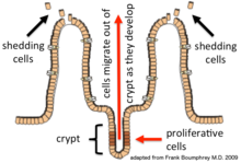
In histology, an intestinal gland is a gland found in between villi in the intestinal epithelium lining of the small intestine and large intestine. The glands and intestinal villi are covered by epithelium, which contains multiple types of cells: enterocytes, goblet cells, enteroendocrine cells, cup cells, tuft cells, and at the base of the gland, Paneth cells and stem cells.

<span class="mw-page-title-main">Transcytosis</span> Type of cellular transport

Transcytosis is a type of transcellular transport in which various macromolecules are transported across the interior of a cell. Macromolecules are captured in vesicles on one side of the cell, drawn across the cell, and ejected on the other side. Examples of macromolecules transported include IgA, transferrin, and insulin. While transcytosis is most commonly observed in epithelial cells, the process is also present elsewhere. Blood capillaries are a well-known site for transcytosis, though it occurs in other cells, including neurons, osteoclasts and M cells of the intestine.

Microfold cells are found in the gut-associated lymphoid tissue (GALT) of the Peyer's patches in the small intestine, and in the mucosa-associated lymphoid tissue (MALT) of other parts of the gastrointestinal tract. These cells are known to initiate mucosal immunity responses on the apical membrane of the M cells and allow for transport of microbes and particles across the epithelial cell layer from the gut lumen to the lamina propria where interactions with immune cells can take place.

<span class="mw-page-title-main">Intraepithelial lymphocyte</span>

Intraepithelial lymphocytes (IEL) are lymphocytes found in the epithelial layer of mammalian mucosal linings, such as the gastrointestinal (GI) tract and reproductive tract. However, unlike other T cells, IELs do not need priming. Upon encountering antigens, they immediately release cytokines and cause killing of infected target cells. In the GI tract, they are components of gut-associated lymphoid tissue (GALT).

Paracellular transport refers to the transfer of substances across an epithelium by passing through the intercellular space between the cells. It is in contrast to transcellular transport, where the substances travel through the cell, passing through both the apical membrane and basolateral membrane.

<span class="mw-page-title-main">Mucosal immunology</span> Field of study

Mucosal immunology is the study of immune system responses that occur at mucosal membranes of the intestines, the urogenital tract, and the respiratory system. The mucous membranes are in constant contact with microorganisms, food, and inhaled antigens. In healthy states, the mucosal immune system protects the organism against infectious pathogens and maintains a tolerance towards non-harmful commensal microbes and benign environmental substances. Disruption of this balance between tolerance and deprivation of pathogens can lead to pathological conditions such as food allergies, irritable bowel syndrome, susceptibility to infections, and more.

The internal surface of the uterus is lined by uterine epithelial cells which undergo dramatic changes during pregnancy. The role of the uterine epithelial cells is to selectively allow the blastocyst to implant at a specific time. All other times of the cycle, these uterine epithelial cells are refractory to blastocyst implantation. Uterine epithelial cells have a similar structure in most species and the changes which occur in the uterine epithelial cells at the time of blastocyst implantation are also conserved among most species.

<span class="mw-page-title-main">Tuft cell</span>

Tuft cells are chemosensory cells in the epithelial lining of the intestines. Similar tufted cells are found in the respiratory epithelium where they are known as brush cells. The name "tuft" refers to the brush-like microvilli projecting from the cells. Ordinarily there are very few tuft cells present but they have been shown to greatly increase at times of a parasitic infection. Several studies have proposed a role for tuft cells in defense against parasitic infection. In the intestine, tuft cells are the sole source of secreted interleukin 25 (IL-25).

Larazotide is a synthetic eight amino acid peptide that functions as a tight junction regulator and reverses leaky junctions to their normally closed state. It is being studied in people with celiac disease.

<span class="mw-page-title-main">Intestinal mucosal barrier</span>

The intestinal mucosal barrier, also referred to as intestinal barrier, refers to the property of the intestinal mucosa that ensures adequate containment of undesirable luminal contents within the intestine while preserving the ability to absorb nutrients. The separation it provides between the body and the gut prevents the uncontrolled translocation of luminal contents into the body proper. Its role in protecting the mucosal tissues and circulatory system from exposure to pro-inflammatory molecules, such as microorganisms, toxins, and antigens is vital for the maintenance of health and well-being. Intestinal mucosal barrier dysfunction has been implicated in numerous health conditions such as: food allergies, microbial infections, irritable bowel syndrome, inflammatory bowel disease, celiac disease, metabolic syndrome, non-alcoholic fatty liver disease, diabetes, and septic shock.

Tight junction proteins are molecules situated at the tight junctions of epithelial, endothelial and myelinated cells. This multiprotein junctional complex has a regulatory function in passage of ions, water and solutes through the paracellular pathway. It can also coordinate the motion of lipids and proteins between the apical and basolateral surfaces of the plasma membrane. Thereby tight junction conducts signaling molecules, that influence the differentiation, proliferation and polarity of cells. So tight junction plays a key role in maintenance of osmotic balance and trans-cellular transport of tissue specific molecules. Nowadays is known more than 40 different proteins, that are involved in these selective TJ channels.

References

  1. Noah, TK; Donahue, B; Shroyer, NF (15 November 2011). "Intestinal development and differentiation". Experimental Cell Research. 317 (19): 2702–10. doi:10.1016/j.yexcr.2011.09.006. PMC   3210330 . PMID   21978911.
  2. Litvak, Y; Byndloss, MX; Bäumler, AJ (30 November 2018). "Colonocyte metabolism shapes the gut microbiota". Science. 362 (6418). Bibcode:2018Sci...362.9076L. doi:10.1126/science.aat9076. PMC   6296223 . PMID   30498100.
  3. Clevers H (2013). "The intestinal crypt, a prototype stem cell compartment". Cell. 154 (2): 274–84. doi: 10.1016/j.cell.2013.07.004 . PMID   23870119.
  4. van der Flier, Laurens G.; Clevers, Hans (1 January 2009). "Stem cells, self-renewal, and differentiation in the intestinal epithelium". Annual Review of Physiology. 71: 241–260. doi:10.1146/annurev.physiol.010908.163145. ISSN   1545-1585. PMID   18808327.
  5. 1 2 Lodish, Harvey; Berk, Arnold; Zipursky, S. Lawrence; Matsudaira, Paul; Baltimore, David; Darnell, James (1 January 2000). "Intestinal Architecture and Development". Molecular Cell Biology (4th ed.). W. H. Freeman. ISBN   978-0716731368.
  6. Khurana (1 January 2005). Textbook Of Medical Physiology. Elsevier India. p. 641. ISBN   9788181478504.
  7. 1 2 Lodish, Harvey; Berk, Arnold; Zipursky, S. Lawrence; Matsudaira, Paul; Baltimore, David; Darnell, James (1 January 2000). "Transport across Epithelia".{{cite journal}}: Cite journal requires |journal= (help)
  8. Laurens G. van der Flier; Hans Clevers (2009). "Stem Cells, Self-Renewal, and Differentiation in the Intestinal Epithelium". Annual Review of Physiology. 71 (1): 241–260. doi:10.1146/annurev.physiol.010908.163145. PMID   18808327.
  9. Sarmento, Bruno (30 September 2015). Concepts and Models for Drug Permeability Studies: Cell and Tissue based In Vitro Culture Models. Woodhead Publishing. pp. 57–58. ISBN   9780081001141.
  10. Bohórquez, Diego; Liddle, Rodger (2015). "Neuroepithelial circuit formed by innervation of sensory enteroendocrine cells". Journal of Clinical Investigation. 125 (2): 782–786. doi:10.1172/JCI78361. PMC   4319442 . PMID   25555217.
  11. Kaelberer, M. Maya; Bohórquez, Diego (2018). "A gut-brain neural circuit for nutrient sensory transduction". Science. 361 (6408): eaat5236. doi:10.1126/science.aat5236. PMC   6417812 . PMID   30237325.
  12. van Es, Johan H.; Clevers, Hans (16 June 2014). "Paneth cells". Current Biology. 24 (12): R547–548. Bibcode:2014CBio...24.R547V. doi: 10.1016/j.cub.2014.04.049 . ISSN   1879-0445. PMID   24937274.
  13. Santaolalla R, Abreu MT (2012). "Innate immunity in the small intestine". Curr Opin Gastroenterol. 28 (2): 124–9. doi:10.1097/MOG.0b013e3283506559. PMC   3502878 . PMID   22241076.
  14. Gerbe, F; Legraverend, C; Jay, P (September 2012). "The intestinal epithelium tuft cells: specification and function". Cellular and Molecular Life Sciences. 69 (17): 2907–17. doi:10.1007/s00018-012-0984-7. PMC   3417095 . PMID   22527717.
  15. Gerbe, F; Legraverend, C; Jay, P (September 2012). "The intestinal epithelium tuft cells: specification and function". Cellular and Molecular Life Sciences. 69 (17): 2907–17. doi:10.1007/s00018-012-0984-7. PMC   3417095 . PMID   22527717.
  16. 1 2 3 Khan, Niamat; Asif, Abdul R. (1 January 2015). "Transcriptional Regulators of Claudins in Epithelial Tight Junctions". Mediators of Inflammation. 2015: 219843. doi: 10.1155/2015/219843 . ISSN   0962-9351. PMC   4407569 . PMID   25948882.
  17. 1 2 3 4 5 6 7 8 9 10 11 12 13 Groschwitz, Katherine R.; Hogan, Simon P. (1 July 2009). "Intestinal Barrier Function: Molecular Regulation and Disease Pathogenesis". The Journal of Allergy and Clinical Immunology. 124 (1): 3–22. doi:10.1016/j.jaci.2009.05.038. ISSN   0091-6749. PMC   4266989 . PMID   19560575.
  18. 1 2 Bennett, M. V.; Barrio, L. C.; Bargiello, T. A.; Spray, D. C.; Hertzberg, E.; Sáez, J. C. (1 March 1991). "Gap junctions: new tools, new answers, new questions". Neuron. 6 (3): 305–320. doi:10.1016/0896-6273(91)90241-q. ISSN   0896-6273. PMID   1848077. S2CID   33441056.
  19. Nekrasova, Oxana; Green, Kathleen J. (1 November 2013). "Desmosome assembly and dynamics". Trends in Cell Biology. 23 (11): 537–546. doi:10.1016/j.tcb.2013.06.004. ISSN   0962-8924. PMC   3913269 . PMID   23891292.
  20. 1 2 3 Rao, Jaladanki N.; Wang, Jian-Ying (1 January 2010). "Intestinal Architecture and Development". Morgan & Claypool Life Sciences.{{cite journal}}: Cite journal requires |journal= (help)
  21. 1 2 3 4 5 Fasano, Alessio (1 January 2011). "Zonulin and Its Regulation of Intestinal Barrier Function: The Biological Door to Inflammation, Autoimmunity, and Cancer". Physiological Reviews. 91 (1): 151–175. doi:10.1152/physrev.00003.2008. ISSN   0031-9333. PMID   21248165. S2CID   1375779.
  22. Allen, Adrian; Flemström, Gunnar (1 January 2005). "Gastroduodenal mucus bicarbonate barrier: protection against acid and pepsin". American Journal of Physiology. Cell Physiology. 288 (1): C1–19. doi:10.1152/ajpcell.00102.2004. ISSN   0363-6143. PMID   15591243. S2CID   6668280.
  23. Geibel, John P. (1 January 2005). "Secretion and absorption by colonic crypts". Annual Review of Physiology. 67: 471–490. doi:10.1146/annurev.physiol.67.031103.153530. ISSN   0066-4278. PMID   15709966.
  24. Binder, Henry J.; Rajendran, Vazhaikkurichi; Sadasivan, Vidyasagar; Geibel, John P. (1 April 2005). "Bicarbonate secretion: a neglected aspect of colonic ion transport". Journal of Clinical Gastroenterology. 39 (4 Suppl 2): S53–58. doi: 10.1097/01.mcg.0000155521.81382.3a . ISSN   0192-0790. PMID   15758660.
  25. Näslund, Erik; Hellström, Per M. (10 September 2007). "Appetite signaling: from gut peptides and enteric nerves to brain". Physiology & Behavior. 92 (1–2): 256–262. doi:10.1016/j.physbeh.2007.05.017. ISSN   0031-9384. PMID   17582445. S2CID   230872.
  26. Cario, E (2010). "Heads up! How the intestinal epithelium safeguards mucosal barrier immunity through the inflammasome and beyond". Current Opinion in Gastroenterology. 26 (6): 583–590. doi: 10.1097/MOG.0b013e32833d4b88 . PMID   20664345. S2CID   12976253.
  27. Cario, E (2005). "Bacterial interactions with cells of the intestinal mucosa: Toll-like receptors and NOD2". Gut. 54 (8): 1182–93. doi:10.1136/gut.2004.062794. PMC   1774880 . PMID   15840688.
  28. Abreu, Maria T.; Fukata, Masayuki; Arditi, Moshe (15 April 2005). "TLR signaling in the gut in health and disease". Journal of Immunology. 174 (8): 4453–4460. doi: 10.4049/jimmunol.174.8.4453 . ISSN   0022-1767. PMID   15814663.
  29. Maloy, Kevin J.; Powrie, Fiona (16 June 2011). "Intestinal homeostasis and its breakdown in inflammatory bowel disease". Nature. 474 (7351): 298–306. doi:10.1038/nature10208. ISSN   1476-4687. PMID   21677746. S2CID   205225483. Archived from the original on 29 October 2019. Retrieved 8 December 2019.
  30. Coskun, Mehmet (25 August 2014). "Intestinal Epithelium in Inflammatory Bowel Disease". Frontiers in Medicine. 1: 24. doi: 10.3389/fmed.2014.00024 . ISSN   2296-858X. PMC   4292184 . PMID   25593900.
  31. Ajouz H, Mukherji D, Shamseddine A. Secondary bile acids: an underrecognized cause of colon cancer. World J Surg Oncol. 2014 May 24;12:164. doi: 10.1186/1477-7819-12-164. PMID 24884764; PMCID: PMC4041630
  32. Bernstein H, Bernstein C. Bile acids as carcinogens in the colon and at other sites in the gastrointestinal system. Exp Biol Med (Maywood). 2023 Jan;248(1):79-89. doi: 10.1177/15353702221131858. Epub 2022 Nov 19. PMID 36408538; PMCID: PMC9989147