Oral mucosa

Last updated
Oral mucosa
Details
Identifiers
Latin tunica mucosa oris
MeSH D009061
TA98 A05.1.01.002
TA2 2785
FMA 59660
Anatomical terminology

The oral mucosa is the mucous membrane lining the inside of the mouth. It comprises stratified squamous epithelium, termed "oral epithelium", and an underlying connective tissue termed lamina propria . [1] The oral cavity has sometimes been described as a mirror that reflects the health of the individual. [2] Changes indicative of disease are seen as alterations in the oral mucosa lining the mouth, which can reveal systemic conditions, such as diabetes or vitamin deficiency, or the local effects of chronic tobacco or alcohol use. [3] The oral mucosa tends to heal faster and with less scar formation compared to the skin. [4] The underlying mechanism remains unknown, but research suggests that extracellular vesicles might be involved. [5]

Contents

Classification

Oral mucosa can be divided into three main categories based on function and histology:

Structure

Schematic illustration of the layers found in keratinized oral mucosa that include a deeper lamina propria and basement membrane in-between and superficial layers of strafied squamous epithelium that include from deepest to most superficial:
1: Stratum basale
2: Stratum spinosum
3: Stratum granulosum
4: Stratum corneum Oral mucosa.png
Schematic illustration of the layers found in keratinized oral mucosa that include a deeper lamina propria and basement membrane in-between and superficial layers of strafied squamous epithelium that include from deepest to most superficial:
1: Stratum basale
2: Stratum spinosum
3: Stratum granulosum
4: Stratum corneum

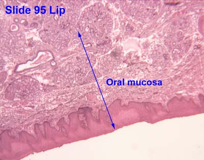
Oral mucosa consists of two layers, the surface stratified squamous epithelium and the deeper lamina propria. In keratinized oral mucosa, the epithelium consists of four layers:

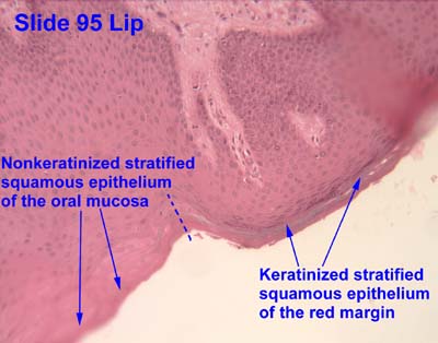
In nonkeratinised epithelium, the two deep layers (basale and spinosum) remain the same but the outer layers are termed the intermediate and superficial layers.

Depending on the region of the mouth, the epithelium may be nonkeratinized or keratinized. Nonkeratinized squamous epithelium covers the soft palate, inner lips, inner cheeks, the floor of the mouth, and ventral surface of the tongue. Keratinized squamous epithelium is present in the gingiva and hard palate as well as areas of the dorsal surface of the tongue. [8] [9]

Keratinization is the differentiation of keratinocytes in the stratum granulosum into nonvital surface cells or squames to form the stratum corneum. The cells terminally differentiate as they migrate to the surface from the stratum basale where the progenitor cells are located to the superficial surface.

Unlike keratinized epithelium, nonkeratinized epithelium normally has no superficial layers showing keratinization. Nonkeratinized epithelium may, however, readily transform into a keratinizing type in response to frictional or chemical trauma, in which case it undergoes hyperkeratinization. This change to hyperkeratinization commonly occurs on the usually nonkeratinized buccal mucosa when the linea alba forms, a white ridge of calloused tissue that extends horizontally at the level where the maxillary and mandibular teeth come together and occlude. Histologically, an excess amount of keratin is noted on the surface of the tissue, and the tissue has all the layers of an orthokeratinized tissue with its granular and keratin layers. In patients who have habits such as clenching or grinding (bruxism) their teeth, a larger area of the buccal mucosa than just the linea alba becomes hyperkeratinized. This larger white, rough, raised lesion needs to be recorded so that changes may be made in the dental treatment plan regarding the patient's parafunctional habits. [10] [11]

Even keratinized tissue can undergo further level of hyperkeratinization; an increase in the amount of keratin is produced as a result of chronic physical trauma to the region. Changes such as hyperkeratinization are reversible if the source of the injury is removed, but it takes time for the keratin to be shed or lost by the tissue. Thus, to check for malignant changes, a baseline biopsy and microscopic study of any whitened tissue may be indicated, especially if in a high-risk cancer category, such with a history of tobacco or alcohol use or are HPV positive. Hyperkeratinized tissue is also associated with the heat from smoking or hot fluids on the hard palate in the form of nicotinic stomatitis. [10]

The lamina propria is a fibrous connective tissue layer that consists of a network of type I and III collagen and elastin fibers in some regions. The main cells of the lamina propria are the fibroblasts, which are responsible for the production of the fibers as well as the extracellular matrix.

The lamina propria, like all forms of connective tissue proper, has two layers: papillary and dense. The papillary layer is the more superficial layer of the lamina propria. It consists of loose connective tissue within the connective tissue papillae, along with blood vessels and nerve tissue. The tissue has an equal amount of fibers, cells, and intercellular substance. The dense layer is the deeper layer of the lamina propria. It consists of dense connective tissue with a large amount of fibers. Between the papillary layer and the deeper layers of the lamina propria is a capillary plexus, which provides nutrition for the all layers of the mucosa and sends capillaries into the connective tissue papillae. [10]

A submucosa may or may not be present deep in the dense layer of the lamina propria, depending on the region of the oral cavity. If present, the submucosa usually contains loose connective tissue and may also contain adipose tissue or salivary glands, as well as overlying bone or muscle within the oral cavity. [10] The oral mucosa has no muscularis mucosae, and clearly identifying the boundary between it and the underlying tissues is difficult. Typically, regions such as the cheeks, lips, and parts of the hard palate contain submucosa (a layer of loose fatty or glandular connective tissue containing the major blood vessels and nerves supplying the mucosa). The submucosa's composition determines the flexibility of the attachment of oral mucosa to the underlying structures. In regions such as the gingiva and parts of the hard palate, oral mucosa is attached directly to the periosteum of underlying bone, with no intervening submucosa. This arrangement is called a mucoperiosteum and provides a firm, inelastic attachment. [12]

A variable number of Fordyce spots or granules are scattered throughout the nonkeratinized tissue. These are a normal variant, visible as small, yellowish bumps on the surface of the mucosa. They correspond to deposits of sebum from misplaced sebaceous glands in the submucosa that are usually associated with hair follicles. [10]

A basal lamina (basement membrane without aid of the microscope) is at the interface between the oral epithelium and lamina propria similar to the epidermis and dermis. [13]

Function

Mechanical stress is continuously placed on the oral environment by actions such as eating, drinking and talking. The mouth is also subject to sudden changes in temperature and pH meaning it must be able to adapt to change quickly. The mouth is the only place in the body which provides the sensation of taste. Due to these unique physiological features, the oral mucosa must fulfil a number of distinct functions.

Clinical significance

Infective

Viral

The majority of viral infections affecting the oral cavity are caused by the human Herpes Virus group. Each human herpes virus may present differently within the oral cavity. They are more likely to affect immunocompromised patients such as children and the elderly.

  • Herpetic Gingivostomatitis: A self-limiting viral infection which is caused by Herpes Simplex Virus-1(HSV-1). It usually presents in young children and is very contagious . It is characterised by the presence of small oral blisters which break down and coalesce into ulcers [18] [19]
  • Herpes Labialis (Cold Sore):Reactivation of latent Herpes Simplex Virus-1 triggered by sunlight, stress, and hormonal changes. It is characterised by the presence of crusting blisters on the upper lip. [18]
  • Chicken Pox: A type of viral infection which is caused by Varicella Zoster Virus and presents in children. Numerous itchy blisters are found on the face and body. Blisters could also be found on inner cheek and palate of the mouth. [18]
  • Herpes Zoster/Shingles: Viral infection caused by reactivation of latent Varicella Zoster Virus and found in adults. Patients can present with acute pain before or after the onset of blisters. If viral reactivation occurs in the facial nerve, it can cause Ramsay-Hunt Syndrome in which patients can develop facial paralysis, blisters around the ears and on the tongue, and loss of tongue sensation. [18]
  • Hand, Foot, and Mouth Disease: A highly contagious viral infection which infects young children and is caused by Coxsackie Virus A16. It is characterised by presence of small blisters all over the limbs and the mouth. [18]
  • Measles: A highly infectious condition which is caused by Measles virus. It most commonly presents in young children who have not been vaccinated. It is characterised by presence of white spots (Koplik's Spots) which rupture to form ulcers on the palate. [18]
  • 6. Papillomas and warts/ verrucae They are caused by various strains of the Human Papilloma Virus (HPV) of which there are more than 100 strains but usually due to HPV types 6 and 11 (2). Papillomas are mostly spiky, finger like projections, or cauliflower head rounded lobular shaped. Usually painless and singular. Verrucae usually appear on the lips. Verrucae Vulgaris is associated with HPV types 2 and 4. HPV types 16 and 18, designated high risk due to association with cancers, do not present like this in the oral mucosa but as white patches (1). Multifocal Epithelial Hyperplasia (Heck Disease) is rare, usually familial, swellings appear in multiples and are more common to some Native American and Inuit groups. Verricuform Xanthoma, a rare lesion which may resemble papilloma, is often white due to hyperkeratosis, usually appear in 50-70 year age group, commonly on the gingivae. They may be mistaken for papilloma or verrucous leukoplakia but are benign (1).

Bacterial

  • Syphilis- Bacterial infection which is usually sexually transmitted. It is caused by bacteria Treponema Pallidum and it has different types of oral presentation at different stages of the disease. [20] [18]

Fungal

Oral fungal infections are most commonly caused by different Candida species such as Candida Albicans, Candida Glabrata and Candida Tropicalis resulting in oral Candidiasis. [18] [21] There are several predisposing factors to fungal infections such as systemic disease for example Diabetes, recent antibiotics, use of steroid inhalers etc . Management includes identifying and addressing contributory factors, the use of topical/systemic anti-fungal agents, oral and denture hygiene instruction. [22]

Different presentations of oral Candidiasis include:

Autoimmune

Hypersensitivity Reaction

Traumatic

Idiopathic

Neoplastic:

●      Oral Submucous Fibrosis:

This is a condition that involves inflammation of the tissues under the surface. This may cause rigid tissues and difficulty opening the mouth. 

Benign soft tissue neoplasms

1.     Peripheral nerve sheath tumours most commonly are traumatic neuromas, a reactive response to trauma (1), neurilemmoma and neurofibroma which are large growing painless tumours usually found on the tongue (3). Neurofibroma may occur as a benign solitary lesion but can present as multiple lesions associated with (Von Reckllinghausen's Disease) neurofibromatosis (4). They can be preceded by café au lait pigmentation spots on the skin, and as they grow can become very disfiguring (4). Malignant change can occur in neurofibromatosis but very rarely in single lesion presentation. Mucosal neuromas can be associated with other conditions such as Multiple Endocrine Neoplasia (MEN) Syndrome and may precede thyroid cancer. (1, 4)

2.     Lipoma and fibrolipoma, are tumour of adipose tissue, or fat, giving them a yellow appearance which varies according to fat content (1, 4). They are usually soft, mobile, slow growing painless and occur mostly in middle age or the elderly.

3.     Granular Cell Tumour is also a tumour arising from neural cells, though it was incorrectly thought to arise from muscle cells and therefore previously called Granular Cell Myoblastoma (4), It also is slow growing large painless and occurs mostly in the tongue (1, 3).

4.     Congenital Epulis, also known as Congenital Granular Cell Tumour (but not related to Granular Cell Tumour) occur mostly on the upper gum, the maxillary alveolar ridge, of newborns, predominantly females (4). Rarely, they occur elsewhere, usually the tongue. They are usually self-resolving (1).

5.     Angiomas – vascular tumours: haemangioma, lymphangioma, venus varix (2). Angiomas are difficult to classify as previously they were considered hamartomas, benign tumour-like malformations (6) but there is debate if they are developmental abnormality, true benign tumour or hamartoma, or can be either (1, 4, 5).  Haemangiomas are common in the oral mucosa but can occur in other structures such as salivary glands (4), and can be congenital (from birth) or develop in childhood. Congenital lesions can resolve spontaneously (involute) but those that develop later usually continue to slowly grow. They are usually dark red-purple or blue, soft, sometimes fluctuant (5) and painless. They commonly blanch on pressure. Usually solitary, they can occur as part of syndromes such as Sturge-weber Syndrome affecting the trigeminal nerve. They are at risk of trauma with subsequent excessive bleeding, thrombosis or calcification (2). Lymphangiomas are far less common in the oral mucosa, usually appearing on the tongue, less commonly the lip at birth or in infancy (1, 2, 4). They are colourless to pale pink and may be nodular projections (1) or resemble 'frog spawn' domes (2). They can cause macroglossia (enlargement of the tongue) (1, 4). Venous Varix, like varicose vein, usually appear in older people on the lower lip as a blue-purple lump (2).

Connective tissue malignancies, sarcomas, are rare in the oral mucosa. Osteosarcoma, chondrosarcoma arise in bone and cartilage, lymphoma in haematological disorders (1). The most common malignancies are carcinomas, overwhelmingly Squamous Cell Carcinoma (SSC) (4). See Oral Cancer.

  1. Rhabdomyosarcoma are fast growing destructive swellings usually in the maxilla. It is the most common oral sarcoma in children and adolescents (1), but rare (4).
  2. Kaposi Sarcoma is related to the Kaposi Sarcoma Herpes Virus (KSHV) or Human Herpes Virus (HHV-8) viral infection (3). Predominating on the hard palate and gingivae it develops initially as a macule ranging in colour from red, blue, purple to brown or black, becoming nodular as it grows (2, 3, 4). The lesions are highly vascular and can ulcerate and bleed easily; death is usually from opportunistic infections (1). It is usually associated with HIV /AIDS but also less commonly with immunosuppression such as organ donor recipients or prevalent in some communities such as Mediterranean Jews (1). There is no cure (4) but the lesions respond well to Highly Active Antiretroviral Treatment (HAART) drugs (2)

See also

Related Research Articles

<span class="mw-page-title-main">Mouth ulcer</span> Sore on the mucous membrane of the oral cavity

A mouth ulcer (aphtha) is an ulcer that occurs on the mucous membrane of the oral cavity. Mouth ulcers are very common, occurring in association with many diseases and by many different mechanisms, but usually there is no serious underlying cause. Rarely, a mouth ulcer that does not heal may be a sign of oral cancer. These ulcers may form individually or multiple ulcers may appear at once. Once formed, an ulcer may be maintained by inflammation and/or secondary infection.

<span class="mw-page-title-main">Lamina propria</span> Thin connective layer forming part of the mucous membranes

The lamina propria is a thin layer of connective tissue that forms part of the moist linings known as mucous membranes or mucosae, which line various tubes in the body, such as the respiratory tract, the gastrointestinal tract, and the urogenital tract.

<span class="mw-page-title-main">Oral candidiasis</span> Fungal infection

Oral candidiasis, also known as oral thrush among other names, is candidiasis that occurs in the mouth. That is, oral candidiasis is a mycosis of Candida species on the mucous membranes of the mouth.

<span class="mw-page-title-main">Leukoplakia</span> Medical condition

Oral leukoplakia is a potentially malignant disorder affecting the oral mucosa. It is defined as "essentially an oral mucosal white lesion that cannot be considered as any other definable lesion." Oral leukoplakia is a white patch or plaque that develops in the oral cavity and is strongly associated with smoking. Leukoplakia is a firmly attached white patch on a mucous membrane which is associated with increased risk of cancer. The edges of the lesion are typically abrupt and the lesion changes with time. Advanced forms may develop red patches. There are generally no other symptoms. It usually occurs within the mouth, although sometimes mucosa in other parts of the gastrointestinal tract, urinary tract, or genitals may be affected.

<span class="mw-page-title-main">Aphthous stomatitis</span> Common condition of mouth ulcers

Aphthous stomatitis, or recurrent aphthous stomatitis (RAS), is a common condition characterized by the repeated formation of benign and non-contagious mouth ulcers (aphthae) in otherwise healthy individuals. The informal term canker sore is also used, mainly in North America, although it may also refer to other types of mouth ulcers. The cause is not completely understood but involves a T cell-mediated immune response triggered by a variety of factors which may include nutritional deficiencies, local trauma, stress, hormonal influences, allergies, genetic predisposition, certain foods, dehydration, some food additives, or some hygienic chemical additives like SDS.

<span class="mw-page-title-main">Stomatitis</span> Medical condition

Stomatitis is inflammation of the mouth and lips. It refers to any inflammatory process affecting the mucous membranes of the mouth and lips, with or without oral ulceration.

<span class="mw-page-title-main">Herpetic gingivostomatitis</span> Medical condition

Gingivostomatitis is a combination of gingivitis and stomatitis, or an inflammation of the oral mucosa and gingiva. Herpetic gingivostomatitis is often the initial presentation during the first ("primary") herpes simplex infection. It is of greater severity than herpes labialis which is often the subsequent presentations. Primary herpetic gingivostomatitis is the most common viral infection of the mouth.

An oral medicine or stomatology doctor/dentist has received additional specialized training and experience in the diagnosis and management of oral mucosal abnormalities including oral cancer, salivary gland disorders, temporomandibular disorders and facial pain, taste and smell disorders; and recognition of the oral manifestations of systemic and infectious diseases. It lies at the interface between medicine and dentistry. An oral medicine doctor is trained to diagnose and manage patients with disorders of the orofacial region, essentially as a "physician of the mouth".

<span class="mw-page-title-main">Head and neck anatomy</span>

This article describes the anatomy of the head and neck of the human body, including the brain, bones, muscles, blood vessels, nerves, glands, nose, mouth, teeth, tongue, and throat.

Leukoedema is a blue, grey or white appearance of mucosae, particularly the buccal mucosa ; it may also occur on the mucosa of the larynx or vagina. It is a harmless and very common condition. Because it is so common, it has been argued that it may in fact represent a variation of the normal appearance rather than a disease, but empirical evidence suggests that leukoedema is an acquired condition caused by local irritation. It is found more commonly in black skinned people and tobacco users. The term is derived from the Greek words λευκός leukós, "white" and οἴδημα oídēma, "swelling".

The junctional epithelium (JE) is that epithelium which lies at, and in health also defines, the base of the gingival sulcus. The probing depth of the gingival sulcus is measured by a calibrated periodontal probe. In a healthy-case scenario, the probe is gently inserted, slides by the sulcular epithelium (SE), and is stopped by the epithelial attachment (EA). However, the probing depth of the gingival sulcus may be considerably different from the true histological gingival sulcus depth.

Oral and maxillofacial pathology refers to the diseases of the mouth, jaws and related structures such as salivary glands, temporomandibular joints, facial muscles and perioral skin. The mouth is an important organ with many different functions. It is also prone to a variety of medical and dental disorders.

Oral submucous fibrosis (OSF) is a chronic, complex, premalignant condition of the oral cavity, characterized by juxta-epithelial inflammatory reaction and progressive fibrosis of the submucosal tissues. As the disease progresses, the oral mucosa becomes fibrotic to the point that the person is unable to open the mouth. The condition is remotely linked to oral cancers and is associated with the chewing of areca nut and/or its byproducts, commonly practiced in South and South-East Asian countries. The incidence of OSF has also increased in western countries due to changing habits and population migration.

Oral pigmentation is asymptomatic and does not usually cause any alteration to the texture or thickness of the affected area. The colour can be uniform or speckled and can appear solitary or as multiple lesions. Depending on the site, depth, and quantity of pigment, the appearance can vary considerably.

<span class="mw-page-title-main">Mouth assessment</span> Part of patient assessment performed on the mouth

A mouth assessment is performed as part of a patient's health assessment. The mouth is the beginning of the digestive system and a substantial part of the respiratory tract. Before an assessment of the mouth, patient is sometimes advised to remove any dentures. The assessment begins with a dental-health questionnaire, including questions about toothache, hoarseness, dysphagia(difficulty swallowing), altered taste or a frequent sore throat, current and previous tobacco use and alcohol consumption and any sores, lesions or bleeding of the gums.

Mucous membrane pemphigoid is a rare chronic autoimmune subepithelial blistering disease characterized by erosive lesions of the mucous membranes and skin. It is one of the pemphigoid diseases that can result in scarring.

Tissue engineering of oral mucosa combines cells, materials and engineering to produce a three-dimensional reconstruction of oral mucosa. It is meant to simulate the real anatomical structure and function of oral mucosa. Tissue engineered oral mucosa shows promise for clinical use, such as the replacement of soft tissue defects in the oral cavity. These defects can be divided into two major categories: the gingival recessions which are tooth-related defects, and the non tooth-related defects. Non tooth-related defects can be the result of trauma, chronic infection or defects caused by tumor resection or ablation. Common approaches for replacing damaged oral mucosa are the use of autologous grafts and cultured epithelial sheets.

<span class="mw-page-title-main">Gastrointestinal wall</span> Digestive system structure

The gastrointestinal wall of the gastrointestinal tract is made up of four layers of specialised tissue. From the inner cavity of the gut outwards, these are:

  1. Mucosa
  2. Submucosa
  3. Muscular layer
  4. Serosa or adventitia
<span class="mw-page-title-main">Anatomical terms of microanatomy</span> Anatomical terminology is used to describe microanatomical (or histological) structures

Anatomical terminology is used to describe microanatomical structures. This helps describe precisely the structure, layout and position of an object, and minimises ambiguity. An internationally accepted lexicon is Terminologia Histologica.

<span class="mw-page-title-main">Gingival grafting</span>

Gingival grafting, also called gum grafting or periodontal plastic surgery, is a generic term for the performance of any of a number of periodontal surgical procedures in which the gum tissue is grafted. The aim may be to cover exposed root surfaces or merely to augment the band of keratinized tissue.

References

  1. Nanci (2013). Ten Cate's Oral Histology. Elsevier. p. 280.
  2. Oral Manifestations of Systemic Diseases at eMedicine
  3. Squier, Christopher A.; Kremer, Mary J. (2001). "Biology of Oral Mucosa and Esophagus". Journal of the National Cancer Institute. Monographs. 2001 (29): 7–15. doi: 10.1093/oxfordjournals.jncimonographs.a003443 . PMID   11694559.
  4. Mak, Karen (2009). "Scarless healing of oral mucosa is characterized by faster resolution of inflammation and control of myofibroblast action compared to skin wounds in the red Duroc pig model". Journal of Dermatological Science. 56 (3): 168–180. doi:10.1016/j.jdermsci.2009.09.005. PMID   19854029.
  5. Sjöqvist, Sebastian (2019). "Exosomes derived from clinical-grade oral mucosal epithelial cell sheets promote wound healing". Journal of Extracellular Vesicles. 8 (1): 1565264. doi:10.1080/20013078.2019.1565264. PMC   6346716 . PMID   30719240.
  6. Chandra (1 January 2004). Textbook of Dental and Oral Histology and Embryology with MCQs. Jaypee Brothers Publishers. pp. 180–. ISBN   978-81-8061-238-1.
  7. "NCI Dictionary of Cancer Terms". National Cancer Institute. 2 February 2011.
  8. 1 2 University of Michigan Health System, Learning Resource Center at http://histology.med.umich.edu/node/2 Archived 2013-05-22 at the Wayback Machine
  9. Luiz Carlos Junquiera et al (2005), Basic Histology, p. 282, ISBN   0-07-144116-6
  10. 1 2 3 4 5 Illustrated Dental Embryology, Histology, and Anatomy, Bath-Balogh and Fehrenbach, Elsevier, 2011, page 106
  11. Ten Cate's Oral Histology, Nanci, Elsevier, 2013, page 285
  12. Squier, Christopher; Brogden, Kim A, eds. (2011). Human Oral Mucosa. doi:10.1002/9781118710470. ISBN   9781118710470.
  13. Nanci (2013). Ten Cate's Oral Mucosa. Elsevier. p. 278.
  14. Squier, C. A.; Kremer, M. J. (2001). "Biology of oral mucosa and esophagus". Journal of the National Cancer Institute. Monographs. 2001 (29): 7–15. doi: 10.1093/oxfordjournals.jncimonographs.a003443 . ISSN   1052-6773. PMID   11694559.
  15. 1 2 3 4 Bergmeier, Lesley, ed. (2018). Oral Mucosa in Health and Disease: A Concise Handbook. Springer International Publishing. ISBN   978-3-319-56064-9.
  16. Pedersen, A. M. L.; Sørensen, C. E.; Proctor, G. B.; Carpenter, G. H.; Ekström, J. (2018). "Salivary secretion in health and disease". Journal of Oral Rehabilitation. 45 (9): 730–746. doi: 10.1111/joor.12664 . ISSN   1365-2842. PMID   29878444. S2CID   46959534.
  17. Nyush, Ayko. "Textbook of Dental & Oral Histology with Embryology & MCQs - 2nd ed. (2010).pdf".{{cite journal}}: Cite journal requires |journal= (help)
  18. 1 2 3 4 5 6 7 8 9 10 11 12 13 14 15 Odell, E. W. (2017). Cawson's essentials of oral pathology and oral medicine. Preceded by (work): Cawson, R. A. (Ninth ed.). [Edinburgh]. ISBN   978-0-7020-4982-8. OCLC   960030340.{{cite book}}: CS1 maint: location missing publisher (link)
  19. "NICE | The National Institute for Health and Care Excellence". NICE. Retrieved 2020-03-05.
  20. Jones, L.; Ong, E. L. C.; Okpokam, A.; Sloan, P.; Macleod, I.; Staines, K. S. (May 2012). "Three cases of oral syphilis – an overview". British Dental Journal. 212 (10): 477–480. doi: 10.1038/sj.bdj.2012.420 . ISSN   1476-5373. PMID   22627222.
  21. Akpan, A.; Morgan, R. (2002-08-01). "Oral candidiasis". Postgraduate Medical Journal. 78 (922): 455–459. doi:10.1136/pmj.78.922.455. ISSN   0032-5473. PMC   1742467 . PMID   12185216.
  22. Drug prescribing for dentistry : dental clinical guidance. Scottish Dental Clinical Effectiveness Programme,, Scotland. National Dental Advisory Committee., NHS Education for Scotland. (Third ed.). Dundee. 2016. ISBN   978-1-905829-28-6. OCLC   948261144.{{cite book}}: CS1 maint: location missing publisher (link) CS1 maint: others (link)
  23. Shirasuna, Kanemitsu (2014). "Oral lichen planus: Malignant potential and diagnosis". Oral Science International. 11 (1): 1–7. doi: 10.1016/S1348-8643(13)00030-X . ISSN   1881-4204.
  24. Margaix-Munoz, M.; Bagan, Jv.; Jimenez, Y.; Sarrion, Mg.; Poveda-Roda, R. (2015). "Graft-versus-host disease affecting oral cavity. A review". Journal of Clinical and Experimental Dentistry. 7 (1): e138–e145. doi:10.4317/jced.51975. PMC   4368002 . PMID   25810826.
  25. Imanguli, Mm; Alevizos, I; Brown, R; Pavletic, Sz; Atkinson, Jc (July 2008). "Oral graft-versus-host disease". Oral Diseases. 14 (5): 396–412. doi:10.1111/j.1601-0825.2008.01448.x. ISSN   1354-523X. PMC   2565862 . PMID   18593456.
  26. 1 2 Kayani, Mahaz; Aslam, Arif M. (2017-06-08). "Bullous pemphigoid and pemphigus vulgaris". BMJ. 357: j2169. doi:10.1136/bmj.j2169. ISSN   0959-8138. PMID   28596152. S2CID   873223.
  27. Kamath, VenkateshVishwanath; Setlur, Krishnanand; Yerlagudda, Komali (2015). "Oral lichenoid lesions - A review and update". Indian Journal of Dermatology. 60 (1): 102. doi: 10.4103/0019-5154.147830 . ISSN   0019-5154. PMC   4318020 . PMID   25657414.
  28. 1 2 Preeti, L; Magesh, Kt; Rajkumar, K; Karthik, Raghavendhar (2011). "Recurrent aphthous stomatitis". Journal of Oral and Maxillofacial Pathology. 15 (3): 252–6. doi: 10.4103/0973-029X.86669 . ISSN   0973-029X. PMC   3227248 . PMID   22144824.