Hard palate

Last updated
Hard palate
Illu mouth.jpg
Mouth (oral cavity)
Blausen 0872 UpperRespiratorySystem.png
Upper respiratory system, with hard palate labeled at right
Details
Artery Greater palatine artery
Nerve Greater palatine nerve, nasopalatine nerve
Identifiers
Latin palatum durum
MeSH D021362
TA98 A05.1.01.103
TA2 2779
FMA 55023
Anatomical terminology

The hard palate is a thin horizontal bony plate made up of two bones of the facial skeleton, located in the roof of the mouth. The bones are the palatine process of the maxilla and the horizontal plate of palatine bone. The hard palate spans the alveolar arch formed by the alveolar process that holds the upper teeth (when these are developed).

Contents

Structure

The hard palate is formed by the palatine process of the maxilla and horizontal plate of palatine bone. It forms a partition between the nasal passages and the mouth. On the anterior portion of the hard palate are the plicae, irregular ridges in the mucous membrane that help facilitate the movement of food backward towards the larynx. This partition is continued deeper into the mouth by a fleshy extension called the soft palate.

On the ventral surface of hard palate, some projections or transverse ridges are present which are called as palatine rugae. [1]

Function

The hard palate is important for feeding and speech. Mammals with a defective hard palate may die shortly after birth due to inability to suckle. It is also involved in mastication in many species. The interaction between the tongue and the hard palate is essential in the formation of certain speech sounds, notably high-front vowels, palatal consonants, and retroflex consonants such as [i] like "see", [j] like "yes", [ç] (realization of /hj/ in English) like "hue", and [ɻ] (/r/, only for some speakers) like "red".

Clinical significance

Cleft palate

In the birth defect called cleft palate, the left and right portions of this plate are not joined, forming a gap between the mouth and nasal passage (a related defect affecting the face is cleft lip).

While a cleft palate has a severe impact upon the ability to nurse and speak, it is now successfully treated through reconstructive surgical procedures at an early age. This is the time where such procedures are available.

Due to the complexity of this birth defect, researchers still do not know exactly what causes the cleft palate to form during foetal development. Recently, these researchers found that even though there is no exact cause, there are several factors that drastically increase the risk of a baby being born with an orofacial cleft palate. As for the environmental risk factors, maternal smoking is the most influential risk factor. Based on a recent study of 103 German patients with cleft palates, it was found that 25.2% of their mothers smoked during pregnancy, a higher proportion than for the population as a whole. [2]

While maternal smoking during pregnancy is a risk, there are also several genetic risk factors. Six single-nucleotide polymorphisms in the PAX7 gene are implicated in the development of facial features. [3] These variations occur at six loci: 1p36, 2p21, 3p11.1, 8q21.3, 13q31.1 and 15q22. [3] When tested in the European and Asian communities, five of the six loci had a significant association at the 95% confidence level. [3] Besides the PAX 7 gene variants, there were also five possible mutations found in the transforming growth factor-alpha gene (TGFA) that could lead to the development of a cleft palate. [4] Even though several risk factors have been linked to cleft palates, more research must be done in order to determine the true causes of the defect.

Palatal abscess

Palatal abscesses may also occur. [5]

Hard palate pigmentation

Long-term use of the drug chloroquine diphosphatase, used in malaria prophylaxis, rheumatoid arthritis and other conditions, was found to cause bluish-grey pigmentation in the hard palate. [6] [7]

See also

Related Research Articles

<span class="mw-page-title-main">Cleft lip and cleft palate</span> Medical condition

A cleft lip contains an opening in the upper lip that may extend into the nose. The opening may be on one side, both sides, or in the middle. A cleft palate occurs when the palate contains an opening into the nose. The term orofacial cleft refers to either condition or to both occurring together. These disorders can result in feeding problems, speech problems, hearing problems, and frequent ear infections. Less than half the time the condition is associated with other disorders.

<span class="mw-page-title-main">Maxilla</span> Upper jaw bone

The maxilla in vertebrates is the upper fixed bone of the jaw formed from the fusion of two maxillary bones. In humans, the upper jaw includes the hard palate in the front of the mouth. The two maxillary bones are fused at the intermaxillary suture, forming the anterior nasal spine. This is similar to the mandible, which is also a fusion of two mandibular bones at the mandibular symphysis. The mandible is the movable part of the jaw.

<span class="mw-page-title-main">Soft palate</span> Flexible part of maxilla

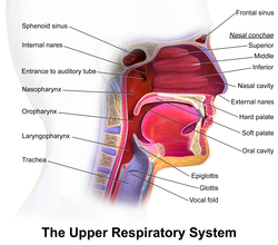
The soft palate is, in mammals, the soft tissue constituting the back of the roof of the mouth. The soft palate is part of the palate of the mouth; the other part is the hard palate. The soft palate is distinguished from the hard palate at the front of the mouth in that it does not contain bone.

<span class="mw-page-title-main">Palatine bone</span> Bone of the facial skeleton

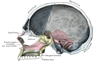
In anatomy, the palatine bones are two irregular bones of the facial skeleton in many animal species, located above the uvula in the throat. Together with the maxillae, they comprise the hard palate.

<span class="mw-page-title-main">Orthognathic surgery</span> Surgery of the jaw

Orthognathic surgery, also known as corrective jaw surgery or simply jaw surgery, is surgery designed to correct conditions of the jaw and lower face related to structure, growth, airway issues including sleep apnea, TMJ disorders, malocclusion problems primarily arising from skeletal disharmonies, and other orthodontic dental bite problems that cannot be treated easily with braces, as well as the broad range of facial imbalances, disharmonies, asymmetries, and malproportions where correction may be considered to improve facial aesthetics and self-esteem.

Van der Woude syndrome (VDWS) is a genetic disorder characterized by the combination of lower lip pits, cleft lip with or without cleft palate (CL/P), and cleft palate only (CPO). The frequency of orofacial clefts ranges from 1:1000 to 1:500 births worldwide, and there are more than 400 syndromes that involve CL/P. VWS is distinct from other clefting syndromes due to the combination of cleft lip and palate (CLP) and CPO within the same family. Other features frequently associated with VWS include hypodontia in 10-81% of cases, narrow arched palate, congenital heart disease, heart murmur and cerebral abnormalities, syndactyly of the hands, polythelia, ankyloglossia, and adhesions between the upper and lower gum pads.

<span class="mw-page-title-main">Alveolar process</span> Bulge on jaws holding teeth

The alveolar process or alveolar bone is the thickened ridge of bone that contains the tooth sockets on the jaw bones. The structures are covered by gums as part of the oral cavity.

<span class="mw-page-title-main">Incisive foramen</span> Opening of the incisive canals on the hard palate immediately behind the incisor teeth

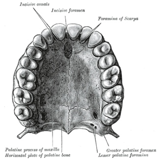
In the human mouth, the incisive foramen is the opening of the incisive canals on the hard palate immediately behind the incisor teeth. It gives passage to blood vessels and nerves. The incisive foramen is situated within the incisive fossa of the maxilla.

<span class="mw-page-title-main">Palatine process of maxilla</span> Thick, horizontal process of the maxilla

In human anatomy of the mouth, the palatine process of maxilla, is a thick, horizontal process of the maxilla. It forms the anterior three quarters of the hard palate, the horizontal plate of the palatine bone making up the rest.

<span class="mw-page-title-main">Pierre Robin sequence</span> Medical condition

Pierre Robin sequence is a congenital defect observed in humans which is characterized by facial abnormalities. The three main features are micrognathia, which causes glossoptosis, which in turn causes breathing problems due to obstruction of the upper airway. A wide, U-shaped cleft palate is commonly also present. PRS is not merely a syndrome, but rather it is a sequence—a series of specific developmental malformations which can be attributed to a single cause.

<span class="mw-page-title-main">Palatal obturator</span>

A palatal obturator is a prosthesis that totally occludes an opening such as an oronasal fistula. They are similar to dental retainers, but without the front wire. Palatal obturators are typically short-term prosthetics used to close defects of the hard/soft palate that may affect speech production or cause nasal regurgitation during feeding. Following surgery, there may remain a residual orinasal opening on the palate, alveolar ridge, or vestibule of the larynx. A palatal obturator may be used to compensate for hypernasality and to aid in speech therapy targeting correction of compensatory articulation caused by the cleft palate. In simpler terms, a palatal obturator covers any fistulas in the roof of the mouth that lead to the nasal cavity, providing the wearer with a plastic/acrylic, removable roof of the mouth, which aids in speech, eating, and proper air flow.

A mucogingival junction is an anatomical feature found on the intraoral mucosa. The mucosa of the cheeks and floor of the mouth are freely moveable and fragile, whereas the mucosa around the teeth and on the palate are firm and keratinized. Where the two tissue types meet is known as a mucogingival junction.

<span class="mw-page-title-main">Premaxilla</span> Cranial bones at the very tip of the upper jaw of many animals

The premaxilla is one of a pair of small cranial bones at the very tip of the upper jaw of many animals, usually, but not always, bearing teeth. In humans, they are fused with the maxilla. The "premaxilla" of therian mammals has been usually termed as the incisive bone. Other terms used for this structure include premaxillary bone or os premaxillare, intermaxillary bone or os intermaxillare, and Goethe's bone.

<span class="mw-page-title-main">VAX1</span> Protein-coding gene in the species Homo sapiens

Ventral anterior homeobox 1 is a protein that in humans is encoded by the VAX1 gene.

<span class="mw-page-title-main">Frontonasal dysplasia</span> Medical condition

Frontonasal dysplasia (FND) is a congenital malformation of the midface. For the diagnosis of FND, a patient should present at least two of the following characteristics: hypertelorism, a wide nasal root, vertical midline cleft of the nose and/or upper lip, cleft of the wings of the nose, malformed nasal tip, encephalocele or V-shaped hair pattern on the forehead. The cause of FND remains unknown. FND seems to be sporadic (random) and multiple environmental factors are suggested as possible causes for the syndrome. However, in some families multiple cases of FND were reported, which suggests a genetic cause of FND.

<span class="mw-page-title-main">Jaw abnormality</span> Medical condition

A jaw abnormality is a disorder in the formation, shape and/or size of the jaw. In general abnormalities arise within the jaw when there is a disturbance or fault in the fusion of the mandibular processes. The mandible in particular has the most differential typical growth anomalies than any other bone in the human skeleton. This is due to variants in the complex symmetrical growth pattern which formulates the mandible.

<span class="mw-page-title-main">Maxillary hypoplasia</span> Medical condition

Maxillary hypoplasia, or maxillary deficiency, is an underdevelopment of the bones of the upper jaw. It is associated with Crouzon syndrome, Angelman syndrome, as well as Fetal alcohol syndrome. It can also be associated with Cleft lip and cleft palate. Some people could develop it due to poor dental extractions.

A facial cleft is an opening or gap in the face, or a malformation of a part of the face. Facial clefts is a collective term for all sorts of clefts. All structures like bone, soft tissue, skin etc. can be affected. Facial clefts are extremely rare congenital anomalies. There are many variations of a type of clefting and classifications are needed to describe and classify all types of clefting. Facial clefts hardly ever occur isolated; most of the time there is an overlap of adjacent facial clefts.

<span class="mw-page-title-main">Human mouth</span> Part of human anatomy

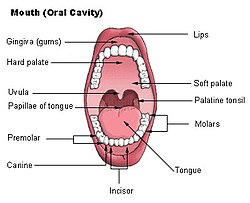
In human anatomy, the mouth is the first portion of the alimentary canal that receives food and produces saliva. The oral mucosa is the mucous membrane epithelium lining the inside of the mouth.

<span class="mw-page-title-main">Alveolar cleft grafting</span>

Alveolar cleft grafting is a surgical procedure, used to repair the defect in the upper jaw that is associated with cleft lip and palate, where the bone defect is filled with bone or bone substitute, and any holes between the mouth and the nose are closed.

References

  1. Patil, Manashvini S.; Patil, Sanjayagouda B.; Acharya, Ashith B. (2008-11-01). "Palatine Rugae and Their Significance in Clinical Dentistry: A Review of the Literature". The Journal of the American Dental Association. 139 (11): 1471–1478. doi:10.14219/jada.archive.2008.0072. ISSN   0002-8177. PMID   18978384.
  2. Reiter, Rudolf; Brosch, Sibylle; Luedeke, Manuel; Fischbein, Elena; Haase, S; Pickhard, A; Assum, G; Schwandt, A; Vogel, W; Maier, C; Hogel, J (April 2012). "Genetic and Environmental Risk Factors for Submucous Cleft Palate". European Journal of Oral Sciences. 120 (2): 97–103. doi:10.1111/j.1600-0722.2012.00948.x. PMID   22409215 . Retrieved 23 November 2019.
  3. 1 2 3 Ludwig, KU; Mangold, E; Herms, S; Nowak, S; Reutter, H; Paul, A; Becker, J; Herberz, R; AlChawa, T; Nasser, E; Bohmer, AC; Mattheisen, M; Alblas, MA; Barth, S; Kluck, N; Lauster, C; Braumann, B; Reich, RH; Hemprich, A; Pötzsch, S; Blaumeiser, B; Daratsianos, N; Kreusch, T; Murray, JC; Marazita, ML; Ruczinski, I; Scott, AF; Beaty, TH; Kramer, FJ; Wienker, TF; Steegers-Theunissen, RP; Rubini, M; Mossey, PA; Hoffman, P; Lange, C; Chichon, S; Propping, P; Knapp, M; Nothen, MM (Aug 5, 2012). "Genome-wide meta-analyses of nonsyndromic cleft lip with or without cleft palate identify six new risk loci". Nat Genet. 44 (9): 968–971. doi:10.1038/ng.2360. PMC   3598617 . PMID   22863734.
  4. Kohli, Sarvraj Singh; Kohli, Virinder Singh (Jan 2012). "A comprehensive review of the genetic basis of cleft lip and palate". Journal of Oral and Maxillofacial Pathology. 16 (1): 64–72. doi: 10.4103/0973-029X.92976 . PMC   3303526 . PMID   22438645.
  5. Houston, GD; Brown, FH (October 1993). "Differential diagnosis of the palatal mass". Compendium (Newtown, Pa.). 14 (10): 1222–4, 1226, 1228–31, quiz 1232. PMID   8118828.
  6. Manger, Karin; von Streitberg, Ulrich; Seitz, Gerhard; Kleyer, Arnd; Manger, Bernhard (2017-11-28). "Hard Palate Hyperpigmentation-a Rare Side Effect of Antimalarials". Arthritis & Rheumatology. 70 (1): 152. doi: 10.1002/art.40327 . ISSN   2326-5191. PMID   28941224.
  7. de Andrade, Bruno-Augusto-Benevenuto; Padron-Alvarado, Nelson-Alejandro; Muñoz-Campos, Edgar-Manuel; Morais, Thayná-Melo-de Lima; Martinez-Pedraza, Ricardo (2017-12-01). "Hyperpigmentation of hard palate induced by chloroquine therapy". Journal of Clinical and Experimental Dentistry. 9 (12): e1487–e1491. doi:10.4317/jced.54387. ISSN   1989-5488. PMC   5794129 . PMID   29410767.