Related Research Articles

The neck is the part of the body on many vertebrates that connects the head with the torso. The neck supports the weight of the head and protects the nerves that carry sensory and motor information from the brain down to the rest of the body. In addition, the neck is highly flexible and allows the head to turn and flex in all directions. The structures of the human neck are anatomically grouped into four compartments: vertebral, visceral and two vascular compartments. Within these compartments, the neck houses the cervical vertebrae and cervical part of the spinal cord, upper parts of the respiratory and digestive tracts, endocrine glands, nerves, arteries and veins. Muscles of the neck are described separately from the compartments. They bound the neck triangles.

The vitreous body is the clear gel that fills the space between the lens and the retina of the eyeball in humans and other vertebrates. It is often referred to as the vitreous humor or simply "the vitreous". Vitreous fluid or "liquid vitreous" is the liquid component of the vitreous gel, found after a vitreous detachment. It is not to be confused with the aqueous humor, the other fluid in the eye that is found between the cornea and lens.

The inguinal canal is a passage in the anterior abdominal wall on each side of the body which in males convey the spermatic cords and in females the round ligament of the uterus. The inguinal canals are larger and more prominent in males.

The inferior alveolar nerve(IAN) (also the inferior dental nerve) is a sensory branch of the mandibular nerve (CN V3) (which is itself the third branch of the trigeminal nerve (CN V)). The nerve provides sensory innervation to the lower/mandibular teeth and their corresponding gingiva as well as a small area of the face (via its mental nerve).
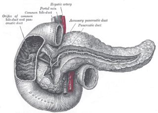
The pancreatic duct, or duct of Wirsung, is a duct joining the pancreas to the common bile duct. This supplies it with pancreatic juice from the exocrine pancreas, which aids in digestion.

The psoas major is a long fusiform muscle located in the lateral lumbar region between the vertebral column and the brim of the lesser pelvis. It joins the iliacus muscle to form the iliopsoas. In animals, this muscle is equivalent to the tenderloin.

The ampulla of Vater, hepatopancreatic ampulla or hepatopancreatic duct is the common duct that is usually formed by a union of the common bile duct and the pancreatic duct within the wall of the duodenum. This common duct usually features a dilation ("ampulla"). The common duct then opens medially into the descending part of the duodenum at the major duodenal papilla. The common duct usually measures 2-10mm in length.
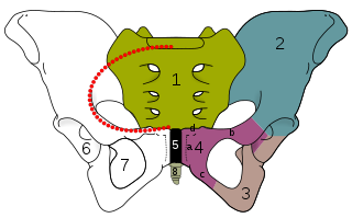
The obturator foramen is the large, bilaterally paired opening of the bony pelvis. It is formed by the pubis and ischium. It is mostly closed by the obturator membrane except for a small opening, the obturator canal, through which the obturator nerve and vessels pass.
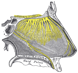
The nasopalatine nerve (also long sphenopalatine nerve) is a nerve of the head. It is a sensory branch of the maxillary nerve (CN V2) that passes through the pterygopalatine ganglion (without synapsing) and then through the sphenopalatine foramen to enter the nasal cavity, and finally out of the nasal cavity through the incisive canal and then the incisive fossa to enter the hard palate. It provides sensory innervation to the posteroinferior part of the nasal septum, and gingiva just posterior to the upper incisor teeth.
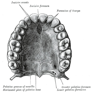
The incisive canals are two bony canals of the anterior hard palate connecting the nasal cavity and the oral cavity. An incisive canal courses through each maxilla. Below, the two incisive canals typically converge medially.
In the human mouth, the incisive foramen is the opening of the incisive canals on the hard palate immediately behind the incisor teeth. It gives passage to blood vessels and nerves. The incisive foramen is situated within the incisive fossa of the maxilla.

In anatomy, the division of the abdomen into regions can employ a nine-region scheme. The hypochondrium refers to the two hypochondriac regions in the upper third of the abdomen; the left hypochondrium and right hypochondrium. They are located on the lateral sides of the abdominal wall respectively, inferior to (below) the thoracic cage, being separated by the epigastrium.

The anterior tympanic artery is a branch of the maxillary artery. It passes through the petrotympanic fissure to entre the middle ear where it contributes to the formation of the circular anastomosis around the tympanic membrane. It provides arterial supply to part of the lining of the middle ear. It is accompanied by the chorda tympani nerve.

The supratrochlear artery is one of the terminal branches of the ophthalmic artery. It arises within the orbit. It exits the orbit alongside the supratrochlear nerve. It contributes arterial supply to the skin, muscles and pericranium of the forehead.

The greater palatine nerve is a branch of the pterygopalatine ganglion. This nerve is also referred to as the anterior palatine nerve, due to its location anterior to the lesser palatine nerve. It carries both general sensory fibres from the maxillary nerve, and parasympathetic fibers from the nerve of the pterygoid canal. It may be anaesthetised for procedures of the mouth and maxillary (upper) teeth.

A nerve fascicle, is a bundle of nerve fibers belonging to a nerve in the peripheral nervous system. A nerve fascicle is also called a fasciculus, as is a nerve tract in the central nervous system.

The clivus, or Blumenbach clivus, is a bony part of the cranium at the base of the skull. It is a shallow depression behind the dorsum sellae of the sphenoid bone. It slopes gradually to the anterior part of the basilar occipital bone at its junction with the sphenoid bone. It extends to the foramen magnum. It is related to the pons and the abducens nerve.
The caroticotympanic artery is a small, sometimes doubled artery which arises from the internal carotid artery. It leaves the carotid canal through a foramen to reach the tympanic cavity. It contributes arterial supply to the osseous part of the pharyngotympanic tube.
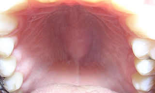
The palatine raphe is a raphe running across the palate, from the palatine uvula to the incisive papilla.
The anterior meniscofemoral ligament is a small fibrous band of the knee joint. It arises from the posterior horn of the lateral meniscus and passes superiorly and medially in front of the posterior cruciate ligament to attach to the lateral surface of medial condyle of the femur.
References
- 1 2 3 4 5 Standring S (2016). Standring S (ed.). Gray's Anatomy: The Anatomical Basis of Clinical Practice (41st ed.). Philadelphia. p. 510. ISBN 978-0-7020-5230-9. OCLC 920806541.
{{cite book}}: CS1 maint: location missing publisher (link) - 1 2 3 4 5 6 7 Solomon EG, Arunachalam KS (December 2012). "The incisive papilla: a significant landmark in prosthodontics". Journal of Indian Prosthodontic Society. 12 (4): 236–247. doi:10.1007/s13191-012-0169-y. PMC 3508097 . PMID 24293921.
- 1 2 Lake S, Iwanaga J, Kikuta S, Oskouian RJ, Loukas M, Tubbs RS (July 2018). "The Incisive Canal: A Comprehensive Review". Cureus. 10 (7): e3069. doi: 10.7759/cureus.3069 . PMC 6166911 . PMID 30280065.