Cementum

Last updated
Cementum
Basic anatomy tooth.jpg
Basic tooth anatomy, showing cementum
Details
Identifiers
Latin caementum
MeSH D003739
TA98 A05.1.03.057
A03.1.03.007
TA2 1612
FMA 55630
Anatomical terminology

Cementum [1] is a specialized calcified substance covering the root of a tooth. The cementum is the part of the periodontium that attaches the teeth to the alveolar bone by anchoring the periodontal ligament. [2] [3]

Contents

Structure

Cementum situated around a human molar Human tooth diagram-en.svg
Cementum situated around a human molar
The cementum is the surface layer of the tooth root, covering the dentin (which is labeled B). Rather than being a passive entity like paint on a wall, cementum is a dynamic entity within the periodontium. It is attached to the alveolar bone (C) by the fibers of the periodontal ligament and to the soft tissue of the gingiva by the gingival fibers (H). Periodontium.svg
The cementum is the surface layer of the tooth root, covering the dentin (which is labeled B). Rather than being a passive entity like paint on a wall, cementum is a dynamic entity within the periodontium. It is attached to the alveolar bone (C) by the fibers of the periodontal ligament and to the soft tissue of the gingiva by the gingival fibers (H).

The cells of cementum are the entrapped cementoblasts, the cementocytes. Each cementocyte lies in its lacuna, similar to the pattern noted in bone. These lacunae also have canaliculi or canals. Unlike those in bone, however, these canals in cementum do not contain nerves, nor do they radiate outward. Instead, the canals are oriented toward the periodontal ligament and contain cementocytic processes that exist to diffuse nutrients from the ligament because it is vascularized.

After the apposition of cementum in layers, the cementoblasts that do not become entrapped in cementum line up along the cemental surface along the length of the outer covering of the periodontal ligament. These cementoblasts can form subsequent layers of cementum if the tooth is injured.

Sharpey fibers are part of the principal collagenous fibers of the periodontal ligament embedded in the cementum and alveolar bone to attach the tooth to the alveolus. [3]

If cementum can be observed on teeth, it can imply that the roots are exposed, showing that the clinical crown (the exposed part of the tooth) is bigger than the anatomical crown (the surface of the tooth covered by enamel). [4] This is often due to gingival recession and may be an indication of periodontal disease. [5]

Cementoenamel junction

The cementum joins the enamel to form the cementoenamel junction (CEJ), which is referred to as the cervical line.

Three possible types of transitional interfaces may be present at the CEJ. The traditional view was that certain interfaces dominated in certain oral cavities. The CEJ may exhibit all of these interfaces in an individual's oral cavity, and there is even considerable variation when one tooth is traced circumferentially. [3]

Dentinocemental junction

When the cementoid reaches the full thickness needed, the cementoid surrounding the cementocytes becomes mineralized, or matured, and is then considered cementum. The dentinocemental junction (DCJ) is formed because of the apposition of cementum over the dentin. [6] [7] This interface is not as defined, either clinically or histologically, as that of the dentinoenamel junction (DEJ), given that cementum and dentin are of common embryological background, unlike that of enamel and dentin. [3]

The dentinocemental junction (DCJ) is a relatively smooth area in the permanent tooth, and attachment of cementum to the dentin is firm but not understood completely. [8]

Types

The different categories of cementum are based on the presence or absence of cementocytes, as well as whether the collagen fibres are extrinsic or intrinsic. It is thought that fibroblasts, and some cementoblasts, secrete extrinsic fibres, but only cementoblasts secrete intrinsic fibres. [9] The extrinsic fibres within acellular extrinsic fibre cementum, travel perpendicular to the surface of the root and allow the tooth to attach to the alveolar bone by the periodontal ligament (PDL), continuous with the cementodentinal junction (CDJ). [10] Acellular cementum only contains extrinsic collagen fibres. Whereas, cellular cementum is quite thick and contains both extrinsic and intrinsic collagen fibres. [10] The first cementum to be formed during tooth development is acellular extrinsic fibre cementum. [11] [12] The acellular layer of cementum is living tissue that does not incorporate cells into its structure and usually predominates on the coronal half of the root; cellular cementum occurs more frequently on the apical half. [8] In summary, the main types of cementum are as follows: acellular afibrillar cementum (AAC), acellular extrinsic fibres cementum (AEFC), cellular intrinsic fibres cementum (CIFC) and mixed stratified cementum (MSC) which displays both cellular and acellular cementum. [10] [13]

Cellular cementum contains cells and is the medium of attachment of collagen fibres to the alveolar bone. It is also responsible for minor repair of any resorption by continued deposition to keep the attachment apparatus intact. [14] Acellular cementum does not contain cells and has a main purpose of adaptive function. [15]

Composition

Cementum is slightly softer than dentin and consists of about 45% to 50% inorganic material (hydroxylapatite) by weight and 50% to 55% organic matter and water by weight. [16] The organic portion is composed primarily of collagen and proteoglycans. [17] Cementum is avascular, receiving its nutrition through its own imbedded cells from the surrounding vascular periodontal ligament. [3]

The cementum is light yellow and slightly lighter in color than dentin. It has the highest fluoride content of all mineralized tissue. Cementum also is permeable to a variety of materials. It is formed continuously throughout life because a new layer of cementum is deposited to keep the attachment intact as the superficial layer of cementum ages. Cementum on the root ends surrounds the apical foramen and may extend slightly onto the inner wall of the pulp canal.

Development

Cementum is secreted by cells called cementoblasts within the root of the tooth and is thickest at the root apex (the end of the root where the nerves and blood vessels enter the tooth). These cementoblasts develop from undifferentiated mesenchymal cells in the connective tissue of the dental follicle or sac. Cementoblasts produces cementum in a rhythmic manner on intervals indicating periods of activity and periods of rest, producing so-called incremental lines of Salter. Incremental lines of Salter are the only incremental line in the tooth that is hypercalcified, due to the fact that there is much less organic portion (collagen fibers) than inorganic portion (hydroxy appetite crystals) of cementum, so when the cementoblasts rest they leave a space for the inorganic portion. Unlike in enamel ameloblast (incremental lines of retzius) and dentin odontoblasts (incremental lines of von ebner) the inorganic portion is much more than the organic portion, so when ameloblast and odontoblasts rest they leave a space for the organic portion and become hypocalcified. [8]

Unlike ameloblasts and odontoblasts, which leave no cellular bodies in their secreted products, during the later steps within the stage of apposition, many of the cementoblasts become entrapped by the cementum they produce, becoming cementocytes. Thus again, cementum is more similar to alveolar bone, with its osteoblasts becoming entrapped osteocytes. [3]

Cementum is capable of repairing itself to a limited degree, but not regenerate. It is not resorbed under normal conditions. [11]

Clinical significance

DNA studies

A 2010 archeological study has found that cementum has five times the amount of mitochondrial DNA compared to dentin, which is commonly sampled. [21] Teeth are increasingly utilized as a source of nuclear DNA to aid identification of human remains. DNA extraction and the results of genetic analysis from the tissue are extremely variable and to some extent unpredictable. However, the quantity of DNA available in dentin is affected by age and dental disease, whereas that in cementum is not. [22]

See also

Related Research Articles

<span class="mw-page-title-main">Human tooth</span> Calcified whitish structure in humans mouths used to break down food

Human teeth function to mechanically break down items of food by cutting and crushing them in preparation for swallowing and digesting. As such, they are considered part of the human digestive system. Humans have four types of teeth: incisors, canines, premolars, and molars, which each have a specific function. The incisors cut the food, the canines tear the food and the molars and premolars crush the food. The roots of teeth are embedded in the maxilla or the mandible and are covered by gums. Teeth are made of multiple tissues of varying density and hardness.

<span class="mw-page-title-main">Tooth enamel</span> Major tissue that makes up part of the tooth in humans and many animals

Tooth enamel is one of the four major tissues that make up the tooth in humans and many animals, including some species of fish. It makes up the normally visible part of the tooth, covering the crown. The other major tissues are dentin, cementum, and dental pulp. It is a very hard, white to off-white, highly mineralised substance that acts as a barrier to protect the tooth but can become susceptible to degradation, especially by acids from food and drink. In rare circumstances enamel fails to form, leaving the underlying dentin exposed on the surface.

<span class="mw-page-title-main">Dentin</span> Calcified tissue of the body; one of the four major components of teeth

Dentin or dentine is a calcified tissue of the body and, along with enamel, cementum, and pulp, is one of the four major components of teeth. It is usually covered by enamel on the crown and cementum on the root and surrounds the entire pulp. By volume, 45% of dentin consists of the mineral hydroxyapatite, 33% is organic material, and 22% is water. Yellow in appearance, it greatly affects the color of a tooth due to the translucency of enamel. Dentin, which is less mineralized and less brittle than enamel, is necessary for the support of enamel. Dentin rates approximately 3 on the Mohs scale of mineral hardness. There are two main characteristics which distinguish dentin from enamel: firstly, dentin forms throughout life; secondly, dentin is sensitive and can become hypersensitive to changes in temperature due to the sensory function of odontoblasts, especially when enamel recedes and dentin channels become exposed.

<span class="mw-page-title-main">Periodontium</span>

The periodontium is the specialized tissues that both surround and support the teeth, maintaining them in the maxillary and mandibular bones. The word comes from the Greek terms περί peri-, meaning "around" and -odont, meaning "tooth". Literally taken, it means that which is "around the tooth". Periodontics is the dental specialty that relates specifically to the care and maintenance of these tissues. It provides the support necessary to maintain teeth in function. It consists of four principal components, namely:

<span class="mw-page-title-main">Cementoenamel junction</span>

Cementoenamel junction (CEJ) is defined as the area of the union of cementum and enamel at the cervical region of the tooth. It is a slightly visible anatomical border identified on a tooth. It is the location where the enamel, which covers the anatomical crown of a tooth, and the cementum, which covers the anatomical root of a tooth, meet. Informally it is known as the neck of the tooth. The border created by these two dental tissues has much significance as it is usually the location where the gingiva attaches to a healthy tooth by fibers called the gingival fibers.

<span class="mw-page-title-main">Periodontal fiber</span> Group of specialized connective tissue fibers

The periodontal ligament, commonly abbreviated as the PDL, are a group of specialized connective tissue fibers that essentially attach a tooth to the alveolar bone within which they sit. It inserts into root cementum on one side and onto alveolar bone on the other.

<span class="mw-page-title-main">Enamel organ</span> Aggregate of cells involved in tooth development

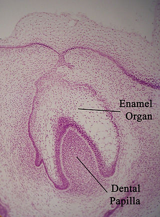
The enamel organ, also known as the dental organ, is a cellular aggregation seen in a developing tooth and it lies above the dental papilla. The enamel organ which is differentiated from the primitive oral epithelium lining the stomodeum. The enamel organ is responsible for the formation of enamel, initiation of dentine formation, establishment of the shape of a tooth's crown, and establishment of the dentoenamel junction.

<span class="mw-page-title-main">Human tooth development</span> Process by which teeth form

Tooth development or odontogenesis is the complex process by which teeth form from embryonic cells, grow, and erupt into the mouth. For human teeth to have a healthy oral environment, all parts of the tooth must develop during appropriate stages of fetal development. Primary (baby) teeth start to form between the sixth and eighth week of prenatal development, and permanent teeth begin to form in the twentieth week. If teeth do not start to develop at or near these times, they will not develop at all, resulting in hypodontia or anodontia.

<span class="mw-page-title-main">Dental papilla</span>

In embryology and prenatal development, the dental papilla is a condensation of ectomesenchymal cells called odontoblasts, seen in histologic sections of a developing tooth. It lies below a cellular aggregation known as the enamel organ. The dental papilla appears after 8–10 weeks intra uteral life. The dental papilla gives rise to the dentin and pulp of a tooth.

<span class="mw-page-title-main">Odontoblast</span> Type of cell that produces dentin in teeth

In vertebrates, an odontoblast is a cell of neural crest origin that is part of the outer surface of the dental pulp, and whose biological function is dentinogenesis, which is the formation of dentin, the substance beneath the tooth enamel on the crown and the cementum on the root.

Periodontology or periodontics is the specialty of dentistry that studies supporting structures of teeth, as well as diseases and conditions that affect them. The supporting tissues are known as the periodontium, which includes the gingiva (gums), alveolar bone, cementum, and the periodontal ligament. A periodontist is a dentist that specializes in the prevention, diagnosis and treatment of periodontal disease and in the placement of dental implants.

<span class="mw-page-title-main">Dental follicle</span> Anatomical entity

The dental follicle, also known as dental sac, is made up of mesenchymal cells and fibres surrounding the enamel organ and dental papilla of a developing tooth. It is a vascular fibrous sac containing the developing tooth and its odontogenic organ. The dental follicle (DF) differentiates into the periodontal ligament. In addition, it may be the precursor of other cells of the periodontium, including osteoblasts, cementoblasts and fibroblasts. They develop into the alveolar bone, the cementum with Sharpey's fibers and the periodontal ligament fibers respectively. Similar to dental papilla, the dental follicle provides nutrition to the enamel organ and dental papilla and also have an extremely rich blood supply.

<span class="mw-page-title-main">Epithelial root sheath</span>

The Hertwig epithelial root sheath (HERS) or epithelial root sheath is a proliferation of epithelial cells located at the cervical loop of the enamel organ in a developing tooth. Hertwig epithelial root sheath initiates the formation of dentin in the root of a tooth by causing the differentiation of odontoblasts from the dental papilla. The root sheath eventually disintegrates with the periodontal ligament, but residual pieces that do not completely disappear are seen as epithelial cell rests of Malassez (ERM). These rests can become cystic, presenting future periodontal infections.

Cementogenesis is the formation of cementum, one of the three mineralized substances of a tooth. Cementum covers the roots of teeth and serves to anchor gingival and periodontal fibers of the periodontal ligament by the fibers to the alveolar bone.

<span class="mw-page-title-main">Gingival sulcus</span> Space between tooth and gums

The gingival sulcus is an area of potential space between a tooth and the surrounding gingival tissue and is lined by sulcular epithelium. The depth of the sulcus is bounded by two entities: apically by the gingival fibers of the connective tissue attachment and coronally by the free gingival margin. A healthy sulcular depth is three millimeters or less, which is readily self-cleansable with a properly used toothbrush or the supplemental use of other oral hygiene aids.

Dental anatomy is a field of anatomy dedicated to the study of human tooth structures. The development, appearance, and classification of teeth fall within its purview. Tooth formation begins before birth, and the teeth's eventual morphology is dictated during this time. Dental anatomy is also a taxonomical science: it is concerned with the naming of teeth and the structures of which they are made, this information serving a practical purpose in dental treatment.

The junctional epithelium (JE) is that epithelium which lies at, and in health also defines, the base of the gingival sulcus. The probing depth of the gingival sulcus is measured by a calibrated periodontal probe. In a healthy-case scenario, the probe is gently inserted, slides by the sulcular epithelium (SE), and is stopped by the epithelial attachment (EA). However, the probing depth of the gingival sulcus may be considerably different from the true histological gingival sulcus depth.

In dentistry, enamel matrix derivative (EMD) is an extract of porcine fetal tooth material used to biomimetically stimulate the soft and hard tissues surrounding teeth to regrow following tissue destruction.

Hard tissue, refers to "normal" calcified tissue, is the tissue which is mineralized and has a firm intercellular matrix. The hard tissues of humans are bone, tooth enamel, dentin, and cementum. The term is in contrast to soft tissue.

A cementicle is a small, spherical or ovoid calcified mass embedded within or attached to the cementum layer on the root surface of a tooth, or lying free within the periodontal ligament. They tend to occur in elderly individuals.

References

  1. "Cementum". DentalFind. 2007-01-01.
  2. Bath-Balogh M, Fehrenbach MJ (10 December 2010). Illustrated Dental Embryology, Histology, and Anatomy. Elsevier Health Sciences. p. 170. ISBN   978-1-4377-2934-4.
  3. 1 2 3 4 5 6 7 8 9 Illustrated Dental Embryology, Histology, and Anatomy, Bath-Balogh and Fehrenbach, Elsevier, 2011, page 170.
  4. Hand AR (2015). Fundamentals of Oral Histology and Physiology. John Wiley & Sons Incorporated. ISBN   978-1-118-34291-6.
  5. "Gum Disease Symptoms | Perio.org". www.perio.org. Retrieved 2019-12-11.
  6. Jang, Andrew T.; Lin, Jeremy D.; Choi, Ryan M.; Choi, Erin M.; Seto, Melanie L.; Ryder, Mark I.; Gansky, Stuart A.; Curtis, Donald A.; Ho, Sunita P. (2014). "Adaptive properties of human cementum and cementum dentin junction with age". Journal of the Mechanical Behavior of Biomedical Materials. 39: 184–196. doi:10.1016/j.jmbbm.2014.07.015. ISSN   1751-6161. PMC   4265544 . PMID   25133753.
  7. "nixon dental" . Retrieved 2023-06-23.
  8. 1 2 3 Bosshardt DD, Selvig KA (February 1997). "Dental cementum: the dynamic tissue covering of the root" (PDF). Periodontology 2000. 13: 41–75. doi:10.1111/j.1600-0757.1997.tb00095.x. PMID   9567923.
  9. Yamamoto T, Hasegawa T, Yamamoto T, Hongo H, Amizuka N (August 2016). "Histology of human cementum: Its structure, function, and development". The Japanese Dental Science Review. 52 (3): 63–74. doi:10.1016/j.jdsr.2016.04.002. PMC   5390338 . PMID   28408958.
  10. 1 2 3 Colard T, Falgayrac G, Bertrand B, Naji S, Devos O, Balsack C, et al. (2016). "Correction: New Insights on the Composition and the Structure of the Acellular Extrinsic Fiber Cementum by Raman Analysis". PLOS ONE. 12 (3): e0174080. doi: 10.1371/journal.pone.0174080 . PMC   5345870 . PMID   28282444.
  11. 1 2 Nanci A (2013). Ten Cate's Oral Histology (8 ed.). Elsevier. pp. 205–207. ISBN   978-0-323-07846-7.
  12. AAP 2010 In-Service Examination, question A-9
  13. Gonçalves, Patricia Furtado; Sallum, Enilson Antonio; Sallum, Antonio Wilson; Casati, Márcio Zaffalon; Toledo, Sérgio de; Junior, Francisco Humberto Nociti (2005-12-31). "Dental cementum reviewed: development, structure, composition, regeneration and potential functions".{{cite journal}}: Cite journal requires |journal= (help)
  14. Ghosh S (2019). Cementum. Germany: SPS. ISBN   978-6202317184.
  15. Ehtisham M (2016). Cementum. LAP Lambert Academic Publishing Mai. ISBN   978-3659879753.
  16. American Academy of Periodontology 2010 In-Service Exam, question A-38
  17. Kumar G (15 Jul 2011). Orban's Oral Histology & Embryology (13th ed.). Elsevier India. p. 152. ISBN   9788131228197 . Retrieved 1 December 2014.
  18. Listgarten MA. "Histology of the Periodontium - Cementum". University of Pennsylvania and Temple University. Archived from the original on 9 July 2013.
  19. Chiego Jr. DJ (14 April 2014). Essentials of Oral Histology and Embryology: A Clinical Approach. Elsevier Health Sciences. p. 133. ISBN   978-0-323-29100-2.
  20. Bath-Balogh M, Fehrenbach MJ (10 December 2010). Illustrated Dental Embryology, Histology, and Anatomy. Elsevier Health Sciences. p. 174. ISBN   978-1-4377-2934-4.
  21. Adler CJ, Haak W, Donlon D, Cooper A (May 2011). "Genographic Consortium. Survival and recovery of DNA from ancient teeth and bones". Journal of Archaeological Science. 38 (5): 956–64. doi:10.1016/j.jas.2010.11.010.
  22. Higgins D, Kaidonis J, Townsend G, Hughes T, Austin JJ (October 2013). "Targeted sampling of cementum for recovery of nuclear DNA from human teeth and the impact of common decontamination measures". Investigative Genetics. 4 (1): 18. doi:10.1080/00450618.2011.583278. PMC   3853689 . PMID   24139166. S2CID   85182819.

Further reading

  1. Listgarten MA. "Histology of the Periodontium - Cementum". University of Pennsylvania and Temple University. Archived from the original on 9 July 2013.
  2. Cherian G (May 2011). "Harvesting cementum from root surface: A new paradigm in the study of cementum and the cemento-dentinal junction". Journal of Advanced Oral Research. 2 (2): 17–20. doi:10.1177/2229411220110203. S2CID   5051968.