Candida tropicalis

Last updated

Candida tropicalis
C tropicalis YC466.png
Scientific classification OOjs UI icon edit-ltr.svg
Domain: Eukaryota
Kingdom: Fungi
Division: Ascomycota
Class: Saccharomycetes
Order: Saccharomycetales
Family: Saccharomycetaceae
Genus: Candida
Species:
C. tropicalis
Binomial name
Candida tropicalis
Synonyms

Candida tropicalis is a species of yeast in the genus Candida . It is a common pathogen in neutropenic hosts, in whom it may spread through the bloodstream to peripheral organs. [1] For invasive disease, treatments include amphotericin B, echinocandins, or extended-spectrum triazole antifungals. [2]

Contents

History and taxonomy

In the history of fungi, the name of genus Candida, derived from the family Debaryomycetaceae, [3] comes from the Latin term "candidus" which has the meaning of “glowing white” and also refers to as smooth and glistering. [4] Genus Candida referred to any asexual yeast without any of the following characteristics: production of acetic acid, pigments of colours red, pink or orange, arthroconidia, unipolar or bipolar budding, enteroblastic-basipetal budding, blastoconidia formation on sympodulae, buds formation on stalks, triangular cells, needle-shaped terminal conidia, and having the ability to grow on inositol as a sole carbon source. [3] Although there are 200 species identified in this genus, [3] the taxonomy remains undefined and incomplete due to several reasons such as changing the words for some representations, the finding of new species and the reclassification of identified old species. [4] This genus no longer includes species that test positive to diazonium blue B (DBB). [3] The defunct genera Oidium and Monilia were used to represent the genus Candida. [4]

In the genus Candida, there are other species that are similar to Candida tropicalis. Candida albicans is taxonomically close to C. tropicalis sharing many pathogenic traits [5] whereas C. maltosa and C. sake are physiologically similar to C. tropicalis but they can be differentiated by the growth at 35 °C (only C. sake showing negative) and assimilation of soluble starch (only C. tropicalis showing positive starch assimilation). [6]

Identification

C. tropicalis is easily identified using phenotypic and molecular methods. [7] The identification of species in the genus Candida relies on morphological and physiological features. Species in the genus are vegetative cells which reproduce asexually by budding, and the structure, shape, septation, color and arrangement of buds is useful for identification. [8] The production and appearance of pseudohyphae and blastoconidia may also be useful for identification. [8] Physiological profiles relating to carbon and nitrogen utilization are of value in determining species, as are the presence certain distinctive biochemical features. [6] Increasingly, molecular genetic methods such as DNA sequencing are used as primary tools for the accurate determination of species identifications in this group. [6]

Growth and morphology

Colonies of C. tropicalis on a Petri dish. Candida tropicalis.jpg
Colonies of C. tropicalis on a Petri dish.

C. tropicalis is a vegetative cell [9] with the shape from round to oval ranging from approximately 2 – 10 micrometers. [3] A mould exhibits dimorphism [8] forming a single-celled yeast or so-called blastoconidia which reproduces by simple budding. [8] Conidia are the asexual units that are produced by budding of the tips or walls of the hyphae. [8] Conidia are types of simple and unicellular bodies that could also take the form of multicellular cells with different shapes, sizes, and colors. [8] Microconidia refer to small and unicellular conidia whereas macroconidia refer to large and multicellular conidia. [8]

There are different media on which C. tropicalis can grow effectively. A common medium used is the Sabouraud’s agar which contains peptone and sugar. This is enough for identifying the species but with a disadvantage of promoting mycelial growth and suppressing conidia formation. [8] Another commonly used medium is the cornmeal agar which is useful in inducing formation of conidia. [8] Potato-glucose, potato-carrot, tomato juice, lima bean and others are also types of media used for growth. [8] The optimal temperature for growth is between 25–35 °C (77–95 °F) [4] and growth is enhanced if sugar or fat is added in the medium. Colonies of C. tropicalis are white, smooth and butyrous with a fringed border. [6] [9]

Physiology

C. tropicalis reproduces asexually by the production of blastoconidia through budding. As blastoconidia increase in number they may elongate in shape producing structures called the pseudohyphae. [3] [4] Under specific conditions of reduced oxygen level in host tissues, submerged colonies in agar medium, or in the presence of 5-10% CO2, true, septate hyphae may form. [9] [3]

Physiological characteristics of C. tropicalis

Growth

Fermentation

Urea hydrolysis

This test is to detect yeast’s ability to produce enzyme urease. Once there's a compatible substrate, urease will split urea to produce ammonia increasing the pH. C. tropicalis is found to be urease negative. [3]

Diazonium Blue B Test

This test uses the azo dye, diazonium blue B which differentially stains cells from species affiliated with the Division, Basidiomycota. For C. tropicalis, this test is negative. [6]

Parasexuality

C. tropicalis diploid cells of opposite mating type can mate to form tetraploid cells. [11] These cells may then undergo chromosome loss during long-term propagation in rich medium resulting in the eventual regeneration of diploid cells. [12] Such diploid cells are again mating competent thus completing a parasexual cycle. Opaque C. tropicalis cells can also form an architecturally complex sexual biofilm. [13]

Habitat and ecology

Candida species are very pervasive yeasts that are distributed worldwide geographically. They are more likely to be found in tropical climate where temperature and humidity will enhance the adaptability of C. tropicalis. [5] They can be found in food such as sauerkraut, molasses, miso, fruit, baker’s yeast and some fruits. [9] They are commonly found on plants and in the digestive system of mammals, especially in the gastrointestinal tract, and in the mucocutaneous membranes of humans. [3] C. tropicalis is considered as an osmotolerant yeast; microorganisms that are able to survive in high salt concentration and able to develop fungal persistence in saline environments. [7]

Storage and transportation

The selection of medium for sample growth is very important accounting for the pros and cons of each type of growth medium. Once decided on the medium, need to add 8 ug of fluconazole per mL to limit bacterial growth and contamination. [3] When the medium is ready, it is optional to add supplement to help and optimize specimen growth. After the growth, features examination including shape, size, bud arrangement, cell wall thickness, temperature of growth, pseudohyphae presence, arthroconidia presence, and capsule presence are all important to take into account. [3] If the species are grown using any type of the medium mentioned, the transportation of specimens for testing should be completed in less than two hours. [3] If there is any delay, the samples should be stored at 37 °C with the exception of contaminated specimens that need be stored in 4 °C. [3]

Pathogenicity

In tropical countries, C. tropicalis is one of the most common colonizer and pathogen causing human disease, [5] especially found on human skin, in the gastrointestinal tract and also in female genitourinary tract. [4] It can be transmitted between health-care workers and patients, [5] especially in environments such as hospitals. [5] C. tropicalis can survive for up to 24 hours therefore be cross-transmitted to a second hand with a probability of 69% and to a third hand with 38% probability. [5] It is the cause responsible for approximately half of the beyond-surface candida infections. [5] C. tropicalis is the second most virulent Candida species [7] that can significantly affect by spreading through the weakened immune system host and can occupy the gastrointestinal tract within 30 minutes of inoculation, all this resulting in increased mortality. [5] [14] [10] Impact of candidiasis, infections cause by C. tropicalis, have increased globally. [14] C. tropicalis is virulent due to its ability to produce biofilm, secrete lytic enzymes, adhere to epithelial and endothelial cells, and undergo transition of bud to hyphae. [15] [10] [7]

Biofilms are complex structures that are formed from the grouping of microorganisms on a local surface, either biotic or abiotic, [15] dependent on the ability of cellular adhesion to substrates. [7] For C. tropicalis to fully enter and cause infection in the host, it needs some helpers. First, once it is attached onto the host tissues, extracellular enzymes called the proteases will be produced to facilitate the penetration of the pathogen and allow it to interfere with the host defense system. [5] proteases will hydrolyze peptide bonds; secreted aspartic proteases (SAP) support C. tropicalis to be attached and penetrate deep into the tissues to affect the organs. [5] phospholipases will hydrolyze phospholipid; assist to break the epithelial cell membrane structure allowing the hyphal tip to enter into the cytoplasm. [5] Many conditions that contribute to C. tropicalis survival and colonization are: a) increase the use of antifungal regimen, b) increased number of immunocompromised patients, c) long-term use of catheters, and d) use of broad-spectrum antibiotics. [14] Although different tests are able to use for identification of species, each of the tests will have different limitations such as sensitivity, specificity, cost and equipment availability. [3]

Human diseases

Chromogenic agar can help in indicating Candida tropicalis infection versus some similar fungi. (CHROMAgar shown) CHROMAgar with N glabratus, P kudriavzevii, Candida albicans and Candida tropicalis, annotated.jpg
Chromogenic agar can help in indicating Candida tropicalis infection versus some similar fungi. (CHROMAgar shown)

Types of disease caused by C. tropicalis will vary depending on the location where the species colonizes. With an infection in the mucous membrane, subject will experience oropharyngeal candidiasis, angular cheilitis, balanoposthitis, oral thrush and vulvovaginal candidiasis. [14] Although provided with oral cavity defenses such as epithelial cells, saliva, salivary immunoglobin (IgA), lysozyme, lactoferrin, histidine-rich polypeptide and lactoperoxidase to suppress C. tropicalis’ overgrowth, [14] C. tropicalis is reported to secrete additional products that can preferably target onto T-cell deficient host. [14] C. tropicalis is a normal flora which is found on the skin and nails on approximately 10% of the patients. [5] Superficial and localized mucosal infections are mostly reported with a higher risk factor when combined with other diseases found in a patient. [5] Patients with C. tropicalis infections are also seen with denture, HIV infection or irradiation for malignancies. [5] 38% of AIDS patients with recurrent disease are more likely to get infected by C. tropicalis, getting oral thrush and oropharyngeal candidiasis. [5] Only filamentous growth of C. tropicalis have the ability to invade and colonize orally in the epithelium, [10] commonly seen in cancer patients and higher risk for someone who subsequently develops disseminated invasive disease. [5] Candiduria is referred to as urinary tract infections caused by C. tropicalis which are often presented as nosocomial infections. [5] Although up to 2% of patients are asymptomatic, those with diabetes mellitus and with leukemia are more likely to be infected. [5]

If an infection involves interdigital candidiasis, paronychia and diaper rash, subject is likely to have cutaneous candidiasis. [14] Otherwise, if an infection involves body fluid and internal organs damages, subject will experience pulmonary candidiasis, invasive and disseminated candidiasis, gastrointestinal candidiasis and candidemia. [14] C. tropicalis colonization is favoured in the gastrointestinal tract; [14] a common risk factor for individuals that are susceptible for invasive candidiasis development. [5] Candidemia is a worldwide bloodstream disease mainly affecting peripheral organs in humans. [5] Usually, candidemia caused by C. tropicalis are associated with cancer patients that have either leukemia or neutropenia. [15] According to the data obtained from 2010, frequency of candidemia is 12-25% in the US, 4.5-9% in Europe, 20-24% in Brazil and 20-60% in South Asia. [5] C. tropicalis can cause nosocomial fungal bloodstream infections along with C. glabrata and C. parapsilosis. [5] Mortality rate of invasive and disseminated infections caused by C. tropicalis is high, ranging from 40% to 70%. [5] Risk factors that contribute to the high rate are leukemia, anti-neoplastic chemotherapy, previous neutropenia, central venous catheters, long stay on intensive care and total parenteral nutrition. [5] Although children infections are not as common seen as in adults, leukemia, secondary neutropenia and bone marrow transplantation [14] are factors favouring C. tropicalis infections. [5] Another infection seen commonly in patients who have leukemia and secondary neutropenia, is chronic disseminated candidiasis (CDC) is another type of disseminated candida infection that mainly develops in the liver, spleen and kidney. [5]

Treatment and prevention

The most important and most essential step to prevent contact with the fungi species is by washing the hands. [4] There are several types of therapy for the different level of infections caused by C. tropicalis. Normally, antifungal agents are used to treat these infections. [4] Amphotericin B deoxycholate is the most common treatment antifungal agent used to treat Candida infections. [4] Topical antifungal agents are commonly taken in 3 forms: oral suspension, ointment and powder. [4] Oral suspension is mainly used to treat thrush whereas ointment is directly applied onto the infected section. [4] Nystatin is a type of antifungal agent used because it is not absorbed by the gastrointestinal tract. [4] These types of agents will function to lower candida species’ phospholipases activities. [14] Flucytosine (5FC) is another type of therapy treatment including 3 agents used; caspofungin, micafungin and anidulafungin. [5] Usage of caspofungin will efficiently target against oropharyngeal and oesophageal candidiasis and invasive candidiasis. [5] Micafungin, compared to amphotericin B, it is more efficient. [5] Anidulafungin results are similar to Caspofungin and Micafungin. [5] echinocandin are a type of non-competitive inhibitors of cell wall 1,3-b-D-glucan synthase complex mainly used to treat fungal infections. [5] [4] [7] Azoles are agents that can deplete ergosterol, the main component of the fungus cell wall membrane, [7] in order to inhibit fungal growth. [4] fluconazole is water-soluble, ready to be taken orally. [5] C. tropicalis can rapidly develop resistance towards fluconazole therefore it's not recommended to retreat fluconazole-treated patients with recurrent candidiasis. [14] Other azoles that are highly active against C. tropicalis are itraconazole, voriconazole, posaconazole, ravuconazole and isavuconazole. [5] Voriconazole is a new generation from fluconazole with a higher potential of broad spectrum activity. [4] All of the mentioned treatments and drug therapies can also be applied onto neonates and premature newborns taking into account the amount of recommended dose. Although there are several ways to treat the different types of C. tropicalis’ infections, the best way to improve treatments results is to improve host immune system. [14]

Related Research Articles

<span class="mw-page-title-main">Candidiasis</span> Fungal infection due to any type of Candida

Candidiasis is a fungal infection due to any species of the genus Candida. When it affects the mouth, in some countries it is commonly called thrush. Signs and symptoms include white patches on the tongue or other areas of the mouth and throat. Other symptoms may include soreness and problems swallowing. When it affects the vagina, it may be referred to as a yeast infection or thrush. Signs and symptoms include genital itching, burning, and sometimes a white "cottage cheese-like" discharge from the vagina. Yeast infections of the penis are less common and typically present with an itchy rash. Very rarely, yeast infections may become invasive, spreading to other parts of the body. This may result in fevers along with other symptoms depending on the parts involved.

<i>Candida albicans</i> Species of fungus

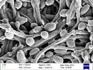
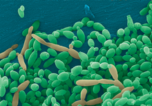
Candida albicans is an opportunistic pathogenic yeast that is a common member of the human gut flora. It can also survive outside the human body. It is detected in the gastrointestinal tract and mouth in 40–60% of healthy adults. It is usually a commensal organism, but it can become pathogenic in immunocompromised individuals under a variety of conditions. It is one of the few species of the genus Candida that cause the human infection candidiasis, which results from an overgrowth of the fungus. Candidiasis is, for example, often observed in HIV-infected patients. C. albicans is the most common fungal species isolated from biofilms either formed on (permanent) implanted medical devices or on human tissue. C. albicans, C. tropicalis, C. parapsilosis, and C. glabrata are together responsible for 50–90% of all cases of candidiasis in humans. A mortality rate of 40% has been reported for patients with systemic candidiasis due to C. albicans. By one estimate, invasive candidiasis contracted in a hospital causes 2,800 to 11,200 deaths yearly in the US. Nevertheless, these numbers may not truly reflect the true extent of damage this organism causes, given new studies indicating that C. albicans can cross the blood–brain barrier in mice.

<i>Candida</i> (fungus) Genus of ascomycete fungi

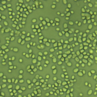
Candida is a genus of yeasts. It is the most common cause of fungal infections worldwide and the largest genus of medically important yeast.

<span class="mw-page-title-main">Oral candidiasis</span> Fungal infection

Oral candidiasis, also known as oral thrush among other names, is candidiasis that occurs in the mouth. That is, oral candidiasis is a mycosis of Candida species on the mucous membranes of the mouth.

<span class="mw-page-title-main">Fungemia</span> Internal, blood-borne infection by fungi, including yeasts.

Fungemia is the presence of fungi or yeasts in the blood. The most common type, also known as candidemia, candedemia, or systemic candidiasis, is caused by Candida species; candidemia is also among the most common bloodstream infections of any kind. Infections by other fungi, including Saccharomyces, Aspergillus and Cryptococcus, are also called fungemia. It is most commonly seen in immunosuppressed or immunocompromised patients with severe neutropenia, cancer patients, or in patients with intravenous catheters. It has been suggested that otherwise immunocompetent patients taking infliximab may also be at a higher risk for fungemia.

<span class="mw-page-title-main">Anidulafungin</span> Antifungal medication

Anidulafungin (INN) is a semisynthetic echinocandin used as an antifungal drug. It was previously known as LY303366. It may also have application in treating invasive Aspergillus infection when used in combination with voriconazole. It is a member of the class of antifungal drugs known as the echinocandins; its mechanism of action is by inhibition of (1→3)-β-D-glucan synthase, an enzyme important to the synthesis of the fungal cell wall.

Candida parapsilosis is a fungal species of yeast that has become a significant cause of sepsis and of wound and tissue infections in immunocompromised people. Unlike Candida albicans and Candida tropicalis, C. parapsilosis is not an obligate human pathogen, having been isolated from nonhuman sources such as domestic animals, insects and soil. C. parapsilosis is also a normal human commensal and it is one of the fungi most frequently isolated from human hands. There are several risk factors that can contribute to C. parapsilosis colonization. Immunocompromised individuals and surgical patients, particularly those undergoing surgery of the gastrointestinal tract, are at high risk for infection with C. parapsilosis. There is currently no consensus on the treatment of invasive candidiasis caused by C. parapsilosis, although the therapeutic approach typically includes the removal of foreign bodies such as implanted prostheses and the administration of systemic antifungal therapy. Amphotericin B and Fluconazole are often used in the treatment of C. parapsilosis infection.

Pichia kudriavzevii is a budding yeast involved in chocolate production. P. kudriavzevii is an emerging fungal nosocomial pathogen primarily found in the immunocompromised and those with hematological malignancies. It has natural resistance to fluconazole, a standard antifungal agent. It is most often found in patients who have had prior fluconazole exposure, sparking debate and conflicting evidence as to whether fluconazole should be used prophylactically. Mortality due to P. kudriavzevii fungemia is much higher than the more common C. albicans. Other Candida species that also fit this profile are C. parapsilosis, C. glabrata, C. tropicalis, C. guillermondii and C. rugosa.

<i>Nakaseomyces glabratus</i> Species of fungus

Nakaseomyces glabratus is a species of haploid yeast of the genus Nakaseomyces, previously known as Candida glabrata. Despite the fact that no sexual life cycle has been documented for this species, N. glabratus strains of both mating types are commonly found. C. glabrata is generally a commensal of human mucosal tissues, but in today's era of wider human immunodeficiency from various causes, N. glabratus is often the second or third most common cause of candidiasis as an opportunistic pathogen. Infections caused by N. glabratus can affect the urogenital tract or even cause systemic infections by entrance of the fungal cells in the bloodstream (Candidemia), especially prevalent in immunocompromised patients.

<span class="mw-page-title-main">Esophageal candidiasis</span> Medical condition

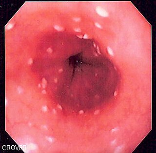
Esophageal candidiasis is an opportunistic infection of the esophagus by Candida albicans. The disease usually occurs in patients in immunocompromised states, including post-chemotherapy and in AIDS. However, it can also occur in patients with no predisposing risk factors, and is more likely to be asymptomatic in those patients. It is also known as candidal esophagitis or monilial esophagitis.

<span class="mw-page-title-main">Echinocandin</span> Group of chemical compounds

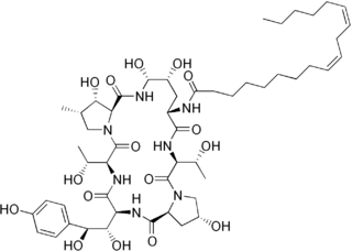
Echinocandins are a class of antifungal drugs that inhibit the synthesis of β-glucan in the fungal cell wall via noncompetitive inhibition of the enzyme 1,3-β glucan synthase. The class has been termed the "penicillin of antifungals," along with the related papulacandins, as their mechanism of action resembles that of penicillin in bacteria. β-glucans are carbohydrate polymers that are cross-linked with other fungal cell wall components, the fungal equivalent to bacterial peptidoglycan. Caspofungin, micafungin, and anidulafungin are semisynthetic echinocandin derivatives with limited clinical use due to their solubility, antifungal spectrum, and pharmacokinetic properties.

Pathogenic fungi are fungi that cause disease in humans or other organisms. Although fungi are eukaryotic, many pathogenic fungi are microorganisms. Approximately 300 fungi are known to be pathogenic to humans; their study is called "medical mycology". Fungal infections are estimated to kill more people than either tuberculosis or malaria—about two million people per year.

<span class="mw-page-title-main">Blastoconidium</span>

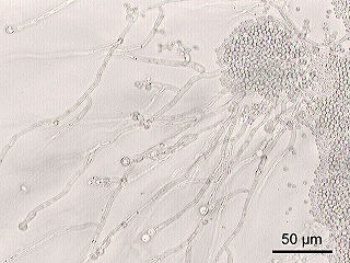
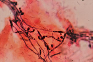
A blastoconidium is an asexual holoblastic conidia formed through the blowing out or budding process of a yeast cell, which is a type of asexual reproduction that results in a bud arising from a parent cell. The production of a blastoconidium can occur along a true hyphae, pseudohyphae, or a singular yeast cell. The word "conidia" comes from the Greek word konis and eidos, konis meaning dust and eidos meaning like. The term "bud" comes from the Greek word blastos, which means bud. Yeasts such as Candida albicans and Cryptococcus neoformans produce these budded cells known as blastoconidia.

<span class="mw-page-title-main">Vaginal yeast infection</span> Medical condition

Vaginal yeast infection, also known as candidal vulvovaginitis and vaginal thrush, is excessive growth of yeast in the vagina that results in irritation. The most common symptom is vaginal itching, which may be severe. Other symptoms include burning with urination, a thick, white vaginal discharge that typically does not smell bad, pain during sex, and redness around the vagina. Symptoms often worsen just before a woman's period.

Mycobiota are a group of all the fungi present in a particular geographic region or habitat type. An analogous term for Mycobiota is funga.

Fungistatics are anti-fungal agents that inhibit the growth of fungus. The term fungistatic may be used as both a noun and an adjective. Fungistatics have applications in agriculture, the food industry, the paint industry, and medicine.

<i>Candida auris</i> Species of fungus

Candida auris is a species of fungus that grows as yeast. It is one of the few species of the genus Candida which cause candidiasis in humans. Often, candidiasis is acquired in hospitals by patients with weakened immune systems. C. auris can cause invasive candidiasis (fungemia) in which the bloodstream, the central nervous system, and internal organs are infected. It has attracted widespread attention because of its multiple drug resistance. Treatment is also complicated because it is easily misidentified as other Candida species.

Invasive candidiasis is an infection (candidiasis) that can be caused by various species of Candida yeast. Unlike Candida infections of the mouth and throat or vagina, invasive candidiasis is a serious, progressive, and potentially fatal infection that can affect the blood (fungemia), heart, brain, eyes, bones, and other parts of the body.

Carol Kumamoto is an American microbiologist who is Professor of Molecular Biology & Microbiology at Tufts University. She investigates the filamentous growth of Candida albicans, a fungal pathogen that causes several diseases. She is also interested in how C. albicans interacts with its host during colonisation and invasive diseases. She is a Fellow of the American Association for the Advancement of Science and the American Academy of Microbiology.

<span class="mw-page-title-main">Oteseconazole</span> Chemical compound

Oteseconazole, a novel orally bioavailable and selective inhibitor of fungal cytochrome P450 enzyme 51 (CYP51), has shown promising efficacy in the treatment of recurrent vulvovaginal candidiasis (RVVC) in patients.

References

  1. Mastromarino, Paola; Vitali, Beatrice; Mosca, Luciana (2013). "Bacterial vaginosis: a review on clinical trials with probiotics" (PDF). New Microbiologica. 36 (3): 229–238. PMID   23912864.
  2. Chai LY, Denning DW, Warn P (2010). "Candida tropicalis in human disease". Crit. Rev. Microbiol. 36 (4): 282–98. doi:10.3109/1040841X.2010.489506. PMID   20883082. S2CID   36366177.
  3. 1 2 3 4 5 6 7 8 9 10 11 12 13 14 Murray, P. R.; Baron, E. J.; Jorgensen, J. H.; Pfaller, M. A.; Yolken, R. H. (2003). Manual of Clinical Microbiology. Washington, US: ASM Press. ISBN   978-1555812553.
  4. 1 2 3 4 5 6 7 8 9 10 11 12 13 14 15 Wilson, C. B.; Nizet, V.; Maldonado, Y.; Remington, J. S.; Klein, J. O. (2015). Remington and Klein's Infectious diseases of the Fetus and Newborn Infant. Philadelphia, US: Saunders. ISBN   9780323241472.
  5. 1 2 3 4 5 6 7 8 9 10 11 12 13 14 15 16 17 18 19 20 21 22 23 24 25 26 27 28 29 30 31 32 33 Chai, L. Y.; Denning, D. W.; Warn, P. (2010). "Candida tropicalis in human disease". Crit. Rev. Microbiol. 26 (4): 282–98. doi:10.3109/1040841X.2010.489506. PMID   20883082. S2CID   36366177.
  6. 1 2 3 4 5 6 7 8 9 10 11 12 Kurtzman, C. P.; Fell, J. W. (1998). The yeasts: A Taxonomic Study. New York, US: Elsevier Science. ISBN   978-0444813121.
  7. 1 2 3 4 5 6 7 8 9 Zuza-Alves, D. L.; Silva-Rocha, W. P.; Chaves, G. M. (2017). "An update on Candida tropicalis based on basic and clinical approaches". Frontiers in Microbiology. 8: 1927. doi: 10.3389/fmicb.2017.01927 . PMC   5645804 . PMID   29081766.
  8. 1 2 3 4 5 6 7 8 9 10 Rippon, J. W. (1988). Medical Mycology: The Pathogenic Fungi and The Pathogenic Actinomycetes. Philadelphia, US: Saunders. ISBN   978-0721624440.
  9. 1 2 3 4 Barnett, J. A.; Payne, R. W.; Yarrow, D. (1991). Yeasts: Characteristics and identification. Cambridge, UK: Cambridge University Press. ISBN   978-0521350563.
  10. 1 2 3 4 5 Silva, S.; Negri, M.; Henriques, M.; Oliveira, R.; Williams, D. W.; Azeredo, J. (2011). "Candida glabrata, Candida parapsilosis and Candida tropicalis: biology, epidemiology, pathogenicity and antifungal resistance". FEMS Microbiology Reviews. 36 (2): 288–305. doi: 10.1111/j.1574-6976.2011.00278.x . PMID   21569057.
  11. Porman AM, Alby K, Hirakawa MP, Bennett RJ. Discovery of a phenotypic switch regulating sexual mating in the opportunistic fungal pathogen Candida tropicalis. Proceedings of the National Academy of Sciences USA 2011 Dec 27;108(52):21158-63. doi: 10.1073/pnas.1112076109.
  12. Seervai RN, Jones SK Jr, Hirakawa MP, Porman AM, Bennett RJ. Parasexuality and ploidy change in Candida tropicalis. Eukaryot Cell. 2013 Dec;12(12):1629-40. doi: 10.1128/EC.00128-13. PMID 24123269
  13. Jones SK Jr, Hirakawa MP, Bennett RJ. Sexual biofilm formation in Candida tropicalis opaque cells. Mol Microbiol. 2014 Apr;92(2):383-98. doi: 10.1111/mmi.12565. PMID 24612417
  14. 1 2 3 4 5 6 7 8 9 10 11 12 13 Kothavade, R. J.; Kura, M. M.; Valand, A. G.; Panthaki, M. H. (2010). "Candida tropicalis: its prevalence, pathogenicity and increasing resistance to fluconazole". Journal of Medical Microbiology. 59 (8): 873–80. doi: 10.1099/jmm.0.013227-0 . PMID   20413622.
  15. 1 2 3 Sardi, J. C. O.; Scorzoni, L.; Bernardi, T.; Fusco-Almeida, A. M.; Mendes Giannini, M. J. S. (2013). "Candida species: current epidemiology, pathogenicity, biofilm formation, natural antifungal products and new therapeutic options". Journal of Medical Microbiology. 62 (Pt 1): 10–24. doi: 10.1099/jmm.0.045054-0 . PMID   23180477.