Streptococcus agalactiae

Last updated

Streptococcus agalactiae
Streptococcus agalactiae.tif
Scientific classification OOjs UI icon edit-ltr.svg
Domain: Bacteria
Phylum: Bacillota
Class: Bacilli
Order: Lactobacillales
Family: Streptococcaceae
Genus: Streptococcus
Species:
S. agalactiae
Binomial name
Streptococcus agalactiae
Lehmann and Neumann, 1896

Streptococcus agalactiae (also known as group B streptococcus or GBS) is a gram-positive coccus (round bacterium) with a tendency to form chains (as reflected by the genus name Streptococcus ). It is a beta-hemolytic, catalase-negative, and facultative anaerobe. [1] [2]

Contents

S. agalactiae is the most common human pathogen of streptococci belonging to group B of the Rebecca Lancefield classification of streptococci. GBS are surrounded by a bacterial capsule composed of polysaccharides (exopolysacharide). The species is subclassified into ten serotypes (Ia, Ib, II–IX) depending on the immunologic reactivity of their polysaccharide capsule. [1] [3] [4]

The plural term group B streptococci (referring to the serotypes) and the singular term group B streptococcus (referring to the single species) are both commonly used synonymously with S. agalactiae even though S. halichoeri and S. pseudoporcinus are also group B Streptococci. These species test positive as group B, but are not frequently carried by humans, and only rarely cause disease. [5]

In general, GBS is a harmless commensal bacterium being part of the human microbiota colonizing the gastrointestinal and genitourinary tract of up to 30% of healthy human adults (asymptomatic carriers). Nevertheless, GBS can cause severe invasive infections especially in newborns, the elderly, and people with compromised immune systems. [6]

b-hemolytic colonies of Streptococcus agalactiae, blood agar 18h at 36degC Streptococcus agalactiae on blood agar.JPG
β-hemolytic colonies of Streptococcus agalactiae, blood agar 18h at 36°C

S. agalactiae is also a common veterinary pathogen, because it can cause bovine mastitis (inflammation of the udder) in dairy cows. The species name agalactiae meaning "of no milk", alludes to this. [7]

Streptococcus agalactiae on granada agar, anaerobic incubation Streptococcus agalactiae on Granada medium.jpg
Streptococcus agalactiae on granada agar, anaerobic incubation
Granadaene Granadaene.svg
Granadaene
Positive CAMP test indicated by the formation of an arrowhead where Streptococcus agalactiae meets the Staphylococcus aureus (white middle streak) CAMP test.JPG
Positive CAMP test indicated by the formation of an arrowhead where Streptococcus agalactiae meets the Staphylococcus aureus (white middle streak)
Gram stain of Streptococcus agalactiae. Gram stain of Streptococcus agalactiae.jpg
Gram stain of Streptococcus agalactiae.

Laboratory identification

GBS grows readily on blood agar plates as colonies surrounded by a narrow zone of β-hemolysis. GBS is characterized by the presence in the cell wall of the antigen group B of Lancefield classification (Lancefield grouping) that can be detected directly in intact bacteria using latex agglutination tests. [8] [9] The CAMP test is also another important test for identification of GBS. The CAMP factor produced by GBS acts synergistically with the staphylococcal β-hemolysin inducing enhanced hemolysis of sheep or bovine erythrocytes. [8] GBS is also able to hydrolyze hippurate and this test can also be used to identify presumptively GBS. [8] Hemolytic GBS strains produce an orange-brick-red non-isoprenoid polyene (ornithine rhamnolipid) pigment (granadaene) [10] when cultivated on granada medium that allows its straightforward identification. [11] GBS can also be identified using MALDI-TOF (Matrix Assisted Laser Desorption/Ionization-Time of Flight) instruments. [12] GBS colonies can additionally be identified tentatively after their appearance in chromogenic agar media, nevertheless GBS-like colonies that develop in chromogenic media should be confirmed as GBS using additional reliable tests (e.g.latex agglutination or the CAMP test) to avoid potential mis-identification. [9] [13] [14] A summary of the laboratory techniques for GBS identification is depicted in Ref 7. [9]

GBS colonization

GBS is a normal component of the intestinal and vaginal microbiota in some women, GBS is an asymptomatic (presenting no symptoms) colonizer of the gastrointestinal tract and vagina in up to 30% of otherwise healthy adults, including pregnant women. [3] [15] GBS colonization may be permanent, intermittent or temporary. In different studies, GBS vaginal colonization rate ranges from 0% to 36%, most studies reporting colonization rates in sexually active women over 20%. [16] It has been estimated that maternal GBS colonization worldwide is 18%, with regional variation from 11% to 35%. [17] These variations in the reported prevalence of asymptomatic GBS colonization could be related to the detection methods used, and differences in populations sampled. [15] [18]

Virulence

As other virulent bacteria, GBS harbors an important number of virulence factors (virulence factors are molecules produced by bacteria that boosts their capacity to infect and damage human tissues), the most important being the capsular polysaccharide (rich in sialic acid) [3] [19] and a pore-forming toxin, β-hemolysin. [19] [20] [21] Today it is considered that GBS pigment and hemolysin are identical or closely related molecules. [22] [23] [24] [25]

Sialic acid is a notable virulence factor in S. agalactiae despite being found normally in humans and many other animals. By expressing an unusually high amount of sialic acid on the bacterial cell surface, S. agalactiae can subvert the innate immune system, convincing leukocytes that the bacteria are human cells. [26] [27]

GBS infection in newborns

GBS colonization of the vagina usually does not cause problems in healthy women, nevertheless during pregnancy it can sometimes cause serious illness for the mother and the newborn. GBS is the leading cause of bacterial neonatal infection in the baby during gestation and after delivery with significant mortality rates in premature infants. GBS infections in the mother can cause chorioamnionitis (a severe infection of the placental tissues) infrequently, postpartum infections (after birth) and it had been related with prematurity and fetal death. [28] GBS urinary tract infections (UTI) may also induce labor and cause premature delivery. [3] In the western world, GBS (in the absence of effective prevention measures) is the major cause of several bacterial infections of the newborn neonatal infection sepsis, pneumonia, and meningitis, which can lead to death or long-term sequelae. [3]

GBS neonatal infection typically originates in the lower reproductive tract of infected mothers. GBS infections in newborns are separated into two clinical syndromes, early-onset disease (EOD) and late-onset disease (LOD). [29] EOD manifests from 0 to 7 living days in the newborn, most of the cases of EOD being apparent within 24h of birth. [3] [30] [31] The most common clinical syndromes of EOD are sepsis without apparent focus, pneumonia, and less frequently meningitis. EOD is acquired vertically (vertical transmission), through exposure of the fetus or the baby to GBS from the vagina of a colonized woman, either intrautero or during birth after rupture of membranes. Infants can be infected during passage through the birth canal, nevertheless newborns that acquire GBS through this route can become only colonized, and these colonized infants habitually do not develop EOD. Roughly 50% of newborns to GBS colonized mothers are also GBS colonized and (without prevention measures) 1–2% of these newborns will develop EOD. [32] In the past, the incidence of EOD ranged from 0.7 to 3.7 per thousand live births in the US [3] and from 0.2 to 3.25 per thousand in Europe. [18] In 2008, after widespread use of antenatal screening and intrapartum antibiotic prophylaxis (IAP), the CDC reported an incidence of 0.28 cases of EOD per thousand live births in the US. [33]

Multistate surveillance 2006-2015 shows a decline in EOD from 0.37 to 0.23 per 1000 live births in the US but LOD remains steady at 0.31 per 1000 live births. [34]

It has been indicated that where there was a policy of providing IAP for GBS colonized mothers the overall risk of EOGBS is 0.3%. [35] Since 2006 to 2015 the incidence of GBS EOD decreased from 0.37 to 0.23 per thousand live births in the US. [36]

Though maternal GBS colonization is the key determinant for EOD, other factors also increase the risk. These factors include onset of labor before 37 weeks of gestation (premature birth), prolonged rupture of membranes (≥18h before delivery), intra-partum fever (>38 °C, >100.4 °F), amniotic infections (chorioamnionitis), young maternal age, and low levels of GBS anticapsular polysaccharide antibodies in the mother. [3] [30] [31] Nevertheless, most babies who develop EOD are born to GBS colonized mothers without any additional risk factor. [30] [31] A previous sibling with EOD is also an important risk factor for development of the infection in subsequent deliveries, probably reflecting a lack of GBS polysaccharides protective antibodies in the mother. Heavy GBS vaginal colonization is also associated with a higher risk for EOD. [30] Overall, the case–fatality rates from EOD have declined, from 50% observed in studies from the 1970s to 2 to 10% in recent years, mainly as a consequence of improvements in therapy and management. Fatal neonatal infections by GBS are more frequent among premature infants. [3] [30] [31] [37]

GBS LOD affects infants from 7 days to 3 months of age and is more likely to cause bacteremia or meningitis. LOD can be acquired from the mother or from environmental sources. Hearing loss and mental impairment can be a long-term sequela of GBS meningitis. [3] [38] [39] In contrast with EOD, the incidence of LOD has remained unchanged at 0.26 per 1000 live births in the US. [40] S. agalactiae neonatal meningitis does not present with the hallmark sign of adult meningitis, a stiff neck; rather, it presents with nonspecific symptoms, such as fever, vomiting and irritability, and can consequently lead to a late diagnosis. [2]

Prevention of neonatal infection

The only reliable way to prevent EOD currently is intrapartum antibiotic prophylaxis (IAP), that is to say administration of antibiotics during delivery. It has been proved that intravenous penicillin or ampicillin administered for at least 4 hours before delivery to GBS colonized women is very effective at preventing vertical transmission of GBS from mother to baby and EOD. Intravenous penicillin remains the agent of choice for IAP, with intravenous ampicillin as an acceptable alternative. [3] [30] [31] For penicillin allergic women, the laboratory requisitions for ordering antepartum GBS screening cultures should indicate clearly the presence of penicillin allergy. [31] Cefazolin, clindamycin, and vancomycin are used to prevent EOD in infants born to penicillin-allergic mothers. [30] [31] Intravenous vancomycin is recommended for IAP in women colonized with a clindamycin-resistant Group B Streptococcus strain and a severe penicillin allergy. [29] [31]

There are two ways to identify female candidates to receive intrapartum antibiotic prophylaxis: a risk-based approach or a culture-based screening approach. The culture-based screening approach identifies candidates to receive IAP using lower vaginal and rectal cultures obtained between 36 and 37 weeks' gestation [30] [31] (32–34 weeks of gestation for women with twins [41] ) and IAP is administered to all GBS colonized women. The risk-based strategy identifies candidates to receive IAP by the aforementioned risk factors known to increase the probability of EOD without considering if the mother is or is not a GBS carrier. [3] [42]

IAP is also recommended for women with intrapartum risk factors if their GBS carrier status is not known at the time of delivery, for women with GBS bacteriuria during their pregnancy, and for women who have had an infant with EOD previously.[ citation needed ]

The risk-based approach for IAP is in general less effective than the culture-based approach because in most of the cases EOD develops among newborns, which are born to mothers without risk factors. [18]

In 2010, the Centers for Disease Control and Prevention (CDC), in collaboration with several professional groups, issued its revised GBS prevention guidelines. [30]

In 2018, the task of revising and updating the GBS prophylaxis guidelines was transferred from the CDC [43] to ACOG (American College of Obstetricians and Gynecologists), the American Academy of Pediatrics and to the American Society for Microbiology. [14] [29] [31]

The ACOG committee issued an update document on Prevention of Group B Streptococcal Early-Onset Disease in Newborns in 2019. [31] This document does not introduce important changes from the CDC guidelines. The key measures necessary for preventing neonatal GBS early onset disease continue to be universal prenatal screening by culture of GBS from swabs collected from the lower vagina and rectum, correct collection and microbiological processing of the samples, and proper implementation of intrapartum antibiotic prophylaxis. The ACOG now recommends performing universal GBS screening between 36 and 37 weeks of gestation. This new recommendation provides a five-week window [44] for valid culture results that includes births that occur up to a gestational age of at least 41 weeks.

The culture-based screening approach is followed in most developed countries [45] such as the United States, [29] [30] [31] France, [46] Spain, [47] Belgium, [48] Canada, Argentina, [49] and Australia. The risk-based strategy is followed in the United Kingdom, [41] [50] and the Netherlands. [18] [51]

Screening for GBS colonization

Though the GBS colonization status of women can change during pregnancy, cultures to detect GBS carried out ≤5 weeks before delivery predict quite accurately the GBS carrier status at delivery.[ citation needed ]

In contrast, if the prenatal culture is performed more than five weeks before delivery it is unreliable for predicting accurately the GBS carrier status at delivery. [30] [31] [44] [52] [53]

The clinical specimens recommended for culture of GBS at 36–37 weeks' gestation, this recommendation provides a 5-week window for valid culture results that includes births that occur up to a gestational age of at least 41 weeks [31] (32–34 weeks of gestation for women with twins [41] ) are swabs collected the lower vagina (near the introitus) and then from the rectum (through the anal sphincter) without use of a speculum. [30] [31] Vaginal-rectal samples should be collected using a flocked swab preferably, since flocked swabs releases samples and microorganisms more effectively than fiber swabs. [14]

Following the recommendations of the Centers for Disease Control and Prevention of United States (CDC) these swabs should be placed into a non-nutritive transport medium and later inoculated into a selective enrichment broth, Todd Hewitt broth with selective antibiotics (enrichment culture). [30] After incubation the enrichment broth is subcultured to blood agar plates and GBS like colonies are identified by the CAMP test or using latex agglutination with GBS antisera. After incubation the enrichment broth can also be subcultured to granada medium agar [11] where GBS grows as pink-red colonies or to chromogenic agars, where GBS grows as colored colonies. [9] [30]

GBS-like colonies that develop in chromogenic media should be confirmed as GBS using additional reliable tests to avoid mis-identification. [9]

Nucleic acid amplification tests (NAAT) such as polymerase chain reaction (PCR) and DNA hybridization probes have been developed for identifying GBS directly from recto-vaginal samples, but they still cannot replace antenatal culture for the most accurate detection of GBS carriers.

[30] [31] [41] Intrapartum NAAT without enrichment has a high false negative rate and the use of intrapartum NAAT without enrichment to rule out the need for IAP. [14]

Red colonies of S.agalactiae in granada agar. Vagino-rectal culture 18h incubation 36degC anaerobiosis S agalactiae vagino-rectal culture GRANADA.png
Red colonies of S.agalactiae in granada agar. Vagino-rectal culture 18h incubation 36°C anaerobiosis
Streptococcus agalactiae colonies in chromogenic medium (ChromID CPS chromogenic agar) Streptococcus agalactiae (Group B Streptococcus) on ChromID CPS chromogenic agar - Detail.jpg
Streptococcus agalactiae colonies in chromogenic medium (ChromID CPS chromogenic agar)

Vaccination

Though IAP for EOD prevention is associated with a large decline in the incidence of the disease, there is, however, no effective strategy for preventing late-onset neonatal GBS disease. [54]

Vaccination is considered an ideal solution to prevent not only EOD and LOD but also GBS infections in adults at risk. [55] Nevertheless, though research and clinical trials for the development of an effective vaccine to prevent GBS infections are underway, no vaccine was available in 2020. [56] [57] [58] The capsular polysaccharide of GBS is not only an important GBS virulence factor but it is also an excellent candidate for the development of an effective vaccine. [18] [59] [60] [61] Protein-based vaccines are also in development. [56] [62] [58]

GBS infection in adults

GBS is also an important infectious agent able to cause invasive infections in adults. Serious life-threatening invasive GBS infections are increasingly recognized in the elderly and individuals compromised by underlying diseases such as diabetes, cirrhosis and cancer. [63] GBS infections in adults include urinary tract infection, skin and soft-tissue infection (skin and skin structure infection) bacteremia, osteomyelitis, meningitis and endocarditis. [6] GBS infection in adults can be serious and related with high mortality. In general penicillin is the antibiotic of choice for treatment of GBS infection. [64] [65] Gentamicin (for synergy with penicillin G or ampicillin) can also be used in patients with life-threatening invasive GBS. [64]

Non-human infections

Streptococcus agalactiae was historically studied as a disease of cattle that harmed milk production, leading to its name "agalactiae" which means "absence of milk". Strains of bovine and human bacteria are generally interchangeable, with evidence of transmission from animals to humans and vice versa. [66]

Cattle

GBS is a major cause of mastitis (an infection of the udder) in dairy cattle and an important source of economic loss for the industry. GBS in cows can either produce an acute febrile disease or a subacute more chronic condition. Both lead to diminishing milk production (hence its name: agalactiae meaning "of no milk"). [67] Outbreaks in herds are common, so this is of major importance for the dairy industry, and programs to reduce the impact of S. agalactiae disease have been enforced in many countries over the last 40 years. [7] [66]

Other animals

GBS also causes severe epidemics in farmed fish, causing sepsis and external and internal hemorrhages, having been reported from wild and captive fish involved in epizootics in many countries. [68] [69] Vaccination is an effective method to prevent pathogenic diseases in aquaculture and different kinds vaccines to prevent GBS infections have been developed recently. [70]

GBS has also been found in many other animals, such as camels, dogs, cats, crocodiles, seals, elephants and dolphins. [71] [72]

Related Research Articles

<i>Streptococcus</i> Genus of bacteria

Streptococcus is a genus of gram-positive coccus or spherical bacteria that belongs to the family Streptococcaceae, within the order Lactobacillales, in the phylum Bacillota. Cell division in streptococci occurs along a single axis, so as they grow, they tend to form pairs or chains that may appear bent or twisted. This differs from staphylococci, which divide along multiple axes, thereby generating irregular, grape-like clusters of cells. Most streptococci are oxidase-negative and catalase-negative, and many are facultative anaerobes.

<span class="mw-page-title-main">Group A streptococcal infection</span> Medical condition

Group A streptococcal infections are a number of infections with Streptococcus pyogenes, a group A streptococcus (GAS). S. pyogenes is a species of beta-hemolytic Gram-positive bacteria that is responsible for a wide range of infections that are mostly common and fairly mild. If the bacteria enter the bloodstream an infection can become severe and life-threatening, and is called an invasive GAS (iGAS).

<i>Streptococcus pyogenes</i> Species of bacterium

Streptococcus pyogenes is a species of Gram-positive, aerotolerant bacteria in the genus Streptococcus. These bacteria are extracellular, and made up of non-motile and non-sporing cocci that tend to link in chains. They are clinically important for humans, as they are an infrequent, but usually pathogenic, part of the skin microbiota that can cause Group A streptococcal infection. S. pyogenes is the predominant species harboring the Lancefield group A antigen, and is often called group A Streptococcus (GAS). However, both Streptococcus dysgalactiae and the Streptococcus anginosus group can possess group A antigen as well. Group A streptococci, when grown on blood agar, typically produce small (2–3 mm) zones of beta-hemolysis, a complete destruction of red blood cells. The name group A (beta-hemolytic) Streptococcus is thus also used.

<span class="mw-page-title-main">Scarlet fever</span> Infectious disease caused by Streptococcus pyogenes

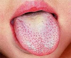
Scarlet fever, also known as scarlatina, is an infectious disease caused by Streptococcus pyogenes, a Group A streptococcus (GAS). It most commonly affects children between five and 15 years of age. The signs and symptoms include a sore throat, fever, headache, swollen lymph nodes, and a characteristic rash. The face is flushed and the rash is red and blanching. It typically feels like sandpaper and the tongue may be red and bumpy. The rash occurs as a result of capillary damage by exotoxins produced by S.pyogenes. On darker-pigmented skin the rash may be hard to discern.

<span class="mw-page-title-main">Streptococcal pharyngitis</span> Medical condition

Streptococcal pharyngitis, also known as streptococcal sore throat, is pharyngitis caused by Streptococcus pyogenes, a gram-positive, group A streptococcus. Common symptoms include fever, sore throat, red tonsils, and enlarged lymph nodes in the front of the neck. A headache and nausea or vomiting may also occur. Some develop a sandpaper-like rash which is known as scarlet fever. Symptoms typically begin one to three days after exposure and last seven to ten days.

<i>Streptococcus pneumoniae</i> Species of bacterium

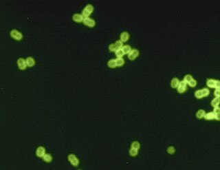
Streptococcus pneumoniae, or pneumococcus, is a Gram-positive, spherical bacteria, alpha-hemolytic member of the genus Streptococcus. They are usually found in pairs (diplococci) and do not form spores and are non motile. As a significant human pathogenic bacterium S. pneumoniae was recognized as a major cause of pneumonia in the late 19th century, and is the subject of many humoral immunity studies.

<span class="mw-page-title-main">Group B streptococcal infection</span> Medical condition

Group B streptococcal infection, also known as Group B streptococcal disease or just Group B strep infection, is the infectious disease caused by the bacterium Streptococcus agalactiae, which is is the most common human pathogen belonging to the group B of the Lancefield classification of streptococci—hence the group B stretococcal (GBS) infection nomenclature. Infection with GBS can cause serious illness and sometimes death, especially in newborns, the elderly, and people with compromised immune systems. The most severe form of group B streptococcal disease is neonatal meningitis in infants, which is frequently lethal and can cause permanent neuro-cognitive impairment.

Screening culture is a type a medical test that is done to find an infection. Screening cultures are often performed to find infections that do not have signs and symptoms.

<span class="mw-page-title-main">Chorioamnionitis</span> Medical condition

Chorioamnionitis, also known as intra-amniotic infection (IAI), is inflammation of the fetal membranes, usually due to bacterial infection. In 2015, a National Institute of Child Health and Human Development Workshop expert panel recommended use of the term "triple I" to address the heterogeneity of this disorder. The term triple I refers to intrauterine infection or inflammation or both and is defined by strict diagnostic criteria, but this terminology has not been commonly adopted although the criteria are used.

<span class="mw-page-title-main">Clobetasone</span> Chemical compound

Clobetasone (INN) is a corticosteroid used in dermatology, for treating such skin inflammation as seen in eczema, psoriasis and other forms of dermatitis, and ophthalmology. Topical clobetasone butyrate has shown minimal suppression of the hypothalamic–pituitary–adrenal axis.

<span class="mw-page-title-main">Pathogenic bacteria</span> Disease-causing bacteria

Pathogenic bacteria are bacteria that can cause disease. This article focuses on the bacteria that are pathogenic to humans. Most species of bacteria are harmless and are often beneficial but others can cause infectious diseases. The number of these pathogenic species in humans is estimated to be fewer than a hundred. By contrast, several thousand species are part of the gut flora present in the digestive tract.

<i>Streptococcus canis</i> Species of bacterium

Streptococcus canis is a group G beta-hemolytic species of Streptococcus. It was first isolated in dogs, giving the bacterium its name. These bacteria are characteristically different from Streptococcus dysgalactiae, which is a human-specific group G species that has a different phenotypic chemical composition. S. canis is important to the skin and mucosal health of cats and dogs, but under certain circumstances, these bacteria can cause opportunistic infections. These infections were known to afflict dogs and cats prior to the formal description of the species in Devriese et al., 1986. However, additional studies revealed cases of infection in other mammal species, including cattle and even humans. Instances of mortality from S. canis in humans are very low with only a few reported cases, while actual instances of infection may be underreported due to mischaracterizations of the bacteria as S. dysgalactiae. This species, in general, is highly susceptible to antibiotics, and plans to develop a vaccine to prevent human infections are currently being considered.

<i>Streptococcus dysgalactiae</i> Species of bacterium

Streptococcus dysgalactiae is a gram positive, beta-haemolytic, coccal bacterium belonging to the family Streptococcaceae. It is capable of infecting both humans and animals, but is most frequently encountered as a commensal of the alimentary tract, genital tract, or less commonly, as a part of the skin flora. The clinical manifestations in human disease range from superficial skin-infections and tonsillitis, to severe necrotising fasciitis and bacteraemia. The incidence of invasive disease has been reported to be rising. Several different animal species are susceptible to infection by S. dysgalactiae, but bovine mastitis and infectious arthritis in lambs have been most frequently reported.

Neonatal sepsis is a type of neonatal infection and specifically refers to the presence in a newborn baby of a bacterial blood stream infection (BSI) in the setting of fever. Older textbooks may refer to neonatal sepsis as "sepsis neonatorum". Criteria with regards to hemodynamic compromise or respiratory failure are not useful clinically because these symptoms often do not arise in neonates until death is imminent and unpreventable. Neonatal sepsis is divided into two categories: early-onset sepsis (EOS) and late-onset sepsis (LOS). EOS refers to sepsis presenting in the first 7 days of life, with LOS referring to presentation of sepsis after 7 days. Neonatal sepsis is the single most common cause of neonatal death in hospital as well as community in developing country.

Perianal cellulitis, also known as perianitis or perianal streptococcal dermatitis, is a bacterial infection affecting the lower layers of the skin (cellulitis) around the anus. It presents as bright redness in the skin and can be accompanied by pain, difficulty defecating, itching, and bleeding. This disease is considered a complicated skin and soft tissue infection (cSSTI) because of the involvement of the deeper soft tissues.

<span class="mw-page-title-main">Neonatal meningitis</span> Medical condition

Neonatal meningitis is a serious medical condition in infants that is rapidly fatal if untreated. Meningitis is an inflammation of the meninges, the protective membranes of the central nervous system, is more common in the neonatal period than any other time in life, and is an important cause of morbidity and mortality globally. Mortality is roughly half in developing countries and ranges from 8%-12.5% in developed countries.

<span class="mw-page-title-main">Enzybiotics</span> Experimental antibacterial therapy

Enzybiotics are an experimental antibacterial therapy. The term is derived from a combination of the words “enzyme” and “antibiotics.” Enzymes have been extensively utilized for their antibacterial and antimicrobial properties. Proteolytic enzymes called endolysins have demonstrated particular effectiveness in combating a range of bacteria and are the basis for enzybiotic research. Endolysins are derived from bacteriophages and are highly efficient at lysing bacterial cells. Enzybiotics are being researched largely to address the issue of antibiotic resistance, which has allowed for the proliferation of drug-resistant pathogens posing great risk to animal and human health across the globe.

<span class="mw-page-title-main">Granada medium</span>

Granada medium is a selective and differential culture medium designed to selectively isolate Streptococcus agalactiae and differentiate it from other microorganisms. Granada Medium was developed by Manuel Rosa-Fraile et al. at the Service of Microbiology in the Hospital Virgen de las Nieves in Granada (Spain).

Granadaene is the trivial name of a non-isoprenoid polyene that constitutes the red pigment characteristic of Streptococcus agalactiae.

<span class="mw-page-title-main">Neonatal infection</span> Human disease

Neonatal infections are infections of the neonate (newborn) acquired during prenatal development or within the first four weeks of life. Neonatal infections may be contracted by mother to child transmission, in the birth canal during childbirth, or after birth. Neonatal infections may present soon after delivery, or take several weeks to show symptoms. Some neonatal infections such as HIV, hepatitis B, and malaria do not become apparent until much later. Signs and symptoms of infection may include respiratory distress, temperature instability, irritability, poor feeding, failure to thrive, persistent crying and skin rashes.

References

  1. 1 2 Whiley RA, Hardie JM (2009). Genus I. Streptococcus Rosenbach 1884. Bergey's Manual of Systematic Bacteriology: Vol 3: The Firmicutes (2nd ed.). Springer. pp. 655–711. ISBN   978-0-387-95041-9.
  2. 1 2 Ryan KJ, Ray CG, et al., eds. (2004). Sherris Medical Microbiology (4th ed.). McGraw Hill. pp.  286–8. ISBN   978-0-8385-8529-0.
  3. 1 2 3 4 5 6 7 8 9 10 11 12 Edwards MS, Nizet V (2011). Group B streptococcal infections. Infectious Diseases of the Fetus and Newborn Infant (7th ed.). Elsevier. pp. 419–469. ISBN   978-0-443-06839-3.
  4. Slotved HC, Kong F, Lambertsen L, Sauer S, Gilbert GL (2007). "Serotype IX, a proposed new Streptococcus agalactiae serotype". J Clin Microbiol. 45 (9): 2929–2936. doi:10.1128/jcm.00117-07. PMC   2045254 . PMID   17634306.
  5. "Guidelines for the Detection and Identification of Group B Streptococcus" (PDF). The American Society for Microbiology. July 23, 2021.
  6. 1 2 Edwards MS, Baker CJ (2010). "Streptococcus agalactiae (group B streptococcus)". In Mandell GL, Bennett JE, Dolin R (eds.). Principles and practice of infectious diseases (7th. ed.). Elsevier. pp. Cap. 202. ISBN   978-0-443-06839-3.
  7. 1 2 Keefe GP (1997). "Streptococcus agalactiae mastitis: a review". Can Vet J. 38 (7): 199–204. PMC   1576741 . PMID   9220132.
  8. 1 2 3 Tille P. (2014). Bailey & Scott's Diagnostic Microbiology (13th ed.). Elsevier. ISBN   978-0-323-08330-0.
  9. 1 2 3 4 5 Rosa-Fraile M, Spellerberg B (2017). "Reliable Detection of Group B Streptococcus in the Clinical Laboratory". J Clin Microbiol. 55 (9): 2590–2598. doi:10.1128/JCM.00582-17. PMC   5648696 . PMID   28659318.
  10. Rosa-Fraile M, Rodríguez-Granger J, Haidour-Benamin A, Cuerva JM, Sampedro A (2006). "Granadaene: Proposed Structure of the Group B Streptococcus Polyenic Pigment". Applied and Environmental Microbiology. 72 (9): 6367–6370. Bibcode:2006ApEnM..72.6367R. doi:10.1128/AEM.00756-06. PMC   1563658 . PMID   16957264.{{cite journal}}: CS1 maint: multiple names: authors list (link)
  11. 1 2 Rosa-Fraile M, Rodriguez-Granger J, Cueto-Lopez M, Sampedro A, Biel Gaye E, Haro M, Andreu A (1999). "Use of Granada medium to detect group B streptococcal colonization in pregnant women". J Clin Microbiol. 37 (8): 2674–2677. doi: 10.1128/JCM.37.8.2674-2677.1999 . PMC   85311 . PMID   10405420.
  12. To KN, Cornwell E, Daniel R, Goonesekera S, Jauneikaite E, Chalker V, Le Doare K (2019). "Evaluation of matrix-assisted laser desorption ionisation time-of-flight mass spectrometry (MALDI-TOF MS) for the Identification of Group B Streptococcus". BMC Res. Notes. 12 (1): 85. doi: 10.1186/s13104-019-4119-1 . PMC   6376729 . PMID   30764872.
  13. El Aila NA, Tency I, Claeys G, Saerens B, Cools P, Verstraelen H, Temmerman M, Verhelst R, Vaneechoutte M (2010). "Comparison of different sampling techniques and of different culture methods for detection of group B streptococcus carriage in pregnant women". BMC Infectious Diseases. 10: 285. doi: 10.1186/1471-2334-10-285 . PMC   2956727 . PMID   20920213.
  14. 1 2 3 4 Filkins L, Hauser J, Robinson-Dunn Tibbetts R, Boyanton B, Revell P. "Guidelines for the Detection and Identification of Group B Streptococcus. March 10, 2020" (PDF). American Society for Microbiology. Archived from the original (PDF) on 27 June 2021. Retrieved 7 January 2021.{{cite web}}: CS1 maint: multiple names: authors list (link)
  15. 1 2 Barcaite E, Bartusevicius A, Tameliene R, Kliucinskas M, Maleckiene L, Nadisauskiene R (2008). "Prevalence of maternal group B streptococcal colonization in European countries". Acta Obstet Gynecol Scand. 87 (3): 260–271. doi:10.1080/00016340801908759. PMID   18307064. S2CID   25897076.
  16. Pignanelli S, Pulcrano G, Schiavone P, Di Santo A, Zaccherini P (2015). "Selectivity evaluation of a new chromogenic medium to detect group B Streptococcus". Indian J Pathol Microbiol. 58 (1): 45–7. doi: 10.4103/0377-4929.151186 . PMID   25673591.
  17. Russell NJ, Seale AC, O'Driscoll M, O'Sullivan C, Bianchi-Jassir F, Gonzalez-Guarin J, Lawn JE, Baker CJ, Bartlett L, Cutland C, Gravett MG, Heath PT, Le Doare K, Madhi SA, Rubens CE, Schrag S, Sobanjo-Ter Meulen A, Vekemans J, Saha SK, Ip M (2017). "Maternal Colonization With Group B Streptococcus and Serotype Distribution Worldwide: Systematic Review and Meta-analyses". Clinical Infectious Diseases. 65 (Suppl 2): S100–S111. doi:10.1093/cid/cix658. PMC   5848259 . PMID   29117327.
  18. 1 2 3 4 5 Rodriguez-Granger J, Alvargonzalez JC, Berardi A, Berner R, Kunze M, Hufnagel M, Melin P, Decheva A, Orefici G, Poyart C, Telford J, Efstratiou A, Killian M, Krizova P, Baldassarri L, Spellerberg B, Puertas A, Rosa-Fraile M (2012). "Prevention of group B streptococcal neonatal disease revisited. The DEVANI European project". Eur J Clin Microbiol Infect Dis. 31 (9): 2097–2114. doi:10.1007/s10096-012-1559-0. PMID   22314410. S2CID   15588906.
  19. 1 2 Rajagopal L (2009). "Understanding the regulation of Group B Streptococcal virulence factors". Future Microbiol. 4 (2): 201–221. doi:10.2217/17460913.4.2.201. PMC   2691590 . PMID   19257847.
  20. Armistead B, Oler E, Adams Waldorf K, Rajagopal (2019). "The Double Life of Group B Streptococcus: Asymptomatic Colonizer and Potent Pathogen»". J. Mol. Biol. 431 (16): 2914–2931. doi:10.1016/j.jmb.2019.01.035. PMC   6646060 . PMID   30711542.{{cite journal}}: CS1 maint: multiple names: authors list (link)
  21. Armistead B, Whidbey C, Iyer LM, Herrero-Foncubierta P, Quach P, Haidour A, Aravind L, Cuerva JM, Jaspan HB and Rajagopal L (2020). "The cyl Genes Reveal the Biosynthetic and Evolutionary Origins of the Group B Streptococcus Hemolytic Lipid, Granadaene". Front. Microbiol. 10: 3123. doi: 10.3389/fmicb.2019.03123 . PMC   6985545 . PMID   32038561.{{cite journal}}: CS1 maint: multiple names: authors list (link)
  22. Rosa-Fraile M, Dramsi S, Spellerberg B (2014). "Group B streptococcal haemolysin and pigment, a tale of twins". FEMS Microbiol. Rev. 38 (5): 932–946. doi:10.1111/1574-6976.12071. PMC   4315905 . PMID   24617549.
  23. Whidbey C, Harrell MI, Burnside K, Ngo L, Becraft AK, Iyer LM, Aravind L, Hitti J, Waldorf KM, Rajagopal L (2013). "A hemolytic pigment of Group B Streptococcus allows bacterial penetration of human placenta". J Exp Med. 210 (6): 1265–1281. doi:10.1084/jem.20122753. PMC   3674703 . PMID   23712433.
  24. Whidbey C, Vornhagen J, Gendrin C, Boldenow E, Samson JM, Doering K, Ngo L, Ezekwe EA Jr, Gundlach JH, Elovitz MA, Liggitt D, Duncan JA, Adams Waldorf KM, Rajagopal L (2015). "A streptococcal lipid toxin induces membrane permeabilization and pyroptosis leading to fetal injury". EMBO Mol. Med. 7 (4): 488–505. doi:10.15252/emmm.201404883. PMC   4403049 . PMID   25750210.
  25. Leclercq SY, Sullivan MJ, Ipe DS, Smith JP, Cripps AW, Ulett GC (2016). "Pathogenesis of Streptococcus urinary tract infection depends on bacterial strain and β-hemolysin/cytolysin that mediates cytotoxicity, cytokine synthesis, inflammation and virulence". Sci. Rep. 6: 29000. Bibcode:2016NatSR...629000L. doi:10.1038/srep29000. PMC   4935997 . PMID   27383371.
  26. Landwehr-Kenzel, Sybille; Henneke, Philipp (2014-10-29). "Interaction of Streptococcus agalactiae and Cellular Innate Immunity in Colonization and Disease". Frontiers in Immunology. 5: 519. doi: 10.3389/fimmu.2014.00519 . ISSN   1664-3224. PMC   4212683 . PMID   25400631.
  27. Severi, Emmanuele; Hood, Derek W.; Thomas, Gavin H. (2007-09-01). "Sialic acid utilization by bacterial pathogens". Microbiology. 153 (9): 2817–2822. doi:10.1099/mic.0.2007/009480-0. ISSN   1350-0872. PMID   17768226.
  28. Muller AE, Oostvogel PM, Steegers EA, Dörr PJ (2006). "Morbidity related to maternal group B streptococcal infections". Acta Obstet Gynecol Scand. 85 (9): 1027–1037. doi: 10.1080/00016340600780508 . PMID   16929406. S2CID   11745321.
  29. 1 2 3 4 Puopolo KM, Lynfield R, Cummings JJ, AAP COMMITTEE ON FETUS AND NEWBORN, AAP COMMITTEE ON INFECTIOUS DISEASES. (2019). "Management of Infants at Risk for Group B Streptococcal Disease" (PDF). Pediatrics. 144 (2): e20191881. doi: 10.1542/peds.2019-1881 . PMID   31285392. S2CID   195843897 . Retrieved 7 January 2021.{{cite journal}}: CS1 maint: multiple names: authors list (link)
  30. 1 2 3 4 5 6 7 8 9 10 11 12 13 14 15 Verani JR, McGee L, Schrag SJ (2010). "Prevention of perinatal group B streptococcal disease: revised guidelines from CDC, 2010" (PDF). MMWR Recomm Rep. 59(RR-10): 1–32.
  31. 1 2 3 4 5 6 7 8 9 10 11 12 13 14 15 16 ACOG. The American College of Obstetricians and Gynecologists (2019). "Prevention of Group B Streptococcal Early-Onset Disease in Newborns: ACOG Committee Opinion, Number 782". Obstetrics & Gynecology. 134 (1): e19–e40. doi: 10.1097/AOG.0000000000003334 . PMID   31241599. S2CID   195659363.
  32. Boyer KM, Gotoff SP (1985). "Strategies for Chemoprophylaxis of GBS Early-Onset Infections". Neonatal Group B Streptococcal Infections. Antibiotics and Chemotherapy. Vol. 35. pp. 267–280. doi:10.1159/000410380. ISBN   978-3-8055-3953-1. PMID   3931544.
  33. CDC. "Group B Strep (GBS)-Clinical Overview" . Retrieved 27 Oct 2015.
  34. Nanduri SA, Petit S, Smelser C, Apostol M, Alden NB, Harrison LH, Lynfield R, Vagnone PS, Burzlaff K, Spina NL, Dufort EM, Schaffner W, Thomas AR, Farley MM, Jain JH, Pondo T, McGee L, Beall BW, Schrag SJ. (2019). "Epidemiology of Invasive Early-Onset and Late-Onset Group B Streptococcal Disease in the United States, 2006 to 2015: Multistate Laboratory and Population-Based Surveillance". JAMA Pediatr. 173 (3): 224–233. doi: 10.1001/jamapediatrics.2018.4826 . PMC   6439883 . PMID   30640366.{{cite journal}}: CS1 maint: multiple names: authors list (link)
  35. Russell NJ, Seale AC, O'Sullivan C, Le Doare K, Heath PT, Lawn JE, Bartlett L, Cutland C, Gravett M, Ip M, Madhi SA, Rubens CE, Saha SK, Schrag S, Sobanjo-Ter Meulen A, Vekemans J, Baker CJ (2017). "Risk of Early-Onset Neonatal Group B Streptococcal Disease With Maternal Colonization Worldwide: Systematic Review and Meta-analyses". Clin Infect Dis. 65, Suppl 2 (suppl_2): S152–S159. doi:10.1093/cid/cix655. PMC   5850448 . PMID   29117325.
  36. Nanduri SA, Petit S, Smelser C, Apostol M, Alden NB, Harrison LH, Lynfield R, Vagnone PS, Burzlaff K, Spina NL, Dufort EM, Schaffner W, Thomas AR, Farley MM, Jain JH, Pondo T, McGee L, Beall BW, Schrag SJ (2019). "Epidemiology of Invasive Early-Onset and Late-Onset Group B Streptococcal Disease in the United States, 2006 to 2015: Multistate Laboratory and Population-Based Surveillance.»". JAMA Pediatr. 173 (3): 224–233. doi:10.1001/jamapediatrics.2018.4826. PMC   6439883 . PMID   30640366.
  37. Edmond KM, Kortsalioudaki C, Scott S, Schrag SJ, Zaidi AK, Cousens S, Heath PT (2012). "Group B streptococcal disease in infants aged younger than 3 months: systematic review and meta-analysis" (PDF). Lancet. 379 (9815): 547–556. doi:10.1016/s0140-6736(11)61651-6. PMID   22226047. S2CID   15438484.
  38. Libster, R; Edwards, KM; Levent, F; Edwards, MS; Rench, MA; Castagnini, LA; Cooper, T; Sparks, RC; Baker, CJ; Shah, PE (July 2012). "Long-term outcomes of group B streptococcal meningitis". Pediatrics. 130 (1): e8–15. doi:10.1542/peds.2011-3453. PMID   22689869. S2CID   1013682.
  39. Kohli-Lynch M, Russell NJ, Seale AC, Dangor Z, Tann CJ, Baker CJ, Bartlett L, Cutland C, Gravett MG, Heath PT, Ip M, Le Doare K, Madhi SA, Rubens CE, Saha SK, Schrag S, Sobanjo-Ter Meulen A, Vekemans J, O'Sullivan C, Nakwa F, Ben Hamouda H, Soua H, Giorgakoudi K, Ladhani S, Lamagni T, Rattue H, Trotter C, Lawn JE (2017). "Neurodevelopmental Impairment in Children After Group B Streptococcal Disease Worldwide: Systematic Review and Meta-analyses". Clinical Infectious Diseases. 65 (Suppl. 2): S190–S199. doi:10.1093/cid/cix663. PMC   5848372 . PMID   29117331.
  40. Baker CJ (2013). "The spectrum of perinatal group B streptococcal disease". Vaccine. 31 (Suppl 4): D3–D6. doi:10.1016/j.vaccine.2013.02.030. PMID   23973344.
  41. 1 2 3 4 Huges RG, Brocklehurst P, Steer PJ, Heath P, Stenson BM (2017). "Prevention of Early-onset Neonatal Group B Streptococcal Disease Green-top Guideline No. 36 September 2017". BJOG. Royal College of Obstetricians and Gynaecologists. 124 (12): e280–e305. doi: 10.1111/1471-0528.14821 . PMID   28901693.
  42. Clifford V, Garland SM, Grimwood K (2011). "Prevention of neonatal group B streptococcus disease in the 21st century". J Paediatr Child Health. 48 (9): 808–815. doi:10.1111/j.1440-1754.2011.02203.x. PMID   22151082. S2CID   36906520.
  43. CDC. "Prevention Guidelines. 2019 Guidelines Update" . Retrieved 4 March 2021.
  44. 1 2 Yancey MK, Schuchat A, Brown LK, Ventura VL, Markenson GR (1996). "The accuracy of late antenatal screening cultures in predicting genital group B streptococcal colonization at delivery". Obstetrics & Gynecology. 88 (5): 811–815. doi:10.1016/0029-7844(96)00320-1. PMID   8885919.
  45. Le Doare K, O'Driscoll M, Turner K, Seedat F, Russell NJ, Seale AC, Heath PT, Lawn JE, Baker CJ, Bartlett L, Cutland C, Gravett MG, Ip M, Madhi SA, Rubens CE, Saha SK, Schrag S, Sobanjo-Ter Meulen A, Vekemans J, Kampmann B, et al. (GBS Intrapartum Antibiotic Investigator Group) (2017). "Intrapartum Antibiotic Chemoprophylaxis Policies for the Prevention of Group B Streptococcal Disease Worldwide: Systematic Review". Clin Infect Dis. 65 (Suppl.2): S143–S151. doi:10.1093/cid/cix654. PMC   5850619 . PMID   29117324.
  46. Agence Nationale d’Accreditation et d’Evaluation en Santé. "Prévention anténatale du risque infectieux bactérien néonatal précoce. 2001" (PDF). Retrieved 22 December 2017.
  47. Alós Cortés JI, Andreu Domingo A, Arribas Mir L, Cabero Roura L, Cueto Lopez M, López Sastre J, Melchor Marcos JC, Puertas Prieto A, de la Rosa Fraile M, Salcedo Abizanda S, Sánchez Luna M, Sánchez Pérez MJ, Torrejón Cardoso R (2012). "Prevención de la infección perinatal por estreptococo del grupo B. Recomendaciones españolas revisadas 2012" (PDF). Rev Esp Quimioter. 25 (1): 79–88. PMID   22488547 . Retrieved 1 December 2019.
  48. Belgian Health Council. "PREVENTION OF PERINATAL GROUP B STREPTOCOCCAL INFECTIONS. Guidelines from the Belgian Health Council, 2003" (PDF). Retrieved 22 December 2017.
  49. Ministerio de Salud de la Nación. Dirección Nacional de Salud Materno Infantil. Argentina. "Recomendaciones para la prevención, diagnóstico y tratamiento de la infección neonatal precoz por Estreptococo β Hemolítico del Grupo B (EGB)" (PDF). Retrieved 2 December 2019.
  50. RCOG and GBSS UK. "Group B Streptococcus (GBS) in pregnancy and newborn babies" (PDF). Retrieved 7 January 2021.
  51. M Trijbels-Smeulders, G A de Jonge, P C M Pasker-de Jong, L J Gerards, A H Adriaanse, R A vanLingen, L A A Kolle´e (2007). "Epidemiology of neonatal group B streptococcal disease in theNetherlands before and after introduction of guidelines for prevention". Archives of Disease in Childhood - Fetal and Neonatal Edition. 92 (4): F271–F276. doi:10.1136/adc.2005.088799. PMC   2675425 . PMID   17227807.{{cite journal}}: CS1 maint: multiple names: authors list (link)
  52. Valkenburg-van den Berg AW, Houtman-Roelofsen RL, Oostvogel PM, Dekker FW, Dorr PJ, Sprij AJ (2010). "Timing of group B streptococcus screening in pregnancy: a systematic review". Gynecologic and Obstetric Investigation. 69 (3): 174–183. doi:10.1159/000265942. PMID   20016190. S2CID   26709882.
  53. Virranniemi M, Raudaskoski T, Haapsamo M, Kauppila J, Renko M, Peltola J, Risteli L, Laatio L (2019). "The effect of screening-to-labor interval on the sensitivity of late-pregnancy culture in the prediction of group B streptococcus colonization at labor: A prospective multicenter cohort study". Acta Obstet Gynecol Scand. 98 (4): 494–499. doi: 10.1111/aogs.13522 . PMID   30578547. S2CID   58106301.{{cite journal}}: CS1 maint: multiple names: authors list (link)
  54. Jordan HT, Farley MM, Craig A, Mohle-Boetani J, Harrison LH, Petit S, Lynfield R, Thomas A, Zansky S, Gershman K, Albanese BA, Schaffner W, Schrag SJ (2008). "Revisiting the need for vaccine prevention of late-onset neonatal group B streptococcal disease: a multistate, population-based analysis". Pediatr Infect Dis J. 27 (12): 1057–1064. doi:10.1097/inf.0b013e318180b3b9. PMID   18989238. S2CID   1533957.
  55. Edwards MS, Rench MA, Rinaudo CD, Fabbrini M, Tuscano G, Buffi G, Bartolini E, Bonacci S, Baker CJ, Margarit I (2016). "Immune Responses to Invasive Group B Streptococcal Disease in Adults". Emerg Infect Dis. 22 (11): 1877–1883. doi:10.3201/eid2211.160914. PMC   5088039 . PMID   27767008.
  56. 1 2 Heath PT (2016). "Status of vaccine research and development of vaccines for GBS". Vaccine. 34 (26): 2876–2879. doi: 10.1016/j.vaccine.2015.12.072 . PMID   26988258.
  57. Davies HG, Carreras-Abad C, Le Doare K, Heath PT (2019). "Group B Streptococcus: Trials and Tribulations" (PDF). Pediatr Infect Dis J. 38 (6S Suppl 1): S72–S76. doi: 10.1097/INF.0000000000002328 . PMID   31205250.
  58. 1 2 Carreras-Abad C, Ramkhelawon L, Heath PT, Le Doare K. (2020). "A Vaccine Against Group B Streptococcus: Recent Advances". Infect Drug Resist. 13: 1263–1272. doi: 10.2147/IDR.S203454 . PMC   7196769 . PMID   32425562.{{cite journal}}: CS1 maint: multiple names: authors list (link)
  59. Baker CJ, Carey VJ, Rench MA, Edwards MS, Hillier SH, Kasper DL, Platt R (2014). "Maternal Antibody at Delivery Protects Neonates From Early Onset Group B Streptococcal Disease". J Infect Dis. 209 (5): 781–788. doi:10.1093/infdis/jit549. PMC   3923540 . PMID   24133184.
  60. Edwards MS, Gonik B (2013). "Preventing the broad spectrum of perinatal morbidity and mortality throughgh group B streptococcal vaccination". Vaccine. 31S: D66–71. doi:10.1016/j.vaccine.2012.11.046. PMID   23200934.
  61. Madhi, Shabir A; Cutland, Clare L; Jose, Lisa; Koen, Anthonet; Govender, Niresha; Wittke, Frederick; Olugbosi, Morounfolu; Meulen, Ajoke Sobanjo-ter; Baker, Sherryl; Dull, Peter M; Narasimhan, Vas; Slobod, Karen (2016). "Safety and immunogenicity of an investigational maternal trivalent group B streptococcus vaccine in healthy women and their infants: a randomised phase 1b/2 trial". The Lancet Infectious Diseases. 16 (8): 923–934. doi:10.1016/S1473-3099(16)00152-3. ISSN   1473-3099. PMID   27139805.
  62. Song JY, Lim JH, Lim S, Yong Z, Seo HS (2018). "Progress toward a group B streptococcal vaccine". Hum Vaccin Immunother. 14 (11): 2669–2681. doi:10.1080/21645515.2018.1493326. PMC   6314413 . PMID   29995578.
  63. Elena Graux, Maya Hites, Delphine Martiny, Evelyne Maillart, Marc Delforge, Pierrette Melin, Nicolas Dauby. (2020). "Invasive group B Streptococcus among non-pregnant adults in Brussels-Capital Region, 2005-2019". Eur J Clin Microbiol Infect Dis. 173 (Sep 17, 1–9): 224–233. doi: 10.1007/s10096-020-04041-0 . PMC   6439883 . PMID   30640366.{{cite journal}}: CS1 maint: multiple names: authors list (link)
  64. 1 2 Edwards MS; . Baker CJ (2005). "Group B streptococcal infections in elderly adults". Clin Infect Dis. 41 (6): 839–847. doi: 10.1086/432804 . PMID   16107984.
  65. Farley MM (2001). "Group B Streptococcal Disease in Nonpregnant Adults". Clinical Infectious Diseases. 33 (4): 556–561. doi: 10.1086/322696 . PMID   11462195.
  66. 1 2 Crestani, Chiara; Forde, Taya L.; Lycett, Samantha J.; Holmes, Mark A.; Fasth, Charlotta; Persson-Waller, Karin; Zadoks, Ruth N. (2021). "The fall and rise of group B Streptococcus in dairy cattle: Reintroduction due to human-to-cattle host jumps?". Microbial Genomics. 7 (9). doi:10.1099/mgen.0.000648. PMC   8715428 . PMID   34486971.
  67. Ruegg PL. (2017). "A 100-Year Review: Mastitis detection, management, and prevention". J. Dairy Sci. 100 (10381–10397): 10381–10397. doi: 10.3168/jds.2017-13023 . PMID   29153171 . Retrieved 22 November 2019.
  68. Evans JJ, Klesius PH, Pasnik DJ, Bohnsack JF (2009). "Human Streptococcus agalactiae isolate in Nile tilapia (Oreochromis niloticus)". Emerg Infect Dis. 15 (5): 774–776. doi:10.3201/eid1505.080222. PMC   2687030 . PMID   19402966.
  69. Liu G, Zhang W, Lu C (2013). "Comparative genomics analysis of Streptococcus". BMC Genomics. 14: 775. doi: 10.1186/1471-2164-14-775 . PMC   3831827 . PMID   24215651.
  70. Liu G, Zhu J, Chen K, Gao T, Yao H, Liu Y, Zhang W, Lu C (2016). "Development of Streptococcus agalactiae vaccines for tilapia". Dis Aquat Org. 122 (2): 163–170. doi: 10.3354/dao03084 . PMID   28000606.
  71. Delannoy CMJ, Crumlish M, Fontaine MC, Pollock J, Foster G, Dagleish MP, Turnbull JF, Zadoks RN (2013). "Human Streptococcus agalactiae strains in aquatic mammal and fish". BMC Microbiology. 13: 41. doi: 10.1186/1471-2180-13-41 . PMC   3585737 . PMID   23419028.
  72. Eisenberg T, Rau J, Westerhüs U, Knauf-Witzens T, Fawzy A, Schlez K, Zschöck M, Prenger-Berninghoff E, Heydel C, Sting R, Glaeser SP, Pulami D, van der Linden M, Ewers C (2017). "Streptococcus agalactiae in elephants. A comparative study with isolates from human and zoo animal and livestock origin". Vet. Microbiol. 204: 141–150. doi:10.1016/j.vetmic.2017.04.018. PMID   28532793.