Staphylococcus saprophyticus

Last updated

Staphylococcus saprophyticus
Ssaphrophyticus-Novobiocin.jpg
S. saprophyticus on Mueller–Hinton agar exhibiting resistance to novobiocin characteristic for species identification
S. saphophyticus.jpg
Gram stain of Gram positive S. saprophyticus sample lab cultured on Tryptic Soy agar (TSA)
Scientific classification OOjs UI icon edit-ltr.svg
Domain: Bacteria
Phylum: Bacillota
Class: Bacilli
Order: Bacillales
Family: Staphylococcaceae
Genus: Staphylococcus
Species:
S. saprophyticus
Binomial name
Staphylococcus saprophyticus
(Fairbrother 1940) Shaw et al. 1951

Staphylococcus saprophyticus is a Gram-positive coccus belonging to the genus Staphylococcus . [1] S. saprophyticus is a common cause of community-acquired urinary tract infections. [2] [3]

Contents

History

Staphylococcus saprophyticus was not recognized as a cause of urinary tract infections until the early 1970s, more than 10 years after its original demonstration in urine specimens. Prior to this, the presence of coagulase-negative staphylococci (CoNS) in urine specimens was dismissed as contamination.[ citation needed ]

Epidemiology and pathogenesis

In humans, S. saprophyticus is found in the normal flora of the female genital tract [3] and perineum. [4] It has been isolated from other sources, too, including meat and cheese products, vegetables, the environment, and human and animal gastrointestinal tracts. [4] S. saprophyticus causes 10–20% of urinary tract infections (UTIs). In females 17–27 years old, it is the second-most common cause of community-acquired UTIs, after Escherichia coli . [5] Sexual activity increases the risk of S. saprophyticus UTIs because bacteria are displaced from the normal flora of the vagina and perineum into the urethra. [3] Most cases occur within 24 hours of sex, [3] earning this infection the nickname "honeymoon cystitis". [6] S. saprophyticus has the capacity to selectively adhere to human urothelium. The adhesin for S. saprophyticus is a lactosamine structure. S. saprophyticus produces no exotoxins. [3]

Clinical features

Patients with urinary tract infections caused by S. saprophyticus usually present with symptomatic cystitis. Symptoms include a burning sensation when passing urine, the urge to urinate more often than usual, a 'dripping effect' after urination, weak bladder, a bloated feeling with sharp razor pains in the lower abdomen around the bladder and ovary areas, and razor-like pains during sexual intercourse. Flank pain may occur due to infection of the upper urinary tract, such as pyelonephritis. Signs and symptoms of renal involvement are also often registered. [7]

Laboratory diagnosis

The urine sediment of a patient with a S. saprophyticus urinary tract infection has a characteristic appearance under the microscope manifesting leukocytes, erythrocytes, and clumping due to cocci adhering to cellular elements. Chemical screening methods for bacteriuria, such as, urine nitrate and glucose do not always detect S. saprophyticus infection. This is because unlike Gram-negative Enterobacteriaceae urinary tract infections, S. saprophyticus does not reduce nitrate and has a longer generation time, thus does not consume glucose as rapidly. Even when such an infection occurs above the neck of the bladder, low numbers of colony-forming units (less than 105 cfu/ml) are often present. [8]

Staphylococcus saprophyticus is identified as belonging to the genus Staphylococcus using the Gram stain and catalase test. It is identitified as a species of coagulase-negative staphylococci (CoNS) using the coagulase test. Lastly, S. saprophyticus is differentiated from S. epidermidis , another species of pathogenic CoNS, by testing for susceptibility to the antibiotic novobiocin. S. saprophyticus is novobiocin-resistant, whereas S. epidermidis is novobiocin-sensitive. [3]

Treatment

Staphylococcus saprophyticus urinary tract infections are usually treated with trimethoprim-sulfamethoxazole or with a quinolone such as to be alone norfloxacin. [3] It has also been shown to be susceptible to ampicillin & ceftriaxone. [9]

The many home remedies or natural treatments for urinary tract infections are not clinically proven, such as cranberry juice, alkalinization, and many types of common herbs and spices. Some show promise, such as to affect the formation of biofilms on surfaces or medical equipment, and in other in vitro situations. [10] [11] [12] [13] [14] [15] [ excessive citations ]

Different subspecies

Two subspecies of S. saprophyticus exist: S. s. bovis and S. s. saprophyticus, the latter has colony diameter of > 5mm, and more commonly found in human UTIs. S. s. saprophyticus is nitrate-reductase negative and pyrrolidonyl-arylamidase negative, while S. saprophyticus bovis has colony diameter of < 5mm, nitrate-reductase positive and pyrolidonyl-arylamidase positive. [16]

Related Research Articles

<span class="mw-page-title-main">Urinary tract infection</span> Infection that affects part of the urinary tract

A urinary tract infection (UTI) is an infection that affects a part of the urinary tract. When it affects the lower urinary tract it is known as a bladder infection (cystitis) and when it affects the upper urinary tract it is known as a kidney infection (pyelonephritis). Symptoms from a lower urinary tract infection include pain with urination, frequent urination, and feeling the need to urinate despite having an empty bladder. Symptoms of a kidney infection include fever and flank pain usually in addition to the symptoms of a lower UTI. Rarely the urine may appear bloody. In the very old and the very young, symptoms may be vague or non-specific.

<span class="mw-page-title-main">Nitrofurantoin</span> Antibacterial drug

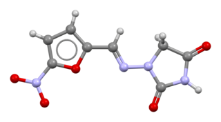
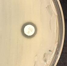
Nitrofurantoin is an antibacterial medication used to treat urinary tract infections, but it is not as effective for kidney infections. It is taken by mouth.

<span class="mw-page-title-main">Coagulase</span> Class of bacterial proteins

Coagulase is a protein enzyme produced by several microorganisms that enables the conversion of fibrinogen to fibrin. In the laboratory, it is used to distinguish between different types of Staphylococcus isolates. Importantly, S. aureus is generally coagulase-positive, meaning that a positive coagulase test would indicate the presence of S. aureus or any of the other 11 coagulase-positive Staphylococci. A negative coagulase test would instead show the presence of coagulase-negative organisms such as S. epidermidis or S. saprophyticus. However, it is now known that not all S. aureus are coagulase-positive. Whereas coagulase-positive Staphylococci are usually pathogenic, coagulase-negative Staphylococci are more often associated with opportunistic infection.

Staphylococcus lugdunensis is a coagulase-negative member of the genus Staphylococcus, consisting of Gram-positive bacteria with spherical cells that appear in clusters.

<i>Staphylococcus haemolyticus</i> Species of bacterium

Staphylococcus haemolyticus is a member of the coagulase-negative staphylococci (CoNS). It is part of the skin flora of humans, and its largest populations are usually found at the axillae, perineum, and inguinal areas. S. haemolyticus also colonizes primates and domestic animals. It is a well-known opportunistic pathogen, and is the second-most frequently isolated CoNS. Infections can be localized or systemic, and are often associated with the insertion of medical devices. The highly antibiotic-resistant phenotype and ability to form biofilms make S. haemolyticus a difficult pathogen to treat. Its most closely related species is Staphylococcus borealis.

Staphylococcus caprae is a Gram-positive, coccus bacteria and a member of the genus Staphylococcus. S. caprae is coagulase-negative. It was originally isolated from goats, but members of this species have also been isolated from human samples.

Staphylococcus hominis is a coagulase-negative member of the bacterial genus Staphylococcus, consisting of Gram-positive, spherical cells in clusters. It occurs very commonly as a harmless commensal on human and animal skin and is known for producing thioalcohol compounds that contribute to body odour. Like many other coagulase-negative staphylococci, S. hominis may occasionally cause infection in patients whose immune systems are compromised, for example by chemotherapy or predisposing illness.

<i>Staphylococcus xylosus</i> Species of bacterium

Staphylococcus xylosus is a species of bacteria belonging to the genus Staphylococcus. It is a Gram-positive bacterium that forms clusters of cells. Like most staphylococcal species, it is coagulase-negative and exists as a commensal on the skin of humans and animals and in the environment.

Staphylococcus warneri is a member of the bacterial genus Staphylococcus, consisting of Gram-positive bacteria with spherical cells appearing in clusters. It is catalase-positive, oxidase-negative, and coagulase-negative, and is a common commensal organism found as part of the skin flora on humans and animals. Like other coagulase-negative staphylococci, S. warneri rarely causes disease, but may occasionally cause infection in patients whose immune system is compromised.

<i>Staphylococcus epidermidis</i> Species of bacterium

Staphylococcus epidermidis is a Gram-positive bacterium, and one of over 40 species belonging to the genus Staphylococcus. It is part of the normal human microbiota, typically the skin microbiota, and less commonly the mucosal microbiota and also found in marine sponges. It is a facultative anaerobic bacteria. Although S. epidermidis is not usually pathogenic, patients with compromised immune systems are at risk of developing infection. These infections are generally hospital-acquired. S. epidermidis is a particular concern for people with catheters or other surgical implants because it is known to form biofilms that grow on these devices. Being part of the normal skin microbiota, S. epidermidis is a frequent contaminant of specimens sent to the diagnostic laboratory.

A nitrite test is a chemical test used to determine the presence of nitrite ion in solution.

Lysostaphin is a Staphylococcus simulans metalloendopeptidase. It can function as a bacteriocin (antimicrobial) against Staphylococcus aureus.

<span class="mw-page-title-main">Bacteriuria</span> Medical condition

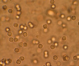
Bacteriuria is the presence of bacteria in urine. Bacteriuria accompanied by symptoms is a urinary tract infection while that without is known as asymptomatic bacteriuria. Diagnosis is by urinalysis or urine culture. Escherichia coli is the most common bacterium found. People without symptoms should generally not be tested for the condition. Differential diagnosis include contamination.

<span class="mw-page-title-main">Staphylococcal infection</span> Medical condition

A staphylococcal infection or staph infection is an infection caused by members of the Staphylococcus genus of bacteria.

<i>Staphylococcus capitis</i> Species of bacterium

Staphylococcus capitis is a coagulase-negative species (CoNS) of Staphylococcus. It is part of the normal flora of the skin of the human scalp, face, neck, scrotum, and ears and has been associated with prosthetic valve endocarditis, but is rarely associated with native valve infection.

<i>Staphylococcus</i> Genus of Gram-positive bacteria

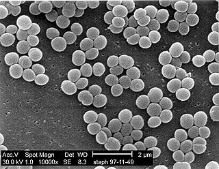
Staphylococcus is a genus of Gram-positive bacteria in the family Staphylococcaceae from the order Bacillales. Under the microscope, they appear spherical (cocci), and form in grape-like clusters. Staphylococcus species are facultative anaerobic organisms.

Staphylococcus gallinarum is a Gram-positive, coagulase-negative member of the bacterial genus Staphylococcus consisting of single, paired, and clustered cocci. Strains of this species were first isolated from chickens and a pheasant. The cells contain cell walls with chemical similarity to those of Staphylococcus epidermidis. Since its initial discovery, S. gallinarum has also been found in the saliva of healthy human adults.

Staphylococcus schleiferi is a Gram-positive, cocci-shaped bacterium of the family Staphylococcaceae. It is facultatively anaerobic, coagulase-variable, and can be readily cultured on blood agar where the bacterium tends to form opaque, non-pigmented colonies and beta (β) hemolysis. There exists two subspecies under the species S. schleiferi: Staphylococcus schleiferi subsp. schleiferi and Staphylococcus schleiferi subsp. coagulans.

Staphylococcus pseudintermedius is a gram positive coccus bacteria of the genus Staphylococcus found worldwide. It is primarily a pathogen for domestic animals, but has been known to affect humans as well. S. pseudintermedius is an opportunistic pathogen that secretes immune modulating virulence factors, has many adhesion factors, and the potential to create biofilms, all of which help to determine the pathogenicity of the bacterium. Diagnoses of Staphylococcus pseudintermedius have traditionally been made using cytology, plating, and biochemical tests. More recently, molecular technologies like MALDI-TOF, DNA hybridization and PCR have become preferred over biochemical tests for their more rapid and accurate identifications. This includes the identification and diagnosis of antibiotic resistant strains.

<span class="mw-page-title-main">Georg Peters</span> German physician

Georg Peters was a German physician, microbiologist and university professor. From 1992 until his fatal mountain accident he headed the Institute of Medical Microbiology at the University of Münster. He was an internationally recognised expert in the field of staphylococci and the infectious diseases caused by them, to which he had devoted himself since the beginning of his scientific career.

References

  1. Schleifer, K. H.; Kloos, W. E. (1975). "Isolation and characterization of Staphylococci from human skin I. Amended descriptions of Staphylococcus epidermidis and Staphylococcus saprophyticus and descriptions of three new species: Staphylococcus cohnii, Staphylococcus haemolyticus, and Staphylococcus xylosus". International Journal of Systematic Bacteriology. 25 (1): 50–61. doi: 10.1099/00207713-25-1-50 . ISSN   0020-7713.
  2. Kuroda, M.; Yamashita, A.; Hirakawa, H.; Kumano, M.; et al. (September 2005). "Whole genome sequence of Staphylococcus saprophyticus reveals the pathogenesis of uncomplicated urinary tract infection". Proc. Natl. Acad. Sci. U.S.A. 102 (37): 13272–7. Bibcode:2005PNAS..10213272K. doi: 10.1073/pnas.0502950102 . PMC   1201578 . PMID   16135568.
  3. 1 2 3 4 5 6 7 Levinson, W. (2010). Review of Medical Microbiology and Immunology (11th ed.). pp. 94–9.
  4. 1 2 Widerström, M.; Wiström, J.; Sjöstedt, A.; Monsen, T. (January 2012). "Coagulase-negative Staphylococci: Update on the molecular epidemiology and clinical presentation, with a focus on Staphylococcus epidermidis and Staphylococcus saprophyticus". European Journal of Clinical Microbiology & Infectious Diseases . 31 (1): 7–20. doi:10.1007/s10096-011-1270-6. PMID   21533877. S2CID   7162887.
  5. Rupp, M. E.; Soper, D. E.; Archer, G. L. (November 1992). "Colonization of the female genital tract with Staphylococcus saprophyticus". Journal of Clinical Microbiology . 30 (11): 2975–9. doi:10.1128/JCM.30.11.2975-2979.1992. PMC   270562 . PMID   1452668.
  6. "Understanding Bladder Infections -- the Basics". WebMD. Retrieved 4 December 2013.
  7. Jordan, P. A.; Iravani, A.; Richard, G. A.; Baer, H. (October 1980). "Urinary tract infection caused by Staphylococcus saprophyticus". The Journal of Infectious Diseases. 142 (4): 510–5. doi:10.1093/infdis/142.4.510. PMID   7192302.
  8. Hovelius, B.; Mårdh, P. A. (May 1984). "Staphylococcus saprophyticus as a common cause of urinary tract infections". Reviews of Infectious Diseases. 6 (3): 328–37. doi:10.1093/clinids/6.3.328. PMID   6377440.
  9. Marrie, T. J.; Kwan, C. (1982). "Antimicrobial susceptibility of Staphylococcus saprophyticus and urethral staphylococci". Antimicrob Agents Chemother. 22 (3): 395–7. doi:10.1128/aac.22.3.395. PMC   183755 . PMID   6982679.
  10. Nostro, A.; Cellini, L.; Di Giulio, M.; D'Arrigo, M.; et al. (September 2012). "Effect of alkaline pH on staphylococcal biofilm formation". APMIS . 120 (9): 733–42. doi:10.1111/j.1600-0463.2012.02900.x. PMID   22882263. S2CID   23267457.
  11. LaPlante, K. L.; Sarkisian, S. A.; Woodmansee, S.; Rowley, D. C.; et al. (September 2012). "Effects of cranberry extracts on growth and biofilm production of Escherichia coli and Staphylococcus species". Phytotherapy Research. 26 (9): 1371–4. doi: 10.1002/ptr.4592 . PMID   22294419. S2CID   5881493.
  12. "Nlss.org.in" (PDF). nlss.org.in. Retrieved 27 February 2019.
  13. Lee, W. H.; Loo, C. Y.; Bebawy, M.; Luk, F.; et al. (July 2013). "Curcumin and its derivatives: Their application in neuropharmacology and neuroscience in the 21st century". Current Neuropharmacology. 11 (4): 338–78. doi:10.2174/1570159X11311040002. PMC   3744901 . PMID   24381528.
  14. Roy, H.; Dare, K.; Ibba, M. (February 2009). "Adaptation of the bacterial membrane to changing environments using aminoacylated phospholipids". Molecular Microbiology. 71 (3): 547–50. doi:10.1111/j.1365-2958.2008.06563.x. PMC   2774118 . PMID   19054327.
  15. "Peterborough Regional Science Fair". peterboroughsciencefair.org. Retrieved 27 February 2019.
  16. Hájek, V.; Meugnier, H.; Bes, M.; Brun, Y.; et al. (July 1996). "Staphylococcus saprophyticus subsp. bovis subsp. nov., isolated from bovine nostrils" (PDF). International Journal of Systematic Bacteriology. 46 (3): 792–6. doi: 10.1099/00207713-46-3-792 . PMID   8782691.