Gram stain

Last updated
Micrograph of a gram-positive coccus and a gram-negative rod. Gram positive coccus and gram negative rod.png
Micrograph of a gram-positive coccus and a gram-negative rod.
A Gram stain of mixed Staphylococcus aureus (S. aureus ATCC 25923, gram-positive cocci, in purple) and Escherichia coli (E. coli ATCC 11775, gram-negative bacilli, in red), the most common Gram stain reference bacteria Gram stain 01.jpg
A Gram stain of mixed Staphylococcus aureus (S. aureus ATCC 25923, gram-positive cocci, in purple) and Escherichia coli (E. coli ATCC 11775, gram-negative bacilli, in red), the most common Gram stain reference bacteria

Gram stain (Gram staining or Gram's method), is a method of staining used to classify bacterial species into two large groups: gram-positive bacteria and gram-negative bacteria. It may also be used to diagnose a fungal infection. [1] The name comes from the Danish bacteriologist Hans Christian Gram, who developed the technique in 1884. [2]

Contents

Gram staining differentiates bacteria by the chemical and physical properties of their cell walls. Gram-positive cells have a thick layer of peptidoglycan in the cell wall that retains the primary stain, crystal violet. Gram-negative cells have a thinner peptidoglycan layer that allows the crystal violet to wash out on addition of ethanol. They are stained pink or red by the counterstain, [3] commonly safranin or fuchsine. Lugol's iodine solution is always added after addition of crystal violet to strengthen the bonds of the stain with the cell membrane.

Gram staining is almost always the first step in the identification of a bacterial group. While Gram staining is a valuable diagnostic tool in both clinical and research settings, not all bacteria can be definitively classified by this technique. This gives rise to gram-variable and gram-indeterminate groups.

History

The method is named after its inventor, the Danish scientist Hans Christian Gram (1853–1938), who developed the technique while working with Carl Friedländer in the morgue of the city hospital in Berlin in 1884. Gram devised his technique not for the purpose of distinguishing one type of bacterium from another but to make bacteria more visible in stained sections of lung tissue. [4] Gram noticed that some bacterial cells possessed noticeable resistance to decolorization. Based off of these observations, Gram developed the initial gram staining procedure, initially making use of Ehrlich’s aniline-gentian violet, Lugol’s iodine, absolute alcohol for decolorization, and Bismarck brown for counterstain. [5] He published his method in 1884, and included in his short report the observation that the typhus bacillus did not retain the stain. [6] Gram did not initially make the distinction between Gram-negative and Gram-positive bacteria using his procedure. [5]

Uses

Gram stain of Candida albicans from a vaginal swab. The small oval chlamydospores are 2-4 um in diameter. Candida Gram stain.jpg
Gram stain of Candida albicans from a vaginal swab. The small oval chlamydospores are 2–4 µm in diameter.

Gram staining is a bacteriological laboratory technique [7] used to differentiate bacterial species into two large groups (gram-positive and gram-negative) based on the physical properties of their cell walls. [8] [ page needed ] Gram staining can also be used to diagnose a fungal infection. [1] Gram staining is not used to classify archaea, since these microorganisms yield widely varying responses that do not follow their phylogenetic groups. [9]

Some organisms are gram-variable (meaning they may stain either negative or positive); some are not stained with either dye used in the Gram technique and are not seen.

Medical

Gram stains are performed on body fluid or biopsy when infection is suspected. Gram stains yield results much more quickly than culturing, and are especially important when infection would make an important difference in the patient's treatment and prognosis; examples are cerebrospinal fluid for meningitis and synovial fluid for septic arthritis. [10] [11]

Staining mechanism

Purple-stained gram-positive (left) and pink-stained gram-negative (right) Gram Staining Bacteria.jpg
Purple-stained gram-positive (left) and pink-stained gram-negative (right)

Gram-positive bacteria have a thick mesh-like cell wall made of peptidoglycan (50–90% of cell envelope), and as a result are stained purple by crystal violet, whereas gram-negative bacteria have a thinner layer (10% of cell envelope), so do not retain the purple stain and are counter-stained pink by safranin. There are four basic steps of the Gram stain:

  1. Applying a primary stain (crystal violet) to a heat-fixed smear of a bacterial culture. Heat fixation kills some bacteria but is mostly used to affix the bacteria to the slide so that they do not rinse out during the staining procedure.
  2. The addition of iodine, which binds to crystal violet and traps it in the cell
  3. Rapid decolorization with ethanol or acetone
  4. Counterstaining with safranin. [12] Carbol fuchsin is sometimes substituted for safranin since it more intensely stains anaerobic bacteria, but it is less commonly used as a counterstain. [13]
Summary of Gram stain
Application ofReagentCell color
Gram-positiveGram-negative
Primary dye crystal violet purplepurple
Trapper iodine purplepurple
Decolorizer alcohol/acetone purplecolorless
Counter stain safranin/carbol fuchsin purplepink or red

Crystal violet (CV) dissociates in aqueous solutions into CV+
and chloride (Cl
) ions. These ions penetrate the cell wall of both gram-positive and gram-negative cells. The CV+
ion interacts with negatively charged components of bacterial cells and stains the cells purple. [14]

Iodide (I
or I
3
) interacts with CV+
and forms large complexes of crystal violet and iodine (CV–I) within the inner and outer layers of the cell. Iodine is often referred to as a mordant, but is a trapping agent that prevents the removal of the CV–I complex and, therefore, colors the cell. [15]

When a decolorizer such as alcohol or acetone is added, it interacts with the lipids of the cell membrane. [16] A gram-negative cell loses its outer lipopolysaccharide membrane, and the inner peptidoglycan layer is left exposed. The CV–I complexes are washed from the gram-negative cell along with the outer membrane. [17] In contrast, a gram-positive cell becomes dehydrated from an ethanol treatment. The large CV–I complexes become trapped within the gram-positive cell due to the multilayered nature of its peptidoglycan. [17] The decolorization step is critical and must be timed correctly; the crystal violet stain is removed from both gram-positive and negative cells if the decolorizing agent is left on too long (a matter of seconds). [18]

After decolorization, the gram-positive cell remains purple and the gram-negative cell loses its purple color. [18] Counterstain, which is usually positively charged safranin or basic fuchsine, is applied last to give decolorized gram-negative bacteria a pink or red color. [3] [19] Both gram-positive bacteria and gram-negative bacteria pick up the counterstain. The counterstain, however, is unseen on gram-positive bacteria because of the darker crystal violet stain.

Examples

Gram-positive bacteria

Gram-stain of gram-positive streptococci surrounded by pus cells Gram-positive bacteria and pus cells.jpg
Gram-stain of gram-positive streptococci surrounded by pus cells

Gram-positive bacteria generally have a single membrane (monoderm) surrounded by a thick peptidoglycan. This rule is followed by two phyla: Bacillota (except for the classes Mollicutes and Negativicutes) and the Actinomycetota. [8] [20] In contrast, members of the Chloroflexota (green non-sulfur bacteria) are monoderms but possess a thin or absent (class Dehalococcoidetes) peptidoglycan and can stain negative, positive or indeterminate; members of the Deinococcota stain positive but are diderms with a thick peptidoglycan. [8] [ page needed ] [20]

The cell wall's strength is enhanced by teichoic acids, glycopolymeric substances embedded within the peptidoglycan. Teichoic acids play multiple roles, such as generating the cell's net negative charge, contributing to cell wall rigidity and shape maintenance, and aiding in cell division and resistance to various stressors, including heat and salt. Despite the density of the peptidoglycan layer, it remains relatively porous, allowing most substances to permeate. For larger nutrients, Gram-positive bacteria utilize exoenzymes, secreted extracellularly to break down macromolecules outside the cell. [21]

Historically, the gram-positive forms made up the phylum Firmicutes, a name now used for the largest group. It includes many well-known genera such as Lactobacillus, Bacillus , Listeria , Staphylococcus , Streptococcus , Enterococcus , and Clostridium . [22] It has also been expanded to include the Mollicutes, bacteria such as Mycoplasma and Thermoplasma that lack cell walls and so cannot be Gram-stained, but are derived from such forms. [23]

Some bacteria have cell walls which are particularly adept at retaining stains. These will appear positive by Gram stain even though they are not closely related to other gram-positive bacteria. These are called acid-fast bacteria, and can only be differentiated from other gram-positive bacteria by special staining procedures. [24]

Gram-negative bacteria

Gram negative Neisseria gonorrhoeae and pus cells Neisseria gonorrhoeae and pus cells Gram stain.jpg
Gram negative Neisseria gonorrhoeae and pus cells

Gram-negative bacteria generally possess a thin layer of peptidoglycan between two membranes (diderm). [25] Lipopolysaccharide (LPS) is the most abundant antigen on the cell surface of most gram-negative bacteria, contributing up to 80% of the outer membrane of E. coli and Salmonella. [26] These LPS molecules, consisting of the O-antigen or O-polysaccharide, core polysaccharide, and lipid A, serve multiple functions including contributing to the cell's negative charge and protecting against certain chemicals. LPS's role is critical in host-pathogen interactions, with the O-antigen eliciting an immune response and lipid A acting as an endotoxin. [21]

Additionally, the outer membrane acts as a selective barrier, regulated by porins, transmembrane proteins forming pores that allow specific molecules to pass. The space between the cell membrane and the outer membrane, known as the periplasm, contains periplasmic enzymes for nutrient processing. A significant structural component linking the peptidoglycan layer and the outer membrane is Braun's lipoprotein, which provides additional stability and strength to the bacterial cell wall. [21]

Most bacterial phyla are gram-negative, including the cyanobacteria, green sulfur bacteria, and most Pseudomonadota (exceptions being some members of the Rickettsiales and the insect-endosymbionts of the Enterobacteriales). [8] [ page needed ] [20]

Gram-variable and gram-indeterminate bacteria

Some bacteria, after staining with the Gram stain, yield a gram-variable pattern: a mix of pink and purple cells are seen. [17] [27] In cultures of Bacillus, Butyrivibrio, and Clostridium, a decrease in peptidoglycan thickness during growth coincides with an increase in the number of cells that stain gram-negative. [27] In addition, in all bacteria stained using the Gram stain, the age of the culture may influence the results of the stain. [27]

Gram-indeterminate bacteria do not respond predictably to Gram staining and, therefore, cannot be determined as either gram-positive or gram-negative. Examples include many species of Mycobacterium , including Mycobacterium bovis, Mycobacterium leprae and Mycobacterium tuberculosis , the latter two of which are the causative agents of leprosy and tuberculosis, respectively. [28] [29] Bacteria of the genus Mycoplasma lack a cell wall around their cell membranes, [10] which means they do not stain by Gram's method and are resistant to the antibiotics that target cell wall synthesis. [30] [31]

Orthographic note

The term Gram staining is derived from the surname of Hans Christian Gram; the eponym (Gram) is therefore capitalized but not the common noun (stain) as is usual for scientific terms. [32] The initial letters of gram-positive and gram-negative, which are eponymous adjectives, can be either capital G or lowercase g, depending on what style guide (if any) governs the document being written. Lowercase style is used by the US Centers for Disease Control and Prevention and other style regimens such as the AMA style. [33] Dictionaries may use lowercase, [34] [35] uppercase, [36] [37] [38] [39] or both. [40] [41] Uppercase Gram-positive or Gram-negative usage is also common in many scientific journal articles and publications. [41] [42] [43] When articles are submitted to journals, each journal may or may not apply house style to the postprint version. Preprint versions contain whichever style the author happened to use. Even style regimens that use lowercase for the adjectives gram-positive and gram-negative still typically use capital for Gram stain.

See also

Related Research Articles

<span class="mw-page-title-main">Cell wall</span> Outermost layer of some cells

A cell wall is a structural layer that surrounds some cell types, found immediately outside the cell membrane. It can be tough, flexible, and sometimes rigid. Primarily, it provides the cell with structural support, shape, protection, and functions as a selective barrier. Another vital role of the cell wall is to help the cell withstand osmotic pressure and mechanical stress. While absent in many eukaryotes, including animals, cell walls are prevalent in other organisms such as fungi, algae and plants, and are commonly found in most prokaryotes, with the exception of mollicute bacteria.

<span class="mw-page-title-main">Gram-positive bacteria</span> Bacteria that give a positive result in the Gram stain test

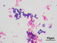
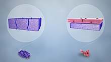
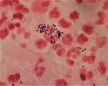
In bacteriology, gram-positive bacteria are bacteria that give a positive result in the Gram stain test, which is traditionally used to quickly classify bacteria into two broad categories according to their type of cell wall.

<span class="mw-page-title-main">Gram-negative bacteria</span> Group of bacteria that do not retain the Gram stain used in bacterial differentiation

Gram-negative bacteria are bacteria that unlike gram-positive bacteria do not retain the crystal violet stain used in the Gram staining method of bacterial differentiation. Their defining characteristic is their cell envelope, which consists of a thin peptidoglycan cell wall sandwiched between an inner (cytoplasmic) membrane and an outer membrane. These bacteria are found in all environments that support life on Earth.

Peptidoglycan or murein is a unique large macromolecule, a polysaccharide, consisting of sugars and amino acids that forms a mesh-like peptidoglycan layer (sacculus) that surrounds the bacterial cytoplasmic membrane. The sugar component consists of alternating residues of β-(1,4) linked N-acetylglucosamine (NAG) and N-acetylmuramic acid (NAM). Attached to the N-acetylmuramic acid is an oligopeptide chain made of three to five amino acids. The peptide chain can be cross-linked to the peptide chain of another strand forming the 3D mesh-like layer. Peptidoglycan serves a structural role in the bacterial cell wall, giving structural strength, as well as counteracting the osmotic pressure of the cytoplasm. This repetitive linking results in a dense peptidoglycan layer which is critical for maintaining cell form and withstanding high osmotic pressures, and it is regularly replaced by peptidoglycan production. Peptidoglycan hydrolysis and synthesis are two processes that must occur in order for cells to grow and multiply, a technique carried out in three stages: clipping of current material, insertion of new material, and re-crosslinking of existing material to new material.

<span class="mw-page-title-main">Staining</span> Technique used to enhance visual contrast of specimens observed under a microscope

Staining is a technique used to enhance contrast in samples, generally at the microscopic level. Stains and dyes are frequently used in histology, in cytology, and in the medical fields of histopathology, hematology, and cytopathology that focus on the study and diagnoses of diseases at the microscopic level. Stains may be used to define biological tissues, cell populations, or organelles within individual cells.

<span class="mw-page-title-main">Teichoic acid</span>

Teichoic acids are bacterial copolymers of glycerol phosphate or ribitol phosphate and carbohydrates linked via phosphodiester bonds.

The periplasm is a concentrated gel-like matrix in the space between the inner cytoplasmic membrane and the bacterial outer membrane called the periplasmic space in gram-negative bacteria. Using cryo-electron microscopy it has been found that a much smaller periplasmic space is also present in gram-positive bacteria, between cell wall and the plasma membrane. The periplasm may constitute up to 40% of the total cell volume of gram-negative bacteria, but is a much smaller percentage in gram-positive bacteria.

The cell envelope comprises the inner cell membrane and the cell wall of a bacterium. In gram-negative bacteria an outer membrane is also included. This envelope is not present in the Mollicutes where the cell wall is absent.

<span class="mw-page-title-main">Acid-fastness</span> Physical property of certain bacterial and eukaryotic cells

Acid-fastness is a physical property of certain bacterial and eukaryotic cells, as well as some sub-cellular structures, specifically their resistance to decolorization by acids during laboratory staining procedures. Once stained as part of a sample, these organisms can resist the acid and/or ethanol-based decolorization procedures common in many staining protocols, hence the name acid-fast.

<span class="mw-page-title-main">Bacterial capsule</span> Polysaccharide layer that lies outside the cell envelope in many bacteria

The bacterial capsule is a large structure common to many bacteria. It is a polysaccharide layer that lies outside the cell envelope, and is thus deemed part of the outer envelope of a bacterial cell. It is a well-organized layer, not easily washed off, and it can be the cause of various diseases.

<span class="mw-page-title-main">Counterstain</span>

A counterstain is a stain with colour contrasting to the principal stain, making the stained structure easily visible using a microscope.

Differential staining is a staining process which uses more than one chemical stain. Using multiple stains can better differentiate between different microorganisms or structures/cellular components of a single organism.

A bacterium, despite its simplicity, contains a well-developed cell structure which is responsible for some of its unique biological structures and pathogenicity. Many structural features are unique to bacteria and are not found among archaea or eukaryotes. Because of the simplicity of bacteria relative to larger organisms and the ease with which they can be manipulated experimentally, the cell structure of bacteria has been well studied, revealing many biochemical principles that have been subsequently applied to other organisms.

<span class="mw-page-title-main">Bacteria</span> Domain of microorganisms

Bacteria are ubiquitous, mostly free-living organisms often consisting of one biological cell. They constitute a large domain of prokaryotic microorganisms. Typically a few micrometres in length, bacteria were among the first life forms to appear on Earth, and are present in most of its habitats. Bacteria inhabit soil, water, acidic hot springs, radioactive waste, and the deep biosphere of Earth's crust. Bacteria play a vital role in many stages of the nutrient cycle by recycling nutrients and the fixation of nitrogen from the atmosphere. The nutrient cycle includes the decomposition of dead bodies; bacteria are responsible for the putrefaction stage in this process. In the biological communities surrounding hydrothermal vents and cold seeps, extremophile bacteria provide the nutrients needed to sustain life by converting dissolved compounds, such as hydrogen sulphide and methane, to energy. Bacteria also live in mutualistic, commensal and parasitic relationships with plants and animals. Most bacteria have not been characterised and there are many species that cannot be grown in the laboratory. The study of bacteria is known as bacteriology, a branch of microbiology.

<span class="mw-page-title-main">Gracilicutes</span> Infrakingdom of bacteria

Gracilicutes is a clade in bacterial phylogeny.

<span class="mw-page-title-main">Lysin</span>

Lysins, also known as endolysins or murein hydrolases, are hydrolytic enzymes produced by bacteriophages in order to cleave the host's cell wall during the final stage of the lytic cycle. Lysins are highly evolved enzymes that are able to target one of the five bonds in peptidoglycan (murein), the main component of bacterial cell walls, which allows the release of progeny virions from the lysed cell. Cell-wall-containing Archaea are also lysed by specialized pseudomurein-cleaving lysins, while most archaeal viruses employ alternative mechanisms. Similarly, not all bacteriophages synthesize lysins: some small single-stranded DNA and RNA phages produce membrane proteins that activate the host's autolytic mechanisms such as autolysins.

Atypical bacteria are bacteria that do not get colored by gram-staining but rather remain colorless: they are neither Gram-positive nor Gram-negative. These include the Chlamydiaceae, Legionella and the Mycoplasmataceae ; the Spirochetes and Rickettsiaceae are also often considered atypical.

Sortases are membrane anchored enzyme that sort these surface proteins onto the bacterial cell surface and anchor them to the peptidoglycan. There are different types of sortases and each catalyse the anchoring of different proteins to cell walls.

<span class="mw-page-title-main">Outer membrane vesicle</span> Vesicles released from the outer membranes of Gram-negative bacteria

Outer membrane vesicles (OMVs) are vesicles released from the outer membranes of Gram-negative bacteria. While Gram-positive bacteria release vesicles as well those vesicles fall under the broader category of bacterial membrane vesicles (MVs). OMVs were the first MVs to be discovered, and are distinguished from outer inner membrane vesicles (OIMVS), which are gram-negative baterial vesicles containing portions of both the outer and inner bacterial membrane. Outer membrane vesicles were first discovered and characterized using transmission-electron microscopy by Indian Scientist Prof. Smriti Narayan Chatterjee and J. Das in 1966-67. OMVs are ascribed the functionality to provide a manner to communicate among themselves, with other microorganisms in their environment and with the host. These vesicles are involved in trafficking bacterial cell signaling biochemicals, which may include DNA, RNA, proteins, endotoxins and allied virulence molecules. This communication happens in microbial cultures in oceans, inside animals, plants and even inside the human body.

<span class="mw-page-title-main">Bacterial secretion system</span> Protein complexes present on the cell membranes of bacteria for secretion of substances

Bacterial secretion systems are protein complexes present on the cell membranes of bacteria for secretion of substances. Specifically, they are the cellular devices used by pathogenic bacteria to secrete their virulence factors to invade the host cells. They can be classified into different types based on their specific structure, composition and activity. Generally, proteins can be secreted through two different processes. One process is a one-step mechanism in which proteins from the cytoplasm of bacteria are transported and delivered directly through the cell membrane into the host cell. Another involves a two-step activity in which the proteins are first transported out of the inner cell membrane, then deposited in the periplasm, and finally through the outer cell membrane into the host cell.

References

  1. 1 2 "Gram Stain: MedlinePlus Medical Test". medlineplus.gov.
  2. Colco, R. (2005). "Gram Staining". Current Protocols in Microbiology . Appendix 3 (1): Appendix 3C. doi:10.1002/9780471729259.mca03cs00. ISBN   978-0471729259. PMID   18770544. S2CID   32452815.
  3. 1 2 Beveridge, T. J.; Davies, J. A. (November 1983). "Cellular responses of Bacillus subtilis and Escherichia coli to the Gram stain". Journal of Bacteriology. 156 (2): 846–58. doi:10.1128/JB.156.2.846-858.1983. PMC   217903 . PMID   6195148.
  4. Austrian, R. (1960). "The Gram stain and the etiology of lobar pneumonia, an historical note". Bacteriological Reviews. 24 (3): 261–265. doi:10.1128/MMBR.24.3.261-265.1960. PMC   441053 . PMID   13685217.
  5. 1 2 Bartholomew, James W.; Mittwer, Tod (March 1952). "THE GRAM STAIN". Bacteriological Reviews. 16 (1): 1–29. doi:10.1128/br.16.1.1-29.1952. ISSN   0005-3678. PMC   180726 . PMID   14925025.
  6. Gram, Hans Christian (1884). "Über die isolierte Färbung der Schizomyceten in Schnitt- und Trockenpräparaten". Fortschritte der Medizin (in German). 2: 185–189..
    English translation in: Brock, T. D. (1999). Milestones in Microbiology 1546–1940 (2nd ed.). ASM Press. pp. 215–218. ISBN   978-1-55581-142-6.
    Translation is also at: Brock, T. D. "Pioneers in Medical Laboratory Science: Christian Gram 1884". HOSLink.com. Retrieved 27 July 2010.
  7. Ryan, K. J.; Ray, C. G., eds. (2004). Sherris Medical Microbiology (4th ed.). McGraw Hill. pp. 232f. ISBN   978-0838585290.
  8. 1 2 3 4 Madigan, M. T.; Martinko, J.; Parker, J. (2004). Brock Biology of Microorganisms (10th ed.). Lippincott Williams & Wilkins. ISBN   978-0-13-066271-2.
  9. Beveridge, T. J. (2001). "Use of the Gram stain in microbiology". Biotechnic & Histochemistry. 76 (3): 111–118. doi:10.1080/714028139. PMID   11475313.
  10. 1 2 Ryan KJ, Ray CG (editors) (2004). Sherris Medical Microbiology (4th ed.). McGraw Hill. pp. 409–12. ISBN   978-0-8385-8529-0.{{cite book}}: |author= has generic name (help)
  11. Søgaard, M.; Nørgaard, M.; Schønheyder, H. (2007). "First notification of positive blood cultures: High accuracy of the Gram stain report". Journal of Clinical Microbiology. 45 (4): 1113–1117. doi:10.1128/JCM.02523-06. PMC   1865800 . PMID   17301283.
  12. Black, Jacquelyn G. (1993). Microbiology: Principles and Explorations. Prentice Hall. p. 65.
  13. "Medical Chemical Corporation". Med-Chem.com. Retrieved 9 March 2016.
  14. Leboffe, Michael (2014). Microbiology Laboratory Theory and Application (3rd ed.). Englewood, Colorado: Morton Publishing Company. p. 105. ISBN   978-1617312809.
  15. "Stain theory – What a mordant is not". StainsFile.info. Archived from the original on 9 March 2016. Retrieved 9 March 2016.
  16. "Gram Stain". Microbugz. Austin Community College. Retrieved 2017-05-26.
  17. 1 2 3 Tim, Sandle (21 October 2015). Pharmaceutical Microbiology: Essentials for Quality Assurance and Quality Control. Elsevier Science. ISBN   9780081000229. OCLC   923807961.
  18. 1 2 Hardy, Jay; Maria, Santa. "Gram's Serendipitous Stain" (PDF). Hardy's Diagnostics. Archived (PDF) from the original on 2017-03-24.
  19. Davies, J. A.; Anderson, G. K.; Beveridge, T. J.; Clark, H. C. (November 1983). "Chemical mechanism of the Gram stain and synthesis of a new electron-opaque marker for electron microscopy, which replaces the iodine mordant of the stain". Journal of Bacteriology. 156 (2): 837–845. doi:10.1128/JB.156.2.837-845.1983. PMC   217902 . PMID   6195147.
  20. 1 2 3 Brenner, Don J.; Krieg, Noel R.; Staley, James T. (26 July 2005) [1984]. Garrity, George M. (ed.). Introductory Essays. Bergey's Manual of Systematic Bacteriology. Vol. 2A (2nd ed.). New York: Springer. p. 304. ISBN   978-0-387-24143-2. British Library no. GBA561951.
  21. 1 2 3 Bruslind, Linda (2019-08-01). General Microbiology. Oregon State University.
  22. Galperin, Michael Y. (27 December 2013). "Genome Diversity of Spore-forming Firmicutes". Microbiology Spectrum. 1 (2): TBS-0015-2012-. doi:10.1128/microbiolspectrum.tbs-0015-2012. ISSN   2165-0497. PMC   4306282 . PMID   26184964.
  23. Hashem, Hams H. "Practical Medical Microbiology". University of Al-Qadisiya.
  24. "The Acid Fast Stain". www2.Highlands.edu. Georgia Highlands College. Archived from the original on 10 June 2017. Retrieved 10 June 2017.
  25. Megrian D, Taib N, Witwinowski J, Gribaldo S (2020). "One or two membranes? Diderm Firmicutes challenge the Gram-positive/Gram-negative divide". Molecular Microbiology . 113 (3): 659–671. doi: 10.1111/mmi.14469 . PMID   31975449. S2CID   210882600.
  26. Avila-Calderón ED, Ruiz-Palma MD, Contreras-Rodríguez A (2021). "Outer Membrane Vesicles of Gram-Negative Bacteria: An Outlook on Biogenesis". Frontiers in Microbiology . 12: 557902. doi: 10.3389/fmicb.2021.557902 . PMC   7969528 . PMID   33746909.
  27. 1 2 3 Beveridge, Terry J. (March 1990). "Mechanism of Gram Variability in Select Bacteria". Journal of Bacteriology. 172 (3): 1609–1620. doi:10.1128/jb.172.3.1609-1620.1990. PMC   208639 . PMID   1689718.
  28. Black, Jacquelyn (2012). Microbiology: Principles and Exploration (8th ed.). John Wiley & Sons. p. 68. ISBN   978-0-470-54109-8.
  29. Reynolds, J.; Moyes, R. B.; Breakwell, D. P. (2009). "Appendix 3". Differential staining of bacteria: Acid fast stain. Vol. Appendix 3. pp. H. doi:10.1002/9780471729259.mca03hs15. ISBN   978-0471729259. PMID   19885935. S2CID   45685776.{{cite book}}: |journal= ignored (help)
  30. Lee EH, Winter HL, van Dijl JM, Metzemaekers JD, Arends JP (December 2012). "Diagnosis and antimicrobial therapy of Mycoplasma hominis meningitis in adults". International Journal of Medical Microbiology. 302 (7–8): 289–92. doi:10.1016/j.ijmm.2012.09.003. PMID   23085510.
  31. Gautier-Bouchardon AV (July 2018). "Antimicrobial Resistance in Mycoplasma spp". Microbiology Spectrum. 6 (4): 425–446. doi:10.1128/microbiolspec.ARBA-0030-2018. ISBN   9781555819798. PMID   30003864. S2CID   51616821.
  32. Waddingham, Anne (28 August 2014). New Hart's Rules: The Oxford Style Guide. Oxford University Press. p. 105. ISBN   978-0199570027.
  33. "Preferred Usage". Emerging Infectious Diseases Style Guide. Centers for Disease Control and Prevention.
  34. "Dorland's Illustrated Medical Dictionary" (32nd ed.). Elsevier. Retrieved 5 June 2020. Use search terms such as gram-negative.
  35. "gram–positive", Merriam-Webster, Encyclopædia Britannica, Inc.
  36. "Gram-positive". CollinsDictionary.com. HarperCollins.
  37. "Gram stain". Lexico.com. Oxford University Press. Archived from the original on June 5, 2020.
  38. "Gram-positive". MedicineNet.
  39. "Gram negative/positive". BusinessDictionary.com. Archived from the original on 2016-10-20. Retrieved 2016-10-20.
  40. "gram-pos·i·tive or Gram-pos·i·tive". The American Heritage Dictionary. Houghton Mifflin.
  41. 1 2 "Gram-positive". Dictionary.com.
  42. Brown, Lisa; Wolf, Julie M.; Prados-Rosales, Rafael; Casadevall, Arturo (2015). "Through the wall: Extracellular vesicles in Gram-positive bacteria, mycobacteria and fungi". Nature Reviews Microbiology. 13 (10): 620–630. doi:10.1038/nrmicro3480. PMC   4860279 . PMID   26324094.
  43. Mueller, Kristen L. (12 June 2015). "Detecting Gram-negative bacteria". Science. 348 (6240): 1218. doi:10.1126/science.348.6240.1218-o.