Isolation (microbiology)

Last updated

In microbiology, the term isolation refers to the separation of a strain from a natural, mixed population of living microbes, as present in the environment, for example in water or soil, or from living beings with skin flora, oral flora or gut flora, in order to identify the microbe(s) of interest. [1] Historically, the laboratory techniques of isolation first developed in the field of bacteriology and parasitology (during the 19th century), before those in virology during the 20th century.

Contents

History

The laboratory techniques of isolating microbes first developed during the 19th century in the field of bacteriology and parasitology using light microscopy. 1860 marked the successful introduction of liquid medium by Louis Pasteur. The liquid culture pasteur developed allowed for the visulization of promoting or inhibiting growth of specific bacteria. This same technique is utilized today through various mediums like Mannitol salt agar, a solid medium. Solid cultures were developed in 1881 when Robert Koch solidified the liquid media through the addition of agar [2]

Proper isolation techniques of virology did not exist prior to the 20th century. The methods of microbial isolation have drastically changed over the past 50 years, from a labor perspective with increasing mechanization, and in regard to the technologies involved, and with it speed and accuracy.

General techniques

Bacteria extracted from soil sample. SoilFlora.jpg
Bacteria extracted from soil sample.

In order to isolate a microbe from a natural, mixed population of living microbes, as present in the environment, for example in water or soil flora, or from living beings with skin flora, oral flora or gut flora, one has to separate it from the mix.

Traditionally microbes have been cultured in order to identify the microbe(s) of interest based on its growth characteristics. Depending on the expected density and viability of microbes present in a liquid sample, physical methods to increase the gradient as for example serial dilution or centrifugation may be chosen. In order to isolate organisms in materials with high microbial content, such as sewage, soil or stool, serial dilutions will increase the chance of separating a mixture.

In a liquid medium with few or no expected organisms, from an area that is normally sterile (such as CSF, blood inside the circulatory system) centrifugation, decanting the supernatant and using only the sediment will increase the chance to grow and isolate bacteria or the usually cell-associated viruses.

If one expects or looks for a particularly fastidious organism, the microbiological culture and isolation techniques will have to be geared towards that microbe. For example, a bacterium that dies when exposed to air, can only be isolated if the sample is carried and processed under airless or anaerobic conditions. A bacterium that dies when exposed to room temperature (thermophilic) requires a pre-warmed transport container, and a microbe that dries and dies when carried on a cotton swab will need a viral transport medium before it can be cultured successfully.

Bacterial and fungal culture

Inoculation

Laboratory technicians inoculate the sample onto certain solid agar plates with the streak plate method or into liquid culture medium, depending what the objective of the isolation is:

Incubation

After the sample is inoculated into or onto the choice media, they are incubated under the appropriate atmospheric settings, such as aerobic, anaerobic or microaerophilic conditions or with added carbon dioxide (5%), at different temperature settings, for example 37 °C in an incubator or in a refrigerator for cold enrichment, under appropriate light, for example strictly without light wrapped in paper or in a dark bottle for scotochromogen mycobacteria, and for different lengths of time, because different bacteria grow at a different speed, varying from hours (Escherichia coli) to weeks (e.g.mycobacteria).

At regular, serial intervals laboratory technicians and microbiologists inspect the media for signs of visible growth and record it. The inspection again has to occur under conditions favoring the isolate's survival, i.e. in an 'anaerobic chamber' for anaerobe bacteria for example, and under conditions that do not threaten the person looking at the plates from being infected by a particularly infectious microbe, i.e. under a biological safety cabinet for Yersinia pestis (plague) or Bacillus anthracis (anthrax) for example.

Identification

When bacteria have visibly grown, they are often still mixed. The identification of a microbe depends upon the isolation of an individual colony, as biochemical testing of a microbe to determine its different physiological features depends on a pure culture. To make a subculture, one again works in aseptic technique in microbiology, lifting a single colony off the agar surface with a loop and streaks the material into the 4 quadrants of an agar plate or all over if the colony was singular and did not look mixed.

Example of gram staining on a gram positive rod. GramPositiveStain.jpg
Example of gram staining on a gram positive rod.

Gram staining allows for visualization of the bacteria's cell wall composition based on the color the bacteria stains after a serious of staining and decolorization steps. [4] This staining process allows for the identification of gram-negative and gram positive bacteria. Gram-negative bacteria will stain a pink color due to the thin layer of peptidoglycan. If a bacteria stains purple, due to the thick layer of peptidoglycan, the bacteria is a gram-positive bacteria. [4]

In clinical microbiology numerous other staining techniques for particular organisms are used (acid fast bacterial stain for mycobacteria). Immunological staining techniques, such as direct immunofluorescence have been developed for medically important pathogens that are slow growing (Auramine-rhodamine stain for mycobacteria) or difficult to grow (such as Legionella pneumophila species) and where the test result would alter standard management and empirical therapy.

Biochemical testing of bacteria involves a set of agars in vials to separate motile from non-motile bacteria. In 1970 a miniaturized version was developed, called the analytical profile index.

Successful identification via e.g. genome sequencing and genomics depends on pure cultures.

Bacteria, culture-independent

While the most rapid method to identify bacteria is by sequencing their 16S rRNA gene, which has been PCR-amplified beforehand, this method does not require isolation. Since most bacteria cannot be grown with conventional methods (particularly environmental or soil bacteria) metagenomics or metatranscriptomics are used, shotgun sequencing or PCR directed sequencing of the genome. Sequencing with mass spectrometry as in Matrix-assisted laser desorption/ionization (MALDI-TOF MS) is used in the analysis of clinical specimens to look for pathogens. Whole genome sequencing is an option for a singular organism that cannot be sufficiently characterized for identification. Small DNA microarrays can also be used for identification.

Related Research Articles

<span class="mw-page-title-main">Bacteriology</span> Subdiscipline of microbiology that studies bacteria

Bacteriology is the branch and specialty of biology that studies the morphology, ecology, genetics and biochemistry of bacteria as well as many other aspects related to them. This subdivision of microbiology involves the identification, classification, and characterization of bacterial species. Because of the similarity of thinking and working with microorganisms other than bacteria, such as protozoa, fungi, and viruses, there has been a tendency for the field of bacteriology to extend as microbiology. The terms were formerly often used interchangeably. However, bacteriology can be classified as a distinct science.

<span class="mw-page-title-main">Agar plate</span> Petri dish with agar used to culture microbes

An agar plate is a Petri dish that contains a growth medium solidified with agar, used to culture microorganisms. Sometimes selective compounds are added to influence growth, such as antibiotics.

<span class="mw-page-title-main">Microbiological culture</span> Method of allowing microorganisms to multiply in a controlled medium

A microbiological culture, or microbial culture, is a method of multiplying microbial organisms by letting them reproduce in predetermined culture medium under controlled laboratory conditions. Microbial cultures are foundational and basic diagnostic methods used as research tools in molecular biology.

<span class="mw-page-title-main">Bacteriological water analysis</span>

Bacteriological water analysis is a method of analysing water to estimate the numbers of bacteria present and, if needed, to find out what sort of bacteria they are. It represents one aspect of water quality. It is a microbiological analytical procedure which uses samples of water and from these samples determines the concentration of bacteria. It is then possible to draw inferences about the suitability of the water for use from these concentrations. This process is used, for example, to routinely confirm that water is safe for human consumption or that bathing and recreational waters are safe to use.

<span class="mw-page-title-main">Blood culture</span> Test to detect bloodstream infections

A blood culture is a medical laboratory test used to detect bacteria or fungi in a person's blood. Under normal conditions, the blood does not contain microorganisms: their presence can indicate a bloodstream infection such as bacteremia or fungemia, which in severe cases may result in sepsis. By culturing the blood, microbes can be identified and tested for resistance to antimicrobial drugs, which allows clinicians to provide an effective treatment.

<span class="mw-page-title-main">Eosin methylene blue</span> Biological stain

Eosin methylene blue is a selective and differential media used for the identification of Gram-negative bacteria, specifically the Enterobacteriaceae. EMB inhibits the growth of most Gram-positive bacteria. EMB is often used to confirm the presence of coliforms in a sample. It contains two dyes, eosin and methylene blue in the ratio of 6:1. EMB is a differential microbiological media, which inhibits the growth of Gram-positive bacteria and differentiates bacteria that ferment lactose from those that do not. Organisms that ferment lactose appear dark/black or green often with "nucleated colonies"—colonies with dark centers. Organisms that do not ferment lactose will appear pink and often mucoid.

<span class="mw-page-title-main">Growth medium</span> Solid, liquid or gel used to grow microorganisms or cells

A growth medium or culture medium is a solid, liquid, or semi-solid designed to support the growth of a population of microorganisms or cells via the process of cell proliferation or small plants like the moss Physcomitrella patens. Different types of media are used for growing different types of cells.

<span class="mw-page-title-main">MacConkey agar</span> Differential media

MacConkey agar is a selective and differential culture medium for bacteria. It is designed to selectively isolate Gram-negative and enteric bacteria and differentiate them based on lactose fermentation. Lactose fermenters turn red or pink on MacConkey agar, and nonfermenters do not change color. The media inhibits growth of Gram-positive organisms with crystal violet and bile salts, allowing for the selection and isolation of gram-negative bacteria. The media detects lactose fermentation by enteric bacteria with the pH indicator neutral red.

The oxidase test is used to determine whether an organism possesses the cytochrome c oxidase enzyme. The test is used as an aid for the differentiation of Neisseria, Moraxella, Campylobacter and Pasteurella species. It is also used to differentiate pseudomonads from related species.

<span class="mw-page-title-main">Streaking (microbiology)</span> Method for isolation of bacterial strains

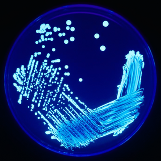
In microbiology, streaking is a technique used to isolate a pure strain from a single species of microorganism, often bacteria. Samples can then be taken from the resulting colonies and a microbiological culture can be grown on a new plate so that the organism can be identified, studied, or tested.

<span class="mw-page-title-main">Medical microbiology</span> Branch of medical science

Medical microbiology, the large subset of microbiology that is applied to medicine, is a branch of medical science concerned with the prevention, diagnosis and treatment of infectious diseases. In addition, this field of science studies various clinical applications of microbes for the improvement of health. There are four kinds of microorganisms that cause infectious disease: bacteria, fungi, parasites and viruses, and one type of infectious protein called prion.

Mycobacterium elephantis, a bacterium of the family Mycobacteriaceae, was discovered and isolated from a deceased elephant near India and may be linked to respiratory dysfunction. Organisms in the genus Mycobacterium are known to be aerobic and non-motile. Organisms within Mycobacterium belong to either the rapid growing group or the slow growing group. M. elephantis is classified as a rapid grower and relates most closely to Mycobacterium confluentis and Mycobacterium phlei.

Plate count agar (PCA), also called standard methods agar (SMA), is a microbiological growth medium commonly used to assess or to monitor "total" or viable bacterial growth of a sample. PCA is not a selective medium.

<span class="mw-page-title-main">Löwenstein–Jensen medium</span> Growth medium used to culture Mycobacterium species

Löwenstein–Jensen medium, more commonly known as LJ medium, is a growth medium specially used for culture of Mycobacterium species, notably Mycobacterium tuberculosis.

Victivallis vadensis is a Gram-negative, coccus-shaped, bacteria found in the human digestive tract. It measures approximately 0.5-1.3 micrometers in diameter, is non-motile and chemoorganotrophic, and does not form spores. Victivallis vadensis is strictly anaerobic, as are 90 percent of the bacteria in the human gastrointestinal system.

<span class="mw-page-title-main">New York City agar</span>

The NYC medium or GC medium agar is used for isolating Gonococci.

<span class="mw-page-title-main">Inoculation needle</span>

An inoculation needle is a laboratory equipment used in the field of microbiology to transfer and inoculate living microorganisms. It is one of the most commonly implicated biological laboratory tools and can be disposable or re-usable. A standard reusable inoculation needle is made from nichrome or platinum wire affixed to a metallic handle. A disposable inoculation needle is often made from plastic resin. The base of the needle is dulled, resulting in a blunted end.

Diagnostic microbiology is the study of microbial identification. Since the discovery of the germ theory of disease, scientists have been finding ways to harvest specific organisms. Using methods such as differential media or genome sequencing, physicians and scientists can observe novel functions in organisms for more effective and accurate diagnosis of organisms. Methods used in diagnostic microbiology are often used to take advantage of a particular difference in organisms and attain information about what species it can be identified as, which is often through a reference of previous studies. New studies provide information that others can reference so that scientists can attain a basic understanding of the organism they are examining.

Propionispira raffinosivorans is a motile, obligate anaerobic, gram-negative bacteria. It was originally isolated from spoiled beer and believed to have some causative effect in beer spoilage. Since then, it has been taxonomically reclassified and proven to play a role in anaerobic beer spoilage, because of its production of acids, such as acetic and propionic acid, during fermentation

<span class="mw-page-title-main">Colonial morphology</span> Examination of microbial colonies

In microbiology, colonial morphology refers to the visual appearance of bacterial or fungal colonies on an agar plate. Examining colonial morphology is the first step in the identification of an unknown microbe. The systematic assessment of the colonies' appearance, focusing on aspects like size, shape, colour, opacity, and consistency, provides clues to the identity of the organism, allowing microbiologists to select appropriate tests to provide a definitive identification.

References

  1. "Isolation and Cultivation of Microorganisms". Biology Discussion. 2016-06-02. Retrieved 2023-04-21.
  2. Bonnet, M.; Lagier, J. C.; Raoult, D.; Khelaifia, S. (2020). "Bacterial culture through selective and non-selective conditions: the evolution of culture media in clinical microbiology". New Microbes and New Infections. 34: 100622. doi:10.1016/j.nmni.2019.100622. ISSN   2052-2975. PMC   6961714 . PMID   31956419.
  3. Alfred Brown, Heidi Smith (2012). Benson's Microbiological Applications: Laboratory Manual in General Microbiology (Thirteenth ed.). McGraw-Hill Education. ISBN   9780073402413.
  4. 1 2 "Gram Stain Protocols". ASM.org. Retrieved 2023-04-21.